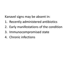
Tenosynovitis
• Definition
• Etiology
• Prognosis
• Pathophysiology
• History
• Physical examination
• Work up
• Treatment
• Postoperative care
• Rare disorders
Tenosynovitis definition
• A group of entities with a common pathology
involving the extrinsic tendons of the hand
and wrist and their corresponding retinacular
sheaths.
• Burman M. Stenosing tendovaginitis of the dorsal and volar
compartments of the wrist. AMA Arch Surg. 1952
Nov;65(5):752-62
• Start as tendon
irritation and pain
• Progress into
catching and locking
when tendon glides
• Synonyms:
– Tendinitis
– Tendovaginitis
• Misleading names
• Tendinosis, most appropriate descriptor
Etiology
Noninfectious causes
• Diabetes mellitus
• Rheumatoid arthritis
• Crystalline deposition
• Overuse syndromes
• Amyloidosis
• Ochronosis
• Psoriatic arthritis
• Systemic lupus erythematosus
• Sarcoidosis
Overuse injury
• De Quervain tenosynovitis
• Volar flexor tenosynovitis (ie, trigger finger)
Nongonococcal infectious
tenosynovitis
• Staphylococcus aureus and Streptococcus species
- most common etiologic agents
• Pasteurella multocida
• Eikenella corrodens - Higher incidence with
human bite wounds
• Anaerobes -
Bacteroides and Fusobacterium species most
common
• Haemophilus species
• Capnocytophaga canimorsus
• Miscellaneous gram-negative organisms
• Mycobacterium tuberculosis
• Other Mycobacterium species
• Clostridium difficile
• Pseudomonas aeruginosa
• Listeria monocytogenes
• Vibrio vulnificus
Gonococcal tenosynovitis
• Neisseria gonorrhoeae (originates as a
mucosal infection of the genital tract, rectum,
or pharynx).
Pyogenic flexor tenosynovitis
• Infectious agent multiplying in
the closed space of the flexor
tendon sheath and culture-
rich synovial fluid medium.
• Natural immune response
mechanisms cause swelling
and migration of
inflammatory cells and
mediators
Epidemiology
• One third of all cases of hand and finger FT are
associated with diabetes mellitus
• 64-95% of patients with RA develop hand or
wrist FT
Prognosis
• Good prognosis
– Present early
– No comorbidities.
• Long-term complications and impairment
– Fulminant infection
– Chronic infection
– Impaired immune status
Complications
• Loss of range of motion (ROM) secondary to
adhesions (most common)
• Soft-tissue necrosis
• Flexor tendon rupture
Risk factors were associated with poorer
outcomes:
1. Age over 45 years
2. Presence of diabetes mellitus, renal failure,
or peripheral vascular disease
3. Ischemic changes at the time of presentation
4. Subcutaneous purulence
5. Polymicrobial infection at the time of surgery
• FT that is diagnosed by magnetic resonance
imaging (MRI) is a strong predictor of early RA
• Among patients with stenosing FT, those with
diabetes have a higher prevalence of multiple
joint involvement than do those without
diabetes
Pathophysiology
Inflammatory flexor tenosynovitis
Inflammatory
stage
Starts
immediately
48 hours to 2
weeks
Release of chemotactic and
vasoactive substances; the
resulting inflammatory cells
create pain, swelling,
erythema, and warmth
Proliferative
stage
Lasts up to 2
weeks
Production of collagen and
ground substances; the
tendon is extremely
vulnerable to injury
Maturation
stage
Lasts up to 12
weeks
Healing phases are
completed. if the
inflammatory response is
reinitiated at this time,
fibrosis can result
Infectious flexor tenosynovitis
• Closed-space infection.
• Tendon sheath
– inner visceral layer
– outer parietal layer.
• Between the two layers is the synovial space,
which is filled with synovial fluid
Accumulation of
pus in flexor
tendon sheath
infections
Pressure
increases (in
excess of 30 mg
Hg)
Inhibiting the
inflammatory
response.
Inhibits blood flow
Tendon ischemia
increases the likelihood
of tendon necrosis
and rupture
History
Gonococcal
• Teenagers and young adults
• More common in women
• Dorsum of the wrist, hand, and ankle
Nongonococcal infectious
tenosynovitis
• Dry, cracked skin or a puncture wound,
laceration, bite, or high-pressure injection
injury
• Flexor hand tendons
De Quervain tenosynovitis
• Pain in the radial aspect of the wrist
• Worse with activity and better with rest
• History of repetitive pinching motion of the
thumb and fingers
• Middle-aged women
• No history of acute trauma
Volar flexor tenosynovitis
• Thumb or ring finger
• Middle-aged women
• Diabetics
• Locking of the involved finger in flexion is
followed by sudden release (hence the name
trigger finger)
• Hand pain radiates to fingers
Physical Examination
Infectious tenosynovitis
Kanavel signs may be absent in:
1. Recently administered antibiotics
2. Early manifestations of the condition
3. Immunocompromised state
4. Chronic infections
Volar flexor tenosynovitis
• Tenderness at the proximal end of the tendon sheath, in
the distal palm (just proximal to the metacarpal head)
• Palpable tendon thickening and nodularity may be present
• Crepitation and catching of the tendon may be
appreciated when the finger is flexed
De Quervain tenosynovitis
• Pain occurs on palpation along
the radial aspect of the wrist
• Pain occurs with passive ROM
of the thumb
• Pain occurs with ulnar
deviation of the wrist with the
thumb cupped in a closed fist
• Swelling is most common initial finding.
• As the tissue expands and impingement
occurs, pain and restricted motion.
• Delayed presentations: fulminant FT with all
Kanavel signs or tendon rupture.
Differential Diagnoses
• Herpetic whitlow
• Pyarthrosis
• Gout
• Pseudogout
• Dactylitis
• Phalanx fracture
• Arthritis
• Osteoarthritis
• Subcutaneous abscess
• Sesamoiditis and angiolipoma
• Hand infections
• Hand injury, high pressure
• Hand injury, soft tissue
• Reactive arthritis
• Rheumatic fever
Workup
Laboratory Studies
• CBC
• ESR
• Rheumatoid factor
• Gonococcal cultures of the urethra or cervix,
rectum, and pharynx
Imaging Studies
• Anteroposterior and lateral radiographs to
rule out bony involvement or a foreign body
• Magnetic resonance imaging (MRI)-accurate
Aspiration and Evaluation of Joint Fluid
• Sterile fluid is common with gonococcal arthritis;
cultures are negative in 50% of patients
• Joint fluid glucose is usually normal.
• White blood cell (WBC) counts are usually below
50,000/μL
• A Gram stain is positive in only 25% of patients
• Cultures should include aerobic, anaerobic,
fungal, acid-fast bacilli (AFB), and atypical AFB
• Nonbirefringent crystals (gout) or birefringent
crystals (calcium pyrophosphate disease [CPPD],
or pseudogout)
Histologic Findings
• Synovial biopsy for inflammatory arthropathy.
• Granulomatous changes observed
in Mycobacterium infections and in cases of
chronic processes
Treatment
Infectious flexor tenosynovitis
• Nonoperatively:- Nonsuppurative
• Surgical intervention:-
– Chronic conditions
– Immunocompromised
– Diabetes
• If medical treatment alone is attempted, then
inpatient observation for at least 48 hours is
indicated.
• Surgical drainage is necessary if no obvious
improvement has occurred within 12-24 hours
• Mycobacterium species infection, extensive
tenosynovectomy
Nonoperative treatment:
• IV antibiotics
• Elevation - Initially, until infection is under
control
• Splinting - In “safe position”
• Rehabilitation - Digital range-of-motion (ROM)
exercises and edema control, initiated once FT
is under control
Michon Classification Scheme
• Proximal incision: A1 pulley
• Distal incision: A5 pulley
• 16-gauge polyethylene catheter or a 3.5-5
French feeding tube
• Irrigated with a minimum of 500 mL of normal
saline
Inflammatory flexor tenosynovitis
• Nonoperative management
• Refractory to at least 3-6 months of good
medical management or in patients with
tendon ruptures, Tenosynovectomy
• Icing and elevation of the affected area
• NSAID
• Short course of oral steroids
• Flexor tendon sheath or carpal tunnel
corticosteroid injections
• Splinting - limited in area to a pain-free ROM
• Rehabilitation - Slow rehabilitation prevents
reinitiation of the inflammatory phase
De Quervain tenosynovitis
• Rest, NSAIDs, and a thumb
spica wrist splint for
patients with minimal
symptoms
• Peritendinous lidocaine-
corticosteroid injection:
initial treatment of choice
• Corticosteroid treatment:
cure rate of greater than
80% & safe
Volar flexor tenosynovitis
• Peritendinous lidocaine-
corticosteroid injection is the
treatment of choice
• 12-month follow-up phase
• Surgical release for trigger
finger has success rates
higher than 90%
Rheumatoid arthritis
• Ice, NSAIDs, rest, splinting,
hydroxychloroquine, gold, penicillamine, and
methotrexate.
• Persistent cases: oral steroid treatment.
• Acute flares: corticosteroid injections
Postoperative Care
Infectious flexor tenosynovitis
• 48 hours after surgery, remove the dressing,
splint, and drains, and inspect the wounds
• Initiate active and passive ROM exercises
• Removable splint is fabricated and elevation is
continued
• Persistent infection, repeat operative
débridement
• Oral antibiotics be continued for 5-14 days,
depending on:
– Intraoperative findings
– Comorbidities
– Organism isolated
– Response to therapy
• Wounds should be left open so they can heal
promptly by secondary intention. Delayed
primary closure is not needed.
Inflammatory flexor tenosynovitis
• Remove the patient’s bandage, splint, and
drain (if used) at 24-48 hours post surgery.
• Intrinsic plus resting splint is fabricated.
• Wounds are fully closed at the time of the
index procedure.
• Sutures can be removed 7-14 days
postoperatively
• At 24-48 hours:
– Hand therapy started consist of gentle, active
ROM exercises, along with swelling and pain
modalities.
• Around 3 weeks:
– Near-full active ROM
– Strengthening exercises
• Rehabilitation course lasting 3-4 months
Rare disorders
• Intersection Syndrome
• Extensor Pollicis Longus Tenosynovitis
• Fourth Compartment Tenosynovitis
• Extensor Carpi Ulnaris Tenosynovitis
Intersection Syndrome
• Occurs when the APL and EPB bellies rub on
the ECRB & ECRL tendons
• Secondary to repetitive flexion and extension
movements during occupation or sporting
activities
Differentials:
1. De Quervain disorder
2. Wartenberg syndrome (neuritis of the dorsal
sensory branch of the radial nerve as it exits
from under the brachioradialis tendon in the
forearm)
3. Tendinitis of the second or third compartment
4. Muscle strain
5. Ganglion cyst
Treatment:
• Cessation of the aggravating activity
• NSAIDs
• Splinting of the wrist in slight extension, including
the thumb to the interphalangeal join
• Local corticosteroid injections
• Surgical decompression of the second dorsal
compartment
Extensor Pollicis Longus Tenosynovitis
• EPL tendon becomes thickened and inflamed.
• Pain and triggering at the level of the Lister
tubercle of the third extensor compartment
• Etiology:
– Drummer palsy
– Inflammatory conditions such as rheumatoid
arthritis
– Inflammation resulting from minimally displaced
distal radial fractures
Treatment:
• Corticosteroid injection
• Surgical release of the tendon sheath
Fourth Compartment Tenosynovitis
• Proliferative tenosynovitis
• Common in patients with rheumatoid arthritis
• Painful dorsal wrist mass that moved with the
extensor tendons and had substantially more
pain with extension of the wrist with the
fingers extended than with the fingers flexed
• Tenosynovectomy to prevent rupture of the
tendon
Extensor Carpi Ulnaris Tenosynovitis
• Racquet sport players
• Ulnar-sided wrist pain
• Pain with forced isometric supination
• Differentials:
– Triangular fibrocartilage complex (TFCC) injury
– Distal radioulnar joint injury
• Corticosteroid injection
• Decompression of the extensor carpi ulnaris
– Synovial thickening: adequate decompression
without releasing the entire retinaculum over the
ECU tendon
– ECU tendon is irritated by chronic subluxation
with wrist pronation-supination: ECU is stabilized
with use of a slip of the extensor retinaculum

Tenosynovitis

  • 1.
  • 2.
    • Definition • Etiology •Prognosis • Pathophysiology • History • Physical examination • Work up • Treatment • Postoperative care • Rare disorders
  • 3.
    Tenosynovitis definition • Agroup of entities with a common pathology involving the extrinsic tendons of the hand and wrist and their corresponding retinacular sheaths. • Burman M. Stenosing tendovaginitis of the dorsal and volar compartments of the wrist. AMA Arch Surg. 1952 Nov;65(5):752-62
  • 4.
    • Start astendon irritation and pain • Progress into catching and locking when tendon glides
  • 6.
    • Synonyms: – Tendinitis –Tendovaginitis • Misleading names • Tendinosis, most appropriate descriptor
  • 7.
  • 8.
    Noninfectious causes • Diabetesmellitus • Rheumatoid arthritis • Crystalline deposition • Overuse syndromes • Amyloidosis • Ochronosis • Psoriatic arthritis • Systemic lupus erythematosus • Sarcoidosis
  • 9.
    Overuse injury • DeQuervain tenosynovitis • Volar flexor tenosynovitis (ie, trigger finger)
  • 10.
    Nongonococcal infectious tenosynovitis • Staphylococcusaureus and Streptococcus species - most common etiologic agents • Pasteurella multocida • Eikenella corrodens - Higher incidence with human bite wounds • Anaerobes - Bacteroides and Fusobacterium species most common • Haemophilus species • Capnocytophaga canimorsus
  • 11.
    • Miscellaneous gram-negativeorganisms • Mycobacterium tuberculosis • Other Mycobacterium species • Clostridium difficile • Pseudomonas aeruginosa • Listeria monocytogenes • Vibrio vulnificus
  • 12.
    Gonococcal tenosynovitis • Neisseriagonorrhoeae (originates as a mucosal infection of the genital tract, rectum, or pharynx).
  • 13.
    Pyogenic flexor tenosynovitis •Infectious agent multiplying in the closed space of the flexor tendon sheath and culture- rich synovial fluid medium. • Natural immune response mechanisms cause swelling and migration of inflammatory cells and mediators
  • 14.
    Epidemiology • One thirdof all cases of hand and finger FT are associated with diabetes mellitus • 64-95% of patients with RA develop hand or wrist FT
  • 15.
    Prognosis • Good prognosis –Present early – No comorbidities. • Long-term complications and impairment – Fulminant infection – Chronic infection – Impaired immune status
  • 16.
    Complications • Loss ofrange of motion (ROM) secondary to adhesions (most common) • Soft-tissue necrosis • Flexor tendon rupture
  • 17.
    Risk factors wereassociated with poorer outcomes: 1. Age over 45 years 2. Presence of diabetes mellitus, renal failure, or peripheral vascular disease 3. Ischemic changes at the time of presentation 4. Subcutaneous purulence 5. Polymicrobial infection at the time of surgery
  • 18.
    • FT thatis diagnosed by magnetic resonance imaging (MRI) is a strong predictor of early RA • Among patients with stenosing FT, those with diabetes have a higher prevalence of multiple joint involvement than do those without diabetes
  • 19.
  • 20.
    Inflammatory flexor tenosynovitis Inflammatory stage Starts immediately 48hours to 2 weeks Release of chemotactic and vasoactive substances; the resulting inflammatory cells create pain, swelling, erythema, and warmth Proliferative stage Lasts up to 2 weeks Production of collagen and ground substances; the tendon is extremely vulnerable to injury Maturation stage Lasts up to 12 weeks Healing phases are completed. if the inflammatory response is reinitiated at this time, fibrosis can result
  • 21.
    Infectious flexor tenosynovitis •Closed-space infection. • Tendon sheath – inner visceral layer – outer parietal layer. • Between the two layers is the synovial space, which is filled with synovial fluid
  • 22.
    Accumulation of pus inflexor tendon sheath infections Pressure increases (in excess of 30 mg Hg) Inhibiting the inflammatory response. Inhibits blood flow Tendon ischemia increases the likelihood of tendon necrosis and rupture
  • 23.
  • 24.
    Gonococcal • Teenagers andyoung adults • More common in women • Dorsum of the wrist, hand, and ankle
  • 25.
    Nongonococcal infectious tenosynovitis • Dry,cracked skin or a puncture wound, laceration, bite, or high-pressure injection injury • Flexor hand tendons
  • 26.
    De Quervain tenosynovitis •Pain in the radial aspect of the wrist • Worse with activity and better with rest • History of repetitive pinching motion of the thumb and fingers • Middle-aged women • No history of acute trauma
  • 29.
    Volar flexor tenosynovitis •Thumb or ring finger • Middle-aged women • Diabetics • Locking of the involved finger in flexion is followed by sudden release (hence the name trigger finger) • Hand pain radiates to fingers
  • 31.
  • 32.
  • 33.
    Kanavel signs maybe absent in: 1. Recently administered antibiotics 2. Early manifestations of the condition 3. Immunocompromised state 4. Chronic infections
  • 34.
    Volar flexor tenosynovitis •Tenderness at the proximal end of the tendon sheath, in the distal palm (just proximal to the metacarpal head) • Palpable tendon thickening and nodularity may be present • Crepitation and catching of the tendon may be appreciated when the finger is flexed
  • 35.
    De Quervain tenosynovitis •Pain occurs on palpation along the radial aspect of the wrist • Pain occurs with passive ROM of the thumb • Pain occurs with ulnar deviation of the wrist with the thumb cupped in a closed fist
  • 36.
    • Swelling ismost common initial finding. • As the tissue expands and impingement occurs, pain and restricted motion. • Delayed presentations: fulminant FT with all Kanavel signs or tendon rupture.
  • 37.
    Differential Diagnoses • Herpeticwhitlow • Pyarthrosis • Gout • Pseudogout • Dactylitis • Phalanx fracture • Arthritis • Osteoarthritis • Subcutaneous abscess
  • 38.
    • Sesamoiditis andangiolipoma • Hand infections • Hand injury, high pressure • Hand injury, soft tissue • Reactive arthritis • Rheumatic fever
  • 39.
  • 40.
    Laboratory Studies • CBC •ESR • Rheumatoid factor • Gonococcal cultures of the urethra or cervix, rectum, and pharynx
  • 41.
    Imaging Studies • Anteroposteriorand lateral radiographs to rule out bony involvement or a foreign body • Magnetic resonance imaging (MRI)-accurate
  • 42.
    Aspiration and Evaluationof Joint Fluid • Sterile fluid is common with gonococcal arthritis; cultures are negative in 50% of patients • Joint fluid glucose is usually normal. • White blood cell (WBC) counts are usually below 50,000/μL • A Gram stain is positive in only 25% of patients • Cultures should include aerobic, anaerobic, fungal, acid-fast bacilli (AFB), and atypical AFB • Nonbirefringent crystals (gout) or birefringent crystals (calcium pyrophosphate disease [CPPD], or pseudogout)
  • 43.
    Histologic Findings • Synovialbiopsy for inflammatory arthropathy. • Granulomatous changes observed in Mycobacterium infections and in cases of chronic processes
  • 44.
  • 45.
    Infectious flexor tenosynovitis •Nonoperatively:- Nonsuppurative • Surgical intervention:- – Chronic conditions – Immunocompromised – Diabetes • If medical treatment alone is attempted, then inpatient observation for at least 48 hours is indicated. • Surgical drainage is necessary if no obvious improvement has occurred within 12-24 hours • Mycobacterium species infection, extensive tenosynovectomy
  • 46.
    Nonoperative treatment: • IVantibiotics • Elevation - Initially, until infection is under control • Splinting - In “safe position” • Rehabilitation - Digital range-of-motion (ROM) exercises and edema control, initiated once FT is under control
  • 47.
  • 48.
    • Proximal incision:A1 pulley • Distal incision: A5 pulley • 16-gauge polyethylene catheter or a 3.5-5 French feeding tube • Irrigated with a minimum of 500 mL of normal saline
  • 49.
    Inflammatory flexor tenosynovitis •Nonoperative management • Refractory to at least 3-6 months of good medical management or in patients with tendon ruptures, Tenosynovectomy
  • 50.
    • Icing andelevation of the affected area • NSAID • Short course of oral steroids • Flexor tendon sheath or carpal tunnel corticosteroid injections • Splinting - limited in area to a pain-free ROM • Rehabilitation - Slow rehabilitation prevents reinitiation of the inflammatory phase
  • 51.
    De Quervain tenosynovitis •Rest, NSAIDs, and a thumb spica wrist splint for patients with minimal symptoms • Peritendinous lidocaine- corticosteroid injection: initial treatment of choice • Corticosteroid treatment: cure rate of greater than 80% & safe
  • 52.
    Volar flexor tenosynovitis •Peritendinous lidocaine- corticosteroid injection is the treatment of choice • 12-month follow-up phase • Surgical release for trigger finger has success rates higher than 90%
  • 53.
    Rheumatoid arthritis • Ice,NSAIDs, rest, splinting, hydroxychloroquine, gold, penicillamine, and methotrexate. • Persistent cases: oral steroid treatment. • Acute flares: corticosteroid injections
  • 54.
  • 55.
    Infectious flexor tenosynovitis •48 hours after surgery, remove the dressing, splint, and drains, and inspect the wounds • Initiate active and passive ROM exercises • Removable splint is fabricated and elevation is continued • Persistent infection, repeat operative débridement
  • 57.
    • Oral antibioticsbe continued for 5-14 days, depending on: – Intraoperative findings – Comorbidities – Organism isolated – Response to therapy • Wounds should be left open so they can heal promptly by secondary intention. Delayed primary closure is not needed.
  • 58.
    Inflammatory flexor tenosynovitis •Remove the patient’s bandage, splint, and drain (if used) at 24-48 hours post surgery. • Intrinsic plus resting splint is fabricated. • Wounds are fully closed at the time of the index procedure. • Sutures can be removed 7-14 days postoperatively
  • 59.
    • At 24-48hours: – Hand therapy started consist of gentle, active ROM exercises, along with swelling and pain modalities. • Around 3 weeks: – Near-full active ROM – Strengthening exercises • Rehabilitation course lasting 3-4 months
  • 61.
    Rare disorders • IntersectionSyndrome • Extensor Pollicis Longus Tenosynovitis • Fourth Compartment Tenosynovitis • Extensor Carpi Ulnaris Tenosynovitis
  • 62.
    Intersection Syndrome • Occurswhen the APL and EPB bellies rub on the ECRB & ECRL tendons • Secondary to repetitive flexion and extension movements during occupation or sporting activities
  • 63.
    Differentials: 1. De Quervaindisorder 2. Wartenberg syndrome (neuritis of the dorsal sensory branch of the radial nerve as it exits from under the brachioradialis tendon in the forearm) 3. Tendinitis of the second or third compartment 4. Muscle strain 5. Ganglion cyst
  • 64.
    Treatment: • Cessation ofthe aggravating activity • NSAIDs • Splinting of the wrist in slight extension, including the thumb to the interphalangeal join • Local corticosteroid injections • Surgical decompression of the second dorsal compartment
  • 65.
    Extensor Pollicis LongusTenosynovitis • EPL tendon becomes thickened and inflamed. • Pain and triggering at the level of the Lister tubercle of the third extensor compartment • Etiology: – Drummer palsy – Inflammatory conditions such as rheumatoid arthritis – Inflammation resulting from minimally displaced distal radial fractures
  • 66.
    Treatment: • Corticosteroid injection •Surgical release of the tendon sheath
  • 67.
    Fourth Compartment Tenosynovitis •Proliferative tenosynovitis • Common in patients with rheumatoid arthritis • Painful dorsal wrist mass that moved with the extensor tendons and had substantially more pain with extension of the wrist with the fingers extended than with the fingers flexed • Tenosynovectomy to prevent rupture of the tendon
  • 68.
    Extensor Carpi UlnarisTenosynovitis • Racquet sport players • Ulnar-sided wrist pain • Pain with forced isometric supination • Differentials: – Triangular fibrocartilage complex (TFCC) injury – Distal radioulnar joint injury
  • 69.
    • Corticosteroid injection •Decompression of the extensor carpi ulnaris – Synovial thickening: adequate decompression without releasing the entire retinaculum over the ECU tendon – ECU tendon is irritated by chronic subluxation with wrist pronation-supination: ECU is stabilized with use of a slip of the extensor retinaculum