LOOSE BODIES IN KNEE
Dr.Harsha Surath
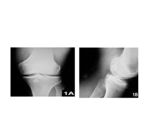
• Loose bodies are fragments of bone or
cartilage that are freely float in joint space.
• Single or in groups
• They effect only one joint
• They classified as stable or unstable.
• Stable loose bodies are in fixed position are
generally well tolerated.
• Unstable loose bodies are free to move about
the joint and cause symptoms.
• Individuals with a degenerative joint disease
(e.g., arthritis or osteochondritis dissecans)
are more likely to develop loose bodies in the
knee
Classification
Individuals who participate in sports are at risk of
developing loose bodies in the knee.
Loose bodies are common and affect men and
women equally
1.Fibrinous
2.Cartilagenous
3.Osteo cartilaginous
Fibrinous
• Due to bleeding with in joint
• from the death of the tissue lining of joints
(synovial membrane) associated
with tuberculosis, osteoarthritis,
and rheumatoid arthritis
• . Cartilaginous loose bodies are fragments of
cartilage and are caused by injury (trauma) to
the joint and osteoarthritis
• Osteocartilaginous loose bodies are fragments
of cartilage and bone caused by fractures,
bone and cartilage inflammation
(osteochondritis dissecans), osteoarthritis,
and benign tumors of the synovial membrane
(synovial chondromatosis).
Clinical features
• knee pain and swelling, with intermittent
locking or catching of the joint.
• The locking disappears spontaneously,
• Hearing a grating sound (crepitus) with joint
movement.
• The joint intermittently "gives way" or "goes
out" causing them to fall
• joint may be locked (unable to fully extend).
• There may be evidence of effusion in the joint.
• Loose bodies are rarely felt by touch
(palpation)
DIAGNOSIS
• Larger loose bodies are typically calcified and
thus easily visible on a plain film x-ray of the
affected joint
• small or no bone may not be visible with an x-
ray and are typically diagnosed using
either CT or arthrography or MRI .
• . Ultrasound scans may be performed to
determine the presence and location of loose
bodies
Treatment
• For small loose bodies, treatment may be directed
at relief of symptoms by NSAIDS
• Large loose bodies may require removal by use of
an arthroscope (arthroscopy
• Very large loose bodies and those located in the
back of the knee need to be removed by open
surgery (arthrotomy
• . In some cases, such as synovial chondromatosis,
part of the synovium may be removed
(partial synovectomy
REHABILITATION
The focus of rehabilitation for loose bodies in
the knee is to control pain and restore
function
The first goal is gait training with an assistive
device
Prolonged immobilization should be avoided for
non-surgical cases .
REFERANCES
• CAMPBELL’S OPERATIVE ORTHOPAEDDICS
12TH EDITION
• THANK YOU

Loose bodies in knee

  • 1.
    LOOSE BODIES INKNEE Dr.Harsha Surath
  • 2.
    • Loose bodiesare fragments of bone or cartilage that are freely float in joint space. • Single or in groups • They effect only one joint • They classified as stable or unstable.
  • 3.
    • Stable loosebodies are in fixed position are generally well tolerated. • Unstable loose bodies are free to move about the joint and cause symptoms. • Individuals with a degenerative joint disease (e.g., arthritis or osteochondritis dissecans) are more likely to develop loose bodies in the knee
  • 5.
    Classification Individuals who participatein sports are at risk of developing loose bodies in the knee. Loose bodies are common and affect men and women equally 1.Fibrinous 2.Cartilagenous 3.Osteo cartilaginous
  • 6.
    Fibrinous • Due tobleeding with in joint • from the death of the tissue lining of joints (synovial membrane) associated with tuberculosis, osteoarthritis, and rheumatoid arthritis
  • 7.
    • . Cartilaginousloose bodies are fragments of cartilage and are caused by injury (trauma) to the joint and osteoarthritis
  • 8.
    • Osteocartilaginous loosebodies are fragments of cartilage and bone caused by fractures, bone and cartilage inflammation (osteochondritis dissecans), osteoarthritis, and benign tumors of the synovial membrane (synovial chondromatosis).
  • 9.
    Clinical features • kneepain and swelling, with intermittent locking or catching of the joint. • The locking disappears spontaneously, • Hearing a grating sound (crepitus) with joint movement. • The joint intermittently "gives way" or "goes out" causing them to fall
  • 10.
    • joint maybe locked (unable to fully extend). • There may be evidence of effusion in the joint. • Loose bodies are rarely felt by touch (palpation)
  • 11.
    DIAGNOSIS • Larger loosebodies are typically calcified and thus easily visible on a plain film x-ray of the affected joint • small or no bone may not be visible with an x- ray and are typically diagnosed using either CT or arthrography or MRI . • . Ultrasound scans may be performed to determine the presence and location of loose bodies
  • 12.
    Treatment • For smallloose bodies, treatment may be directed at relief of symptoms by NSAIDS • Large loose bodies may require removal by use of an arthroscope (arthroscopy • Very large loose bodies and those located in the back of the knee need to be removed by open surgery (arthrotomy • . In some cases, such as synovial chondromatosis, part of the synovium may be removed (partial synovectomy
  • 13.
    REHABILITATION The focus ofrehabilitation for loose bodies in the knee is to control pain and restore function The first goal is gait training with an assistive device Prolonged immobilization should be avoided for non-surgical cases .
  • 14.
    REFERANCES • CAMPBELL’S OPERATIVEORTHOPAEDDICS 12TH EDITION
  • 15.