SlideShare a Scribd company logo
Radiological imaging of Brain Arteriovenous Malformations(AVM).
Dr/ ABD ALLAH NAZEER. MD.
Cerebral arteriovenous malformations (CAVM's) are a common form
of cerebral vascular malformation and are composed of a nidus of vessels
through which arteriovenous shunting occur. Three types are described:
parenchymal (pial) AVM
dural AVM
mixed AVM
Epidemiology
Although arteriovenous malformations are thought to represent a congenital
abnormality, they are thought to develop over time and are rarely found
incidentally in the very young. Having said this, a third of AVMs which are
diagnosed due to hemorrhage are identified before the age of 20 years of
age. Overall AVMs are diagnosed at a mean age of 31 years.
Overall AVMs are thought to occur in approximately 4% of the population, but
become symptomatic in only 12% of affected individuals. There is no gender
predilection.
Arteriovenous malformations tend to be solitary in the vast majority of cases
(>95%). When multiple, syndromic associations include
Wyburn-Mason syndrome
Osler-Weber-Rendu syndrome
Clinical presentation
CAVMs are the most common symptomatic vascular malformations. Possible
presentations include
incidental finding in asymptomatic patients: 15%
seizures: 20%
headaches
ischemic events due to vascular steal from normal brain
hemorrhage: 65%5, 2-3% per year
parenchymal
subarachnoid
intraventricular
Pathology
The origin of arteriovenous malformations remains uncertain, although they are
thought to be congenital and perhaps involves dysregulation of vascular
endothelium growth factor (VEGF).
AVMs comprise of a number of components
feeding arteries
nidus
shunting arterioles: the true culprit
interconnected venous loops
draining veins
The nidus is fed by one or more arteries and drained by one or more veins. The feeding
arteries are enlarged due to the increased flow and flow related arterial aneurysms are
encountered . Venous aneurysms are also seen. It may contain dystrophic calcification,
small amount of gliotic tissue and blood in different stages of evolution.
Location: supratentorial: ~85%
superficial (two-thirds)
deep (one-third)
infratentorial: ~15%
Incidence
solitary AVMs (98%)
multiple AVMs (2%)
associated with syndromes
Associated abnormalities
flow related angiopathy secondary to endothelial hyperplasia
flow related aneurysm
intranidal: located in the nidus
intrapedicular: located in the feeding vessel
remote aneurysm: hemodynamically unrelated to malformation
Classification and grading
AVMs can be divided into two types :
compact: nidus contains little if any neuronal tissue, which is non-functioning
diffuse: no well formed nidus is present, with functional neuronal tissue interspersed
amongst the anomalous vessels.
Radiographic features
CT: Diagnosis can be difficult on non contrast CT. The nidus is blood density and
therefore usually somewhat hyperdense compared to adjacent brain. Enlarged
draining veins may be seen.
Following contrast administration, and especially with CTA the diagnosis is usually
self evident with feeding arteries, nidus and draining veins visible. The exact
anatomy of feeding vessels and draining veins is often difficult to delineate, and
thus angiography remains necessary.
MRI:
Fast flow generates flow voids easily seen on T2 weighted images. Complications
including previous hemorrhage and adjacent edema may be evident.
MRA: phase contrast MR angiography is often useful to subtract the hematoma
components when an AVM complicated by an acute hemorrhage needs to be
imaged DSA - angiography
Remains the gold standard, able to exquisitely delineate the location and number
of feeding vessels and the pattern of drainage. Ideally angiography is performed
in a bi-plane system with a high rate of acquisition, as the shunts can be very
rapid.
On angiogram AVM appears as tightly packed mass of enlarged feeding arteries
that supply central nidus. One or more dilated veins drain the nidus and there is
abnormal opacification of veins occurs in arterial phase, represents shunting.
Superficial AVM with parietal hematoma.
CTA MIP images demonstrated an AVM in the parieto-temporal lobe surrounding the hematoma
AVM in the right temporal lobe (arrows). AVM in the left parietal lobe (arrow).
Superficial AVM with intra-cerebral hematoma.
Deep AVM.
Deep AVM.
An AVM in the midbrain and right thalamus (arrow).
Proliferative type of brain AVM.
Proliferative type of left ganglionic AVM.
Large right hemispheric vascular lesion .
Developmental VA of the brain stem.
Pial AVM.
Dural AVF with left occipital and intra-ventricular hemorrhage.
Moyamoya with peri-mesencephlic AVM and associated IV hemorrhage
Grade 3 thalamic AVM with intra-nidal aneurysm.
(a) T2 axial, (b) T2 coronal, (c) T1 sagittal and, (d) MR angiogram sequence of cerebral magnetic resonance
imaging reveals a left frontal well defined flow void of size 60 × 66 mm communicating with the superior
sagittal sinus through dilated cortical vein with feeder supply from left anterior cerebral artery.
128 Multi slice cerebral computed tomographic angiography sagittal (a) and coronal (b)
view demonstrates a large left frontal arteriovenous fistula supplied by the distal left
anterior cerebral artery draining through a dilated cortical vein into the superior sagittal
sinus. A large variceal dilatation of the proximal venous end measuring nearly 6.5 cm in
diameter with wall calcification is shown in association with the arteriovenous fistula.
Thank You.

More Related Content

What's hot

IMAGING IN CEREBRAL VENOUS THROMBOSIS
IMAGING IN CEREBRAL VENOUS THROMBOSIS IMAGING IN CEREBRAL VENOUS THROMBOSIS
IMAGING IN CEREBRAL VENOUS THROMBOSIS
Shivshankar Badole
 
Venous Doppler upper limb
Venous Doppler upper limb Venous Doppler upper limb
Venous Doppler upper limb
Milan Silwal
 
Presentation1.pptx, supratentorial brain tumour
Presentation1.pptx, supratentorial brain tumourPresentation1.pptx, supratentorial brain tumour
Presentation1.pptx, supratentorial brain tumour
Abdellah Nazeer
 
Presentation1.pptx, radiological imaging of peri natal acute ischemia and hyp...
Presentation1.pptx, radiological imaging of peri natal acute ischemia and hyp...Presentation1.pptx, radiological imaging of peri natal acute ischemia and hyp...
Presentation1.pptx, radiological imaging of peri natal acute ischemia and hyp...
Abdellah Nazeer
 
Diagnostic Imaging of Brain Tumors
Diagnostic Imaging of Brain TumorsDiagnostic Imaging of Brain Tumors
Diagnostic Imaging of Brain Tumors
Mohamed M.A. Zaitoun
 
Peripheral arterial doppler
Peripheral  arterial dopplerPeripheral  arterial doppler
Peripheral arterial doppler
Anish Choudhary
 
Imaging in SAH
Imaging  in  SAHImaging  in  SAH
Imaging in SAH
Sarath Menon
 
Radiology of demyelinating diseases
Radiology of demyelinating diseases Radiology of demyelinating diseases
Radiology of demyelinating diseases
NeurologyKota
 
Radiology Spotters
Radiology Spotters Radiology Spotters
Radiology Spotters
Anish Choudhary
 
Radiological features of intracranial tumors 1
Radiological features of intracranial tumors 1Radiological features of intracranial tumors 1
Radiological features of intracranial tumors 1
Dr Praveen kumar tripathi
 
Doppler ultrasound in deep vein thrombosis
Doppler ultrasound in deep vein thrombosisDoppler ultrasound in deep vein thrombosis
Doppler ultrasound in deep vein thrombosis
Samir Haffar
 
Diagnostic Imaging of Cerebellopontine Angle Masses
Diagnostic Imaging of Cerebellopontine Angle MassesDiagnostic Imaging of Cerebellopontine Angle Masses
Diagnostic Imaging of Cerebellopontine Angle Masses
Mohamed M.A. Zaitoun
 
Aortic aneurysm imaging
Aortic aneurysm imagingAortic aneurysm imaging
Aortic aneurysm imaging
Sandeep Ponnaganti
 
Diagnostic Imaging of Stroke
Diagnostic Imaging of StrokeDiagnostic Imaging of Stroke
Diagnostic Imaging of Stroke
Mohamed M.A. Zaitoun
 
Neurosonogram.. Dr.Padmesh
Neurosonogram.. Dr.PadmeshNeurosonogram.. Dr.Padmesh
Neurosonogram.. Dr.Padmesh
Dr Padmesh Vadakepat
 
Diagnostic Imaging of Cerebral Trauma
Diagnostic Imaging of Cerebral TraumaDiagnostic Imaging of Cerebral Trauma
Diagnostic Imaging of Cerebral Trauma
Mohamed M.A. Zaitoun
 
Presentation2.pptx. posterior fossa tumour
Presentation2.pptx. posterior fossa tumourPresentation2.pptx. posterior fossa tumour
Presentation2.pptx. posterior fossa tumour
Abdellah Nazeer
 
Imaging of Renal Tumors
Imaging of Renal TumorsImaging of Renal Tumors
Imaging of Renal Tumors
Dr. Yash Kumar Achantani
 
MRI imaging hypothalamus Dr Ahmed Esawy
MRI imaging hypothalamus Dr Ahmed Esawy MRI imaging hypothalamus Dr Ahmed Esawy
MRI imaging hypothalamus Dr Ahmed Esawy
AHMED ESAWY
 
MRI imaging of brain tumors. A practical approach.
MRI imaging of brain tumors. A practical approach. MRI imaging of brain tumors. A practical approach.
MRI imaging of brain tumors. A practical approach.
hazem youssef
 

What's hot (20)

IMAGING IN CEREBRAL VENOUS THROMBOSIS
IMAGING IN CEREBRAL VENOUS THROMBOSIS IMAGING IN CEREBRAL VENOUS THROMBOSIS
IMAGING IN CEREBRAL VENOUS THROMBOSIS
 
Venous Doppler upper limb
Venous Doppler upper limb Venous Doppler upper limb
Venous Doppler upper limb
 
Presentation1.pptx, supratentorial brain tumour
Presentation1.pptx, supratentorial brain tumourPresentation1.pptx, supratentorial brain tumour
Presentation1.pptx, supratentorial brain tumour
 
Presentation1.pptx, radiological imaging of peri natal acute ischemia and hyp...
Presentation1.pptx, radiological imaging of peri natal acute ischemia and hyp...Presentation1.pptx, radiological imaging of peri natal acute ischemia and hyp...
Presentation1.pptx, radiological imaging of peri natal acute ischemia and hyp...
 
Diagnostic Imaging of Brain Tumors
Diagnostic Imaging of Brain TumorsDiagnostic Imaging of Brain Tumors
Diagnostic Imaging of Brain Tumors
 
Peripheral arterial doppler
Peripheral  arterial dopplerPeripheral  arterial doppler
Peripheral arterial doppler
 
Imaging in SAH
Imaging  in  SAHImaging  in  SAH
Imaging in SAH
 
Radiology of demyelinating diseases
Radiology of demyelinating diseases Radiology of demyelinating diseases
Radiology of demyelinating diseases
 
Radiology Spotters
Radiology Spotters Radiology Spotters
Radiology Spotters
 
Radiological features of intracranial tumors 1
Radiological features of intracranial tumors 1Radiological features of intracranial tumors 1
Radiological features of intracranial tumors 1
 
Doppler ultrasound in deep vein thrombosis
Doppler ultrasound in deep vein thrombosisDoppler ultrasound in deep vein thrombosis
Doppler ultrasound in deep vein thrombosis
 
Diagnostic Imaging of Cerebellopontine Angle Masses
Diagnostic Imaging of Cerebellopontine Angle MassesDiagnostic Imaging of Cerebellopontine Angle Masses
Diagnostic Imaging of Cerebellopontine Angle Masses
 
Aortic aneurysm imaging
Aortic aneurysm imagingAortic aneurysm imaging
Aortic aneurysm imaging
 
Diagnostic Imaging of Stroke
Diagnostic Imaging of StrokeDiagnostic Imaging of Stroke
Diagnostic Imaging of Stroke
 
Neurosonogram.. Dr.Padmesh
Neurosonogram.. Dr.PadmeshNeurosonogram.. Dr.Padmesh
Neurosonogram.. Dr.Padmesh
 
Diagnostic Imaging of Cerebral Trauma
Diagnostic Imaging of Cerebral TraumaDiagnostic Imaging of Cerebral Trauma
Diagnostic Imaging of Cerebral Trauma
 
Presentation2.pptx. posterior fossa tumour
Presentation2.pptx. posterior fossa tumourPresentation2.pptx. posterior fossa tumour
Presentation2.pptx. posterior fossa tumour
 
Imaging of Renal Tumors
Imaging of Renal TumorsImaging of Renal Tumors
Imaging of Renal Tumors
 
MRI imaging hypothalamus Dr Ahmed Esawy
MRI imaging hypothalamus Dr Ahmed Esawy MRI imaging hypothalamus Dr Ahmed Esawy
MRI imaging hypothalamus Dr Ahmed Esawy
 
MRI imaging of brain tumors. A practical approach.
MRI imaging of brain tumors. A practical approach. MRI imaging of brain tumors. A practical approach.
MRI imaging of brain tumors. A practical approach.
 

Viewers also liked

AVM, Vascular Malformation
AVM, Vascular MalformationAVM, Vascular Malformation
AVM, Vascular Malformation
elango mk
 
0914
09140914
0914
calaf0618
 
Brain Aneurysms & AV Malformations
Brain Aneurysms & AV MalformationsBrain Aneurysms & AV Malformations
Brain Aneurysms & AV Malformations
Sherry Knowles
 
Arteriovenous Malformations
Arteriovenous MalformationsArteriovenous Malformations
Arteriovenous Malformations
Ahmed Shammasi
 
Arteriovenous Malformation (AVM) of Brain
Arteriovenous Malformation (AVM) of BrainArteriovenous Malformation (AVM) of Brain
Arteriovenous Malformation (AVM) of Brain
Dhaval Shukla
 
Mri patho avm
Mri patho avmMri patho avm
Mri patho avm
e04randy
 
Presentation1.pptx, intra cranial vascular malformation.
Presentation1.pptx, intra cranial vascular malformation.Presentation1.pptx, intra cranial vascular malformation.
Presentation1.pptx, intra cranial vascular malformation.
Abdellah Nazeer
 
Chapter 22
Chapter 22Chapter 22
Chapter 22
francklin castillo
 
31 aneurysms and vascular malformations
31 aneurysms and vascular malformations31 aneurysms and vascular malformations
31 aneurysms and vascular malformations
Dr. Muhammad Bin Zulfiqar
 
Case record...Multiple dural arteriovenous fistulas
Case record...Multiple dural arteriovenous fistulasCase record...Multiple dural arteriovenous fistulas
Case record...Multiple dural arteriovenous fistulas
Professor Yasser Metwally
 
Cerebral Vascular Disease
Cerebral Vascular DiseaseCerebral Vascular Disease
Cerebral Vascular Disease
shatrughana yadav
 
Neurology advanced hsa jy gauvrit
Neurology advanced hsa jy gauvritNeurology advanced hsa jy gauvrit
Neurology advanced hsa jy gauvrit
JFIM
 
Cerebral aneurysms
Cerebral aneurysmsCerebral aneurysms
Cerebral aneurysms
Cesare Colombo
 
brain AVMs
brain AVMsbrain AVMs
Imaging: AV Malformation
Imaging: AV MalformationImaging: AV Malformation
Dural arteriovenous fistula & AVM
Dural arteriovenous fistula & AVMDural arteriovenous fistula & AVM
Dural arteriovenous fistula & AVM
airwave12
 
Intracranial vascular disease
Intracranial vascular diseaseIntracranial vascular disease
Intracranial vascular disease
Seyed Amirhossein Javadi
 
Presentation1.pptx, diagnostic pitfalls mimicking meniscal tear and post oper...
Presentation1.pptx, diagnostic pitfalls mimicking meniscal tear and post oper...Presentation1.pptx, diagnostic pitfalls mimicking meniscal tear and post oper...
Presentation1.pptx, diagnostic pitfalls mimicking meniscal tear and post oper...
Abdellah Nazeer
 
Cerebral aneurysm
Cerebral aneurysmCerebral aneurysm
Cerebral aneurysm
Mohamed Al-Banna
 
Cerebral aneurysm
Cerebral aneurysmCerebral aneurysm
Cerebral aneurysm
Mohd Hafizi Mamat
 

Viewers also liked (20)

AVM, Vascular Malformation
AVM, Vascular MalformationAVM, Vascular Malformation
AVM, Vascular Malformation
 
0914
09140914
0914
 
Brain Aneurysms & AV Malformations
Brain Aneurysms & AV MalformationsBrain Aneurysms & AV Malformations
Brain Aneurysms & AV Malformations
 
Arteriovenous Malformations
Arteriovenous MalformationsArteriovenous Malformations
Arteriovenous Malformations
 
Arteriovenous Malformation (AVM) of Brain
Arteriovenous Malformation (AVM) of BrainArteriovenous Malformation (AVM) of Brain
Arteriovenous Malformation (AVM) of Brain
 
Mri patho avm
Mri patho avmMri patho avm
Mri patho avm
 
Presentation1.pptx, intra cranial vascular malformation.
Presentation1.pptx, intra cranial vascular malformation.Presentation1.pptx, intra cranial vascular malformation.
Presentation1.pptx, intra cranial vascular malformation.
 
Chapter 22
Chapter 22Chapter 22
Chapter 22
 
31 aneurysms and vascular malformations
31 aneurysms and vascular malformations31 aneurysms and vascular malformations
31 aneurysms and vascular malformations
 
Case record...Multiple dural arteriovenous fistulas
Case record...Multiple dural arteriovenous fistulasCase record...Multiple dural arteriovenous fistulas
Case record...Multiple dural arteriovenous fistulas
 
Cerebral Vascular Disease
Cerebral Vascular DiseaseCerebral Vascular Disease
Cerebral Vascular Disease
 
Neurology advanced hsa jy gauvrit
Neurology advanced hsa jy gauvritNeurology advanced hsa jy gauvrit
Neurology advanced hsa jy gauvrit
 
Cerebral aneurysms
Cerebral aneurysmsCerebral aneurysms
Cerebral aneurysms
 
brain AVMs
brain AVMsbrain AVMs
brain AVMs
 
Imaging: AV Malformation
Imaging: AV MalformationImaging: AV Malformation
Imaging: AV Malformation
 
Dural arteriovenous fistula & AVM
Dural arteriovenous fistula & AVMDural arteriovenous fistula & AVM
Dural arteriovenous fistula & AVM
 
Intracranial vascular disease
Intracranial vascular diseaseIntracranial vascular disease
Intracranial vascular disease
 
Presentation1.pptx, diagnostic pitfalls mimicking meniscal tear and post oper...
Presentation1.pptx, diagnostic pitfalls mimicking meniscal tear and post oper...Presentation1.pptx, diagnostic pitfalls mimicking meniscal tear and post oper...
Presentation1.pptx, diagnostic pitfalls mimicking meniscal tear and post oper...
 
Cerebral aneurysm
Cerebral aneurysmCerebral aneurysm
Cerebral aneurysm
 
Cerebral aneurysm
Cerebral aneurysmCerebral aneurysm
Cerebral aneurysm
 

Similar to Presentation1.pptx, radiological imaging of brain av malformation.

Intracranial vascular malformations
Intracranial vascular malformationsIntracranial vascular malformations
Intracranial vascular malformations
Milan Silwal
 
Av ms
Av msAv ms
Vascular Lesions of the Brain
Vascular Lesions of the BrainVascular Lesions of the Brain
Vascular Lesions of the Brain
Liew Boon Seng
 
Cerebral/Pial Arteriovenous Malformation (AVM)
Cerebral/Pial Arteriovenous Malformation (AVM)Cerebral/Pial Arteriovenous Malformation (AVM)
Cerebral/Pial Arteriovenous Malformation (AVM)
macshrestha
 
Cerebral AVM
Cerebral AVMCerebral AVM
Cerebral AVM
Julio Fernández
 
Vascular malformations of the brain
Vascular malformations of the brainVascular malformations of the brain
Vascular malformations of the brain
Mohamed M.A. Zaitoun
 
10.-Nervous-System.pdf
10.-Nervous-System.pdf10.-Nervous-System.pdf
10.-Nervous-System.pdf
InnaDuco
 
Avm.23.11
Avm.23.11Avm.23.11
Avm.23.11
Le Jang
 
Walif Chbeir: Cerebral Cavernous Malformation
Walif Chbeir: Cerebral Cavernous MalformationWalif Chbeir: Cerebral Cavernous Malformation
Walif Chbeir: Cerebral Cavernous Malformation
Walif Chbeir
 
Vascular brain lesions for radiology by Dr Soumitra Halder
Vascular brain lesions for radiology by Dr Soumitra HalderVascular brain lesions for radiology by Dr Soumitra Halder
Vascular brain lesions for radiology by Dr Soumitra Halder
Soumitra Halder
 
BRAIN ARTERIOVENOUS MALFORMATION
BRAIN ARTERIOVENOUS MALFORMATIONBRAIN ARTERIOVENOUS MALFORMATION
BRAIN ARTERIOVENOUS MALFORMATION
suresh Bishokarma
 
cerebral vascular malformation
cerebral vascular malformationcerebral vascular malformation
cerebral vascular malformation
MojtabaKhazaei2
 
Radiological pathology of spontaneous cerebral hemorrhage
Radiological pathology of spontaneous cerebral hemorrhageRadiological pathology of spontaneous cerebral hemorrhage
Radiological pathology of spontaneous cerebral hemorrhage
Professor Yasser Metwally
 
Cerebral AVM
Cerebral AVMCerebral AVM
Cerebral AVM
shashank nahar
 
spinal angiography with spnal av anomalies
spinal angiography with spnal av anomaliesspinal angiography with spnal av anomalies
spinal angiography with spnal av anomalies
NeurologyKota
 
Anterior cerebral circulation aneurysm./
Anterior cerebral circulation aneurysm./Anterior cerebral circulation aneurysm./
Anterior cerebral circulation aneurysm./
Anas Ahmed
 
Cerebro vascular anomalies
Cerebro vascular anomaliesCerebro vascular anomalies
Cerebro vascular anomalies
Shweta Sharma
 
Spinal AVM AIIMS.pptx
Spinal AVM AIIMS.pptxSpinal AVM AIIMS.pptx
Spinal AVM AIIMS.pptx
PrasannaKumarVelchur
 
Moyamoya disease
Moyamoya diseaseMoyamoya disease
Moyamoya disease
Ayman Al-Malt
 
SPINAL CORD ARTERIOVENOUS MALFORMATIONS
SPINAL CORD ARTERIOVENOUS MALFORMATIONSSPINAL CORD ARTERIOVENOUS MALFORMATIONS
SPINAL CORD ARTERIOVENOUS MALFORMATIONS
Dr Praveen kumar tripathi
 

Similar to Presentation1.pptx, radiological imaging of brain av malformation. (20)

Intracranial vascular malformations
Intracranial vascular malformationsIntracranial vascular malformations
Intracranial vascular malformations
 
Av ms
Av msAv ms
Av ms
 
Vascular Lesions of the Brain
Vascular Lesions of the BrainVascular Lesions of the Brain
Vascular Lesions of the Brain
 
Cerebral/Pial Arteriovenous Malformation (AVM)
Cerebral/Pial Arteriovenous Malformation (AVM)Cerebral/Pial Arteriovenous Malformation (AVM)
Cerebral/Pial Arteriovenous Malformation (AVM)
 
Cerebral AVM
Cerebral AVMCerebral AVM
Cerebral AVM
 
Vascular malformations of the brain
Vascular malformations of the brainVascular malformations of the brain
Vascular malformations of the brain
 
10.-Nervous-System.pdf
10.-Nervous-System.pdf10.-Nervous-System.pdf
10.-Nervous-System.pdf
 
Avm.23.11
Avm.23.11Avm.23.11
Avm.23.11
 
Walif Chbeir: Cerebral Cavernous Malformation
Walif Chbeir: Cerebral Cavernous MalformationWalif Chbeir: Cerebral Cavernous Malformation
Walif Chbeir: Cerebral Cavernous Malformation
 
Vascular brain lesions for radiology by Dr Soumitra Halder
Vascular brain lesions for radiology by Dr Soumitra HalderVascular brain lesions for radiology by Dr Soumitra Halder
Vascular brain lesions for radiology by Dr Soumitra Halder
 
BRAIN ARTERIOVENOUS MALFORMATION
BRAIN ARTERIOVENOUS MALFORMATIONBRAIN ARTERIOVENOUS MALFORMATION
BRAIN ARTERIOVENOUS MALFORMATION
 
cerebral vascular malformation
cerebral vascular malformationcerebral vascular malformation
cerebral vascular malformation
 
Radiological pathology of spontaneous cerebral hemorrhage
Radiological pathology of spontaneous cerebral hemorrhageRadiological pathology of spontaneous cerebral hemorrhage
Radiological pathology of spontaneous cerebral hemorrhage
 
Cerebral AVM
Cerebral AVMCerebral AVM
Cerebral AVM
 
spinal angiography with spnal av anomalies
spinal angiography with spnal av anomaliesspinal angiography with spnal av anomalies
spinal angiography with spnal av anomalies
 
Anterior cerebral circulation aneurysm./
Anterior cerebral circulation aneurysm./Anterior cerebral circulation aneurysm./
Anterior cerebral circulation aneurysm./
 
Cerebro vascular anomalies
Cerebro vascular anomaliesCerebro vascular anomalies
Cerebro vascular anomalies
 
Spinal AVM AIIMS.pptx
Spinal AVM AIIMS.pptxSpinal AVM AIIMS.pptx
Spinal AVM AIIMS.pptx
 
Moyamoya disease
Moyamoya diseaseMoyamoya disease
Moyamoya disease
 
SPINAL CORD ARTERIOVENOUS MALFORMATIONS
SPINAL CORD ARTERIOVENOUS MALFORMATIONSSPINAL CORD ARTERIOVENOUS MALFORMATIONS
SPINAL CORD ARTERIOVENOUS MALFORMATIONS
 

More from Abdellah Nazeer

Muculoskeletal Pediatic Imaging..pptx
Muculoskeletal Pediatic Imaging..pptxMuculoskeletal Pediatic Imaging..pptx
Muculoskeletal Pediatic Imaging..pptx
Abdellah Nazeer
 
Presentation1, Ultrasound of the bowel loops and the lymph nodes..pptx
Presentation1, Ultrasound of the bowel loops and the lymph nodes..pptxPresentation1, Ultrasound of the bowel loops and the lymph nodes..pptx
Presentation1, Ultrasound of the bowel loops and the lymph nodes..pptx
Abdellah Nazeer
 
Presentation1 Short cases MD..pptx
Presentation1 Short cases MD..pptxPresentation1 Short cases MD..pptx
Presentation1 Short cases MD..pptx
Abdellah Nazeer
 
Presentation1, MD MCQ Cases..pptx
Presentation1, MD MCQ Cases..pptxPresentation1, MD MCQ Cases..pptx
Presentation1, MD MCQ Cases..pptx
Abdellah Nazeer
 
Presentation1, Short Cases Quiz..pptx
Presentation1, Short Cases Quiz..pptxPresentation1, Short Cases Quiz..pptx
Presentation1, Short Cases Quiz..pptx
Abdellah Nazeer
 
Presentation1, radiological imaging of lateral hindfoot impingement.
Presentation1, radiological imaging of lateral hindfoot impingement.Presentation1, radiological imaging of lateral hindfoot impingement.
Presentation1, radiological imaging of lateral hindfoot impingement.
Abdellah Nazeer
 
Presentation2, radiological anatomy of the liver and spleen.
Presentation2, radiological anatomy of the liver and spleen.Presentation2, radiological anatomy of the liver and spleen.
Presentation2, radiological anatomy of the liver and spleen.
Abdellah Nazeer
 
Presentation1, artifacts and pitfalls of the wrist and elbow joints.
Presentation1, artifacts and pitfalls of the wrist and elbow joints.Presentation1, artifacts and pitfalls of the wrist and elbow joints.
Presentation1, artifacts and pitfalls of the wrist and elbow joints.
Abdellah Nazeer
 
Presentation1, artifact and pitfalls of the knee, hip and ankle joints.
Presentation1, artifact and pitfalls of the knee, hip and ankle joints.Presentation1, artifact and pitfalls of the knee, hip and ankle joints.
Presentation1, artifact and pitfalls of the knee, hip and ankle joints.
Abdellah Nazeer
 
Presentation1, radiological imaging of artifact and pitfalls in shoulder join...
Presentation1, radiological imaging of artifact and pitfalls in shoulder join...Presentation1, radiological imaging of artifact and pitfalls in shoulder join...
Presentation1, radiological imaging of artifact and pitfalls in shoulder join...
Abdellah Nazeer
 
Presentation1, radiological imaging of internal abdominal hernia.
Presentation1, radiological imaging of internal abdominal hernia.Presentation1, radiological imaging of internal abdominal hernia.
Presentation1, radiological imaging of internal abdominal hernia.
Abdellah Nazeer
 
Presentation11, radiological imaging of ovarian torsion.
Presentation11, radiological imaging of ovarian torsion.Presentation11, radiological imaging of ovarian torsion.
Presentation11, radiological imaging of ovarian torsion.
Abdellah Nazeer
 
Presentation1, musculoskeletal anatomy.
Presentation1, musculoskeletal anatomy.Presentation1, musculoskeletal anatomy.
Presentation1, musculoskeletal anatomy.
Abdellah Nazeer
 
Presentation1, new mri techniques in the diagnosis and monitoring of multiple...
Presentation1, new mri techniques in the diagnosis and monitoring of multiple...Presentation1, new mri techniques in the diagnosis and monitoring of multiple...
Presentation1, new mri techniques in the diagnosis and monitoring of multiple...
Abdellah Nazeer
 
Presentation1, radiological application of diffusion weighted mri in neck mas...
Presentation1, radiological application of diffusion weighted mri in neck mas...Presentation1, radiological application of diffusion weighted mri in neck mas...
Presentation1, radiological application of diffusion weighted mri in neck mas...
Abdellah Nazeer
 
Presentation1, radiological application of diffusion weighted images in breas...
Presentation1, radiological application of diffusion weighted images in breas...Presentation1, radiological application of diffusion weighted images in breas...
Presentation1, radiological application of diffusion weighted images in breas...
Abdellah Nazeer
 
Presentation1, radiological application of diffusion weighted images in abdom...
Presentation1, radiological application of diffusion weighted images in abdom...Presentation1, radiological application of diffusion weighted images in abdom...
Presentation1, radiological application of diffusion weighted images in abdom...
Abdellah Nazeer
 
Presentation1, radiological application of diffusion weighted imges in neuror...
Presentation1, radiological application of diffusion weighted imges in neuror...Presentation1, radiological application of diffusion weighted imges in neuror...
Presentation1, radiological application of diffusion weighted imges in neuror...
Abdellah Nazeer
 
Presentation1, mr physics.
Presentation1, mr physics.Presentation1, mr physics.
Presentation1, mr physics.
Abdellah Nazeer
 
Presentation1. ct physics.
Presentation1. ct physics.Presentation1. ct physics.
Presentation1. ct physics.
Abdellah Nazeer
 

More from Abdellah Nazeer (20)

Muculoskeletal Pediatic Imaging..pptx
Muculoskeletal Pediatic Imaging..pptxMuculoskeletal Pediatic Imaging..pptx
Muculoskeletal Pediatic Imaging..pptx
 
Presentation1, Ultrasound of the bowel loops and the lymph nodes..pptx
Presentation1, Ultrasound of the bowel loops and the lymph nodes..pptxPresentation1, Ultrasound of the bowel loops and the lymph nodes..pptx
Presentation1, Ultrasound of the bowel loops and the lymph nodes..pptx
 
Presentation1 Short cases MD..pptx
Presentation1 Short cases MD..pptxPresentation1 Short cases MD..pptx
Presentation1 Short cases MD..pptx
 
Presentation1, MD MCQ Cases..pptx
Presentation1, MD MCQ Cases..pptxPresentation1, MD MCQ Cases..pptx
Presentation1, MD MCQ Cases..pptx
 
Presentation1, Short Cases Quiz..pptx
Presentation1, Short Cases Quiz..pptxPresentation1, Short Cases Quiz..pptx
Presentation1, Short Cases Quiz..pptx
 
Presentation1, radiological imaging of lateral hindfoot impingement.
Presentation1, radiological imaging of lateral hindfoot impingement.Presentation1, radiological imaging of lateral hindfoot impingement.
Presentation1, radiological imaging of lateral hindfoot impingement.
 
Presentation2, radiological anatomy of the liver and spleen.
Presentation2, radiological anatomy of the liver and spleen.Presentation2, radiological anatomy of the liver and spleen.
Presentation2, radiological anatomy of the liver and spleen.
 
Presentation1, artifacts and pitfalls of the wrist and elbow joints.
Presentation1, artifacts and pitfalls of the wrist and elbow joints.Presentation1, artifacts and pitfalls of the wrist and elbow joints.
Presentation1, artifacts and pitfalls of the wrist and elbow joints.
 
Presentation1, artifact and pitfalls of the knee, hip and ankle joints.
Presentation1, artifact and pitfalls of the knee, hip and ankle joints.Presentation1, artifact and pitfalls of the knee, hip and ankle joints.
Presentation1, artifact and pitfalls of the knee, hip and ankle joints.
 
Presentation1, radiological imaging of artifact and pitfalls in shoulder join...
Presentation1, radiological imaging of artifact and pitfalls in shoulder join...Presentation1, radiological imaging of artifact and pitfalls in shoulder join...
Presentation1, radiological imaging of artifact and pitfalls in shoulder join...
 
Presentation1, radiological imaging of internal abdominal hernia.
Presentation1, radiological imaging of internal abdominal hernia.Presentation1, radiological imaging of internal abdominal hernia.
Presentation1, radiological imaging of internal abdominal hernia.
 
Presentation11, radiological imaging of ovarian torsion.
Presentation11, radiological imaging of ovarian torsion.Presentation11, radiological imaging of ovarian torsion.
Presentation11, radiological imaging of ovarian torsion.
 
Presentation1, musculoskeletal anatomy.
Presentation1, musculoskeletal anatomy.Presentation1, musculoskeletal anatomy.
Presentation1, musculoskeletal anatomy.
 
Presentation1, new mri techniques in the diagnosis and monitoring of multiple...
Presentation1, new mri techniques in the diagnosis and monitoring of multiple...Presentation1, new mri techniques in the diagnosis and monitoring of multiple...
Presentation1, new mri techniques in the diagnosis and monitoring of multiple...
 
Presentation1, radiological application of diffusion weighted mri in neck mas...
Presentation1, radiological application of diffusion weighted mri in neck mas...Presentation1, radiological application of diffusion weighted mri in neck mas...
Presentation1, radiological application of diffusion weighted mri in neck mas...
 
Presentation1, radiological application of diffusion weighted images in breas...
Presentation1, radiological application of diffusion weighted images in breas...Presentation1, radiological application of diffusion weighted images in breas...
Presentation1, radiological application of diffusion weighted images in breas...
 
Presentation1, radiological application of diffusion weighted images in abdom...
Presentation1, radiological application of diffusion weighted images in abdom...Presentation1, radiological application of diffusion weighted images in abdom...
Presentation1, radiological application of diffusion weighted images in abdom...
 
Presentation1, radiological application of diffusion weighted imges in neuror...
Presentation1, radiological application of diffusion weighted imges in neuror...Presentation1, radiological application of diffusion weighted imges in neuror...
Presentation1, radiological application of diffusion weighted imges in neuror...
 
Presentation1, mr physics.
Presentation1, mr physics.Presentation1, mr physics.
Presentation1, mr physics.
 
Presentation1. ct physics.
Presentation1. ct physics.Presentation1. ct physics.
Presentation1. ct physics.
 

Presentation1.pptx, radiological imaging of brain av malformation.

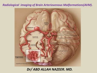
  • 1. Radiological imaging of Brain Arteriovenous Malformations(AVM). Dr/ ABD ALLAH NAZEER. MD.
  • 2. Cerebral arteriovenous malformations (CAVM's) are a common form of cerebral vascular malformation and are composed of a nidus of vessels through which arteriovenous shunting occur. Three types are described: parenchymal (pial) AVM dural AVM mixed AVM Epidemiology Although arteriovenous malformations are thought to represent a congenital abnormality, they are thought to develop over time and are rarely found incidentally in the very young. Having said this, a third of AVMs which are diagnosed due to hemorrhage are identified before the age of 20 years of age. Overall AVMs are diagnosed at a mean age of 31 years. Overall AVMs are thought to occur in approximately 4% of the population, but become symptomatic in only 12% of affected individuals. There is no gender predilection. Arteriovenous malformations tend to be solitary in the vast majority of cases (>95%). When multiple, syndromic associations include Wyburn-Mason syndrome Osler-Weber-Rendu syndrome
  • 3. Clinical presentation CAVMs are the most common symptomatic vascular malformations. Possible presentations include incidental finding in asymptomatic patients: 15% seizures: 20% headaches ischemic events due to vascular steal from normal brain hemorrhage: 65%5, 2-3% per year parenchymal subarachnoid intraventricular Pathology The origin of arteriovenous malformations remains uncertain, although they are thought to be congenital and perhaps involves dysregulation of vascular endothelium growth factor (VEGF). AVMs comprise of a number of components feeding arteries nidus shunting arterioles: the true culprit interconnected venous loops draining veins
  • 4. The nidus is fed by one or more arteries and drained by one or more veins. The feeding arteries are enlarged due to the increased flow and flow related arterial aneurysms are encountered . Venous aneurysms are also seen. It may contain dystrophic calcification, small amount of gliotic tissue and blood in different stages of evolution. Location: supratentorial: ~85% superficial (two-thirds) deep (one-third) infratentorial: ~15% Incidence solitary AVMs (98%) multiple AVMs (2%) associated with syndromes Associated abnormalities flow related angiopathy secondary to endothelial hyperplasia flow related aneurysm intranidal: located in the nidus intrapedicular: located in the feeding vessel remote aneurysm: hemodynamically unrelated to malformation Classification and grading AVMs can be divided into two types : compact: nidus contains little if any neuronal tissue, which is non-functioning diffuse: no well formed nidus is present, with functional neuronal tissue interspersed amongst the anomalous vessels.
  • 5. Radiographic features CT: Diagnosis can be difficult on non contrast CT. The nidus is blood density and therefore usually somewhat hyperdense compared to adjacent brain. Enlarged draining veins may be seen. Following contrast administration, and especially with CTA the diagnosis is usually self evident with feeding arteries, nidus and draining veins visible. The exact anatomy of feeding vessels and draining veins is often difficult to delineate, and thus angiography remains necessary. MRI: Fast flow generates flow voids easily seen on T2 weighted images. Complications including previous hemorrhage and adjacent edema may be evident. MRA: phase contrast MR angiography is often useful to subtract the hematoma components when an AVM complicated by an acute hemorrhage needs to be imaged DSA - angiography Remains the gold standard, able to exquisitely delineate the location and number of feeding vessels and the pattern of drainage. Ideally angiography is performed in a bi-plane system with a high rate of acquisition, as the shunts can be very rapid. On angiogram AVM appears as tightly packed mass of enlarged feeding arteries that supply central nidus. One or more dilated veins drain the nidus and there is abnormal opacification of veins occurs in arterial phase, represents shunting.
  • 6.
  • 7. Superficial AVM with parietal hematoma.
  • 8. CTA MIP images demonstrated an AVM in the parieto-temporal lobe surrounding the hematoma
  • 9. AVM in the right temporal lobe (arrows). AVM in the left parietal lobe (arrow).
  • 10. Superficial AVM with intra-cerebral hematoma.
  • 13. An AVM in the midbrain and right thalamus (arrow).
  • 14. Proliferative type of brain AVM.
  • 15.
  • 16. Proliferative type of left ganglionic AVM.
  • 17. Large right hemispheric vascular lesion .
  • 18.
  • 19.
  • 20. Developmental VA of the brain stem.
  • 22. Dural AVF with left occipital and intra-ventricular hemorrhage.
  • 23.
  • 24. Moyamoya with peri-mesencephlic AVM and associated IV hemorrhage
  • 25. Grade 3 thalamic AVM with intra-nidal aneurysm.
  • 26.
  • 27.
  • 28. (a) T2 axial, (b) T2 coronal, (c) T1 sagittal and, (d) MR angiogram sequence of cerebral magnetic resonance imaging reveals a left frontal well defined flow void of size 60 × 66 mm communicating with the superior sagittal sinus through dilated cortical vein with feeder supply from left anterior cerebral artery.
  • 29. 128 Multi slice cerebral computed tomographic angiography sagittal (a) and coronal (b) view demonstrates a large left frontal arteriovenous fistula supplied by the distal left anterior cerebral artery draining through a dilated cortical vein into the superior sagittal sinus. A large variceal dilatation of the proximal venous end measuring nearly 6.5 cm in diameter with wall calcification is shown in association with the arteriovenous fistula.