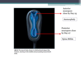
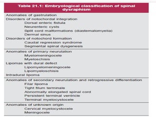
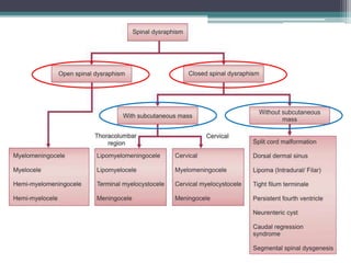
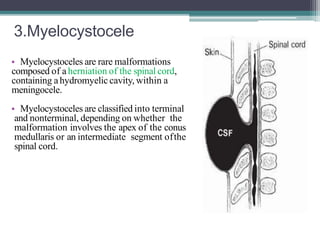
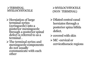
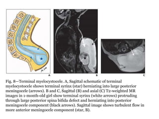
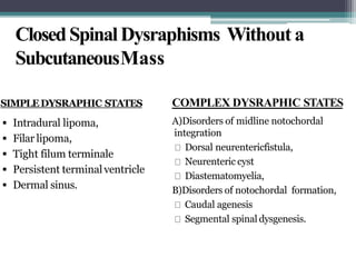
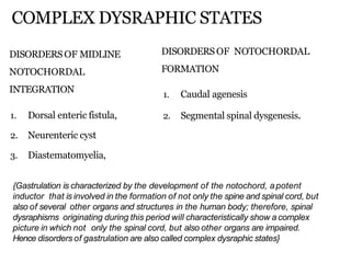
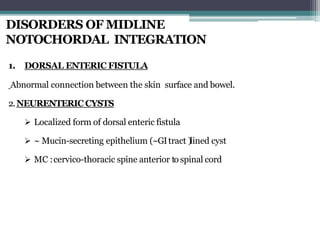
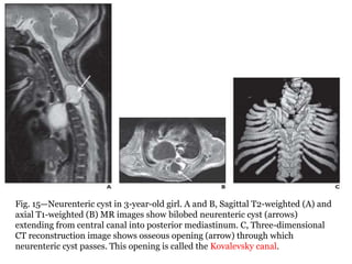
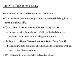
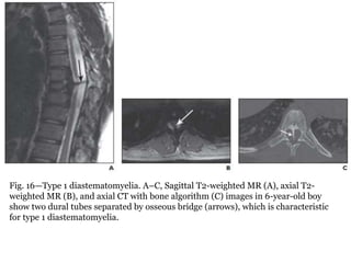
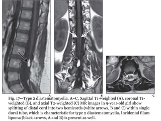
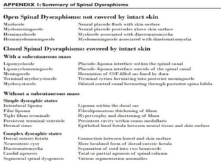
SlideShare a Scribd company logo
Dr.Pooja
Junior Resident
Department of Radiodiagnosis
Azeezia Medical College
Imaging in spinal dysraphism
Congenital malformations of the spine and spinal cord are
generally referred to as spinal dysraphisms
*Neural tube defects
* Spina bifida
In the spine, the most common congenital lesions
 diverse forms of spinal dysraphism
diverse forms of caudal spinal anomalies
IMAGING MODALITIES
 MRI :IOC
 PLAIN RADIOGRAPH:
 bony defects ,
 widened spinal canal ,
 scoliosis,
 segmentation anomalies like blockvertebra, butterfly
vertebrae ,bony spur (diastematomyelia)
 USG: antenatal Dx –open widened neural arch ,
meningomyelocele sac , hydrocephalus .
 CT :bonyspur
Spinal CordDevelopment
Summarized in three basic embryologic stages
The first stage : Gastrulation (the 2or 3week)
a bilaminar embryonic disk to a trilaminar disk.
The secondstage:primaryneurulation(weeks3–4)
the notochord and overlying ectoderm interact neural plate.
bends and folds  neural tube,
which then closes bidirectional in a zipperlike manner
The final stage :secondary neurulation (weeks 5–6),
 a secondary neural tube is formed by the caudal cell mass.
cavitation,  the tip of the conus medullaris and filum terminale
retrogressive differentiation.
1.GASTRULATION
2.Primary neurulation.
Formation of the Neural Plate
Shaping of the Neural Plate
Bending of the Neural Plate
Fusion
Anterior
neuropore
close by Day 25
Posterior
neuropore close
by Day 27
Anencephaly
Spina Bifida
Disjunction.
Upon completion of disjunction,
the cutaneous ectoderm fuses in
the midline, dorsal to the closed
neural tube.
Failure leads to Myelomeningocele.
Canalization and retrogressive
differentiation (synonym:
secondary neurulation).
Diagrammatic representation of
proposed embryogenesis.
3.SECONDARY NERULATION
OPEN SPINAL DYSRAPHISM (OSD)
 There is a defect in the overlying skin, and the neural tissue is
exposed to the environment.
 MYELOCELEAND MYELOMENINGOCELE
 Embryological defect : Complete nondisjunction of cutaneous ectoderm from
neural ectoderm.
 Neural folds do not fuse in the midline to form neural tube.
 Remain in continuity with the cutaneous ectoderm.
 This exposed part of the spinal cord is NEURAL PLACODE.
 MC location : Lumbosacral region >thoraco- lumbar region
 Imaging usually not done(clinically obvious)
 The main differentiating feature between a myelomeningocele and
myelocele is the position of the neural placode relative to the skin surface
 The neural placode protrudes above the skin surface with a myelomeningocele
and is flush with the skin surface with a myelocele
MYELOMENINGOCELE MYELOCELE
Myelomeningocele. Axial
schematic of myelomeningocele
shows neural placode (star)
protruding above skin surface
due to expansion of underlying
subarachnoid space (arrow).
Myelomeningocele.
Axial T2- weighted
MR image
Myelomeningocele.
Sagittal T2- weighted MR
image).
Myelocele.Axial schematic of
myeloceleshows neural placode
(arrow) flush with skin surface.
Myelocele. Axial T2-weighted MR
image in 1-day-old girl shows
exposed neural placode (arrow) that
is flush with skin surface, consistent
with myelocele. There is no
expansion of underlying
subarachnoid space
HEMIMYELOMENINGOCELE AND HEMIMYELOCELE
 Hemimyelomeningoceles and hemimyeloceles can also occur
but are extremely rare .
 These conditions occur when a myelomeningocele or myelocele
is associated with diastematomyelia (cord splitting) and one
hemicord fails to neurulate.
• Placode ulceration and infection are leading causes of mortality in
the untreated newborn, affected patients are operated on soon after
birth.
• MRI investigation should be performed whenever possible to
obtain:
(a)anatomic characterization of the various components of the
malformation, especially regarding the relationships
between the placode and nerve roots.
(b)presurgical evaluation of the entity and morphology of the
malformation sequence (hydromyelia, Chiari-II
malformation,and associated hydrocephalus)
(c)identification of rare caseswith associated cord splitting
(hemimyelomeningoceles andhemimyeloceles)
• All patients with OSDsalso harbor a Chiari-II
malformation,which is an element, rather than an associated
feature, of the diseasecommonly called
“myelomeningocele,” aswell asof the other forms of OSDs
CLOSED SPINAL DYSRAPHISM
CLOSEDSPINALDYSRAPHISMS
WITHASUBCUTANEOUSMASS
1. Lipoma with dorsaldefects
2. Myelocystocele(Terminal or cervical)
3. Meningocele
4. Cervical myelomeningocele
1.Lipomas with a duraldefect
• lipomyeloceles and lipomyelomeningoceles.
• defect in primary neurulation whereby mesenchymal tissue
enters the neural tube and forms lipomatous tissue
• Characterized clinically by the presence of a
subcutaneous fatty mass above the intergluteal crease.
• Diff both based on lipoma – placode interface
• Lipomyelocele : interface within spinal canal
• Lipomyelomenigocele: Outside the spinal canal due to sub-
arachnoid space expansion.
Fig. 4—Lipomyelocele. A, Axial schematic of lipomyelocele shows
placode–lipoma interface (arrow) lies within spinal canal. B, Axial T2-
weighted MR image in 3-year-old girl shows placode–lipoma interface
(arrow) within spinal canal, characteristic for lipomyelocele. C, Sagittal
T1-weighted MR image in 3-year-old girl with lipomyelocele shows
subcutaneous fatty mass (black arrow) and placode–lipoma interface
(white arrow) within spinal canal.
Fig. 5—Lipomyelomeningocele. A, Axial schematic of
lipomyelomeningocele shows placode–lipoma interface (arrow) lies
outside of spinal canal due to expansion of subarachnoid space. B, Axial
T1-weighted MR image in 18-month-old boy shows
lipomyelomeningocele (arrow) that is differentiated from lipomyelocele
by location of placode–lipoma interface outside of spinal canal due to
expansion of subarachnoid space.
thehairytuftoverlying subcutaneouslipomas
2. MENINGOCELE :herniation of CSF filled sac lined by dura and arachnoid matter .
Spinal cord not located within a meningocele but may be tethered to the neck of the sac
Posterior meningocele : posterior spina bifida  Lumbar/sacral
Anterior : Pre-sacral mostly
Fig. 6—Posterior meningocele. A, Sagittal T1-weighted MR image in in 12-month-old
girl shows posterior herniation of CSF-filled sac (arrow) in occipital region, consistent
with posterior meningocele. B, Sagittal T2-weighted MR image in 5-year-old boy
shows large posterior meningocele (arrow) in cervical region. C, Sagittal T2-weighted
MR image in 30-month-old girl shows small posterior meningocele (arrow) in lumbar
region.
ANTERIOR MENINGOCELE
3.Myelocystocele
• Myelocystoceles are rare malformations
composed of a herniation of the spinal cord,
containing ahydromyelic cavity, within a
meningocele.
• Myelocystoceles are classified into terminal
and nonterminal, depending on whether the
malformation involves the apex of the conus
medullaris or an intermediate segment ofthe
spinal cord.
TERMINAL
MYELOCYSTOCELE
MYELOCYSTOCELE
(NON TERMINAL)
 Herniation of large
terminal syrinx
(syringocele) into a
posterior meningocele
through a posterior spinal
defect is referred to as a
terminal .
 The terminal syrinx and
meningocele components
do not usually
communicate with each
other
 Dilated central canal
herniates through a
posterior spina bifida
defect.
 covered with skin
 MC -cervical or
cervicothoracic regions
Fig. 8—Terminal myelocystocele. A, Sagittal schematic of terminal
myelocystocele shows terminal syrinx (star) herniating into large posterior
meningocele (arrows). B and C, Sagittal (B) and axial (C) T2-weighted MR
images in 1-month-old girl show terminal syrinx (white arrows) protruding
through large posterior spina bifida defect and herniating into posterior
meningocele component (black arrows). Sagittal image shows turbulent flow in
more anterior meningocele component (star, B).
NON TERMINAL MYELOCYSTOCELE
—Schematic of nonterminal myelocystocele
shows herniation of dilated central canal
through posterior spinal defect.
ClosedSpinalDysraphisms Without a
SubcutaneousMass
COMPLEX DYSRAPHIC STATES
A)Disorders of midline notochordal
integration
Dorsal neurentericfistula,
Neurenteric cyst
Diastematomyelia,
B)Disorders of notochordal formation,
Caudal agenesis
Segmental spinal dysgenesis.
SIMPLEDYSRAPHIC STATES
 Intradural lipoma,
 Filar lipoma,
 Tight filum terminale
 Persistent terminalventricle
 Dermal sinus.
1. LIPOMA
2Types :Intradural lipoma and Filar lipoma
Embryological defect :focal premature disjunction of epidermalfrom
neural ectoderm.
1.INTRADURALLIPOMA
Lipoma within the duralsac
MC :Lumbosacralspine
a/w tethered-cord syndrome
2.FILAR LIPOMA
Fibrolipomatous thickening of the filum terminale is referred to asa
filar lipoma.
MR :T1hyperintense signal +thickened filum terminale
Intradural lipoma
Filarlipoma
, Sagittal (A) and axial (B) T1-weighted MR
images I with filar lipoma (arrows),
which has characteristic T1 hyperintensity and
marked thickening of filum terminale
.Sagittal T1-weighted (A) and sagittal T2- weighted fat-saturated (B) MR images show
large intradural lipoma (arrows), which is
hyperintense on T1-weighted image and hypointense on
T2-weighted fat-saturated image. Lipoma is attached to
conus medullaris, which is low lying.
3. TIGHT FILUM TERMINALE
 hypertrophy and shortening of the filum terminale
 EMBRYOLOGY:incomplete involution of
the distal spinalcord during embryogenesis.
 This condition causes tethering of the spinal cord
and impaired ascent of the conus medullaris.
 The conus medullaris is low lying relative to its
normal position, which is usually above the L2–
L3disc level
Sagittal T2-weighted MR image in 12-month-old boy shows tight filum terminale, characterized
by thickening and shortening of filum terminale (black arrow) with low-lying conus medullaris.
Incidental cross-fused renal ectopia (white arrow) is also present.
Persistence of a small,
ependymal lined cavity within
the conus medullaris.
It is the widest part of the
central canal at the levelof
conus
point of union between the
portion of the central canal
made by neurulation and the
portion made by canalization
of the caudal cell mass
Imaging :Location –above the
filum terminale and lack of
contrast enhancment Persistent terminal ventricle.
A and B, Sagittal T2-weighted (A) and sagittal
T1-weighted contrast-enhanced (B) MR
images show persistent terminal ventricle as
cystic structure (arrows) at inferior aspect of
conus medullaris, which does not enhance
4.PERSISTENTTERMINAL VENTRICLE
Epithelial lined fistula that
connects neural tissue or
meninges to the skinsurface.
If the superficial ectoderm
fails to separate from the
neural ectoderm at onepoint.
MC :Lumbo sacralregion
C/F :midline dimple , hairy
naevus , hyperpigmented
patch /capillary hemangioma
Infectious complication if not
surgically treated
5.DERMAL SINUS
Fig. 14—Dermal sinus. A and B, Sagittal schematic (A) and sagittal T2-weighted
MR image (B) in 9-year-old girl show intradural dermoid (stars) with tract
extending from central canal to skin surface (black arrows). Note tenting of dural
sac at origin of dermal sinus (white arrows). C, Axial T2-weighted MR image from
same patient as in B shows posterior location of hyperintense dermoid (arrow).
COMPLEX DYSRAPHIC STATES
DISORDERS OF MIDLINE
NOTOCHORDAL
INTEGRATION
1. Dorsal enteric fistula,
2. Neurenteric cyst
3. Diastematomyelia,
DISORDERS OF NOTOCHORDAL
FORMATION
1. Caudal agenesis
2. Segmental spinal dysgenesis.
{Gastrulation is characterized by the development of the notochord, apotent
inductor that is involved in the formation of not only the spine and spinal cord, but
also of several other organs and structures in the human body; therefore, spinal
dysraphisms originating during this period will characteristically show a complex
picture in which not only the spinal cord, but also other organs are impaired.
Hence disorders of gastrulation are also called complex dysraphic states}
DISORDERS OF MIDLINE
NOTOCHORDAL INTEGRATION
1. DORSAL ENTERIC FISTULA
Abnormal connection between the skin surface and bowel.
2.NEURENTERIC CYSTS
 Localized form of dorsal enteric fistula
 ~ Mucin-secreting epithelium (~GItract )lined cyst
 MC :cervico-thoracic spine anterior to spinal cord
Fig. 15—Neurenteric cyst in 3-year-old girl. A and B, Sagittal T2-weighted (A) and
axial T1-weighted (B) MR images show bilobed neurenteric cyst (arrows)
extending from central canal into posterior mediastinum. C, Three-dimensional
CT reconstruction image shows osseous opening (arrow) through which
neurenteric cyst passes. This opening is called the Kovalevsky canal.
3.DIASTEMATOMYELIA
 Separation of the spinal cord into two hemicords
 The two hemicords are usually symmetric, although thelength of
separation is variable.
 Type 1: Dual Dural-Arachnoid Tubes (Pang Type I) :
 the two hemicords are located within individual dural sacs
separated by an osseous or cartilaginousseptum
 In Type 2 : Single Dural-Arachnoid Tube (Pang Type II) :
 Single dural tube containing two hemicords, sometimes with an
intervening fibrous septum
 C/F:Hairy tuft , scoliosis , tethered cordsyndrome.
Fig. 16—Type 1 diastematomyelia. A–C, Sagittal T2-weighted MR (A), axial T2-
weighted MR (B), and axial CT with bone algorithm (C) images in 6-year-old boy
show two dural tubes separated by osseous bridge (arrows), which is characteristic
for type 1 diastematomyelia.
Fig. 17—Type 2 diastematomyelia. A–C, Sagittal T1-weighted (A), coronal T1-
weighted (B), and axial T2-weighted (C) MR images in 9-year-old girl show
splitting of distal cord into two hemicords (white arrows, B and C) within single
dural tube, which is characteristic for type 2 diastematomyelia. Incidental filum
lipoma (black arrows, A and B) is present as well.
II.DISORDERS OFNOTOCHORDAL FORMATION
1. CAUDALAGENESIS
Total or partial agenesis of the spinalcolumn
A/w anal imperforation, genital anomalies, renal dysplasia or aplasia,
pulmonary hypoplasia, or limb abnormalities.
2Types
Type 1: high positionof conus +abrupt termination of conus
medullaris(D11/12)+ sig neuro deficit
Type II:low position(L1) +tethering of conus medullaris
CAUDALAGENESIS
,
Fig. 18A —Caudal agenesis.
Sagittal T2-weighted (A) and
sagittal T1-weighted (B) MR
images in 6-month-old girl show
agenesis of sacrum.
Conus medullaris is high in
position and wedge shaped (arrow)
due to abrupt termination.
These findings are characteristic of
type 1 caudal agenesis.
Distal cord syrinx (arrowhead) is
present as well.
CAUDALREGRESSION SYNDROME
• Partial agenesis of the thoracolumbosacral spine
• Imperforate anus
• Malformed genitalia
• Bilateral renal dysplasia or aplasia
• Pulmonary hypoplasia
• Extreme external rotation and fusion of the lower
extremities (sirenomelia)
• Sacral agenesis arises early in gestation, probably
before the 10th week of gestation
• a/w diabetes mellitus,
CLASSIFICATION OF LUMBOSACRALAGENESIS
1. Type I :Total sacral agenesis +somelumbar vertebrae missing
2. Type II:Total SA +lumbar vertebrae not involved, severely shortening
transverse pelvic diameter
3. Type III: Subtotal SA +at least S-1 ispresent.
4. Type IV:Hemisacrum
1. IVA Total hemisacrum; all sacral segments present on one side, but entire
opposite side ismissing
2. IVB Subtotal hemisacrum, unilateral; all sacral segments present on one side,
only part of opposite sideis missing
3. IVC Subtotal hemisacrum, bilateral; part of each side is missing but to different
extents
5. Type V: Coccygeal agenesis
6. VATotal
7. VB Subtotal
2.SEGMENTALSPINAL DYSGENESIS
Segmental agenesis or dysgenesis of
the thoracic or lumbar spine
+ segmental abnormality of the spinal
cord / nerve roots
+ congenital paraparesis / paraplegia,
+ congenital lower limb deformities.
Three-dimensional CT reconstruction image
(A) in 4-year-old girl and schematic illustration
(B) show multiple segmentation anomalies in
lumbar spine (superior to inferior
beginning at level of arrow):
partial sagittal partition,
butterfly vertebra,
hemivertebra,
tripedicular vertebra
widely separated butterfly
vertebra
Imaging in spinal dysraphism

More Related Content

What's hot

Radiology Spots PPT- 3 by Dr Chandni Wadhwani
 Radiology Spots PPT- 3 by Dr Chandni Wadhwani Radiology Spots PPT- 3 by Dr Chandni Wadhwani
Radiology Spots PPT- 3 by Dr Chandni Wadhwani
Chandni Wadhwani
 
Imaging of white matter diseases
Imaging of white matter diseasesImaging of white matter diseases
Imaging of white matter diseases
Navni Garg
 
radiology Spotters mixed bag
radiology Spotters mixed bagradiology Spotters mixed bag
radiology Spotters mixed bag
Anish Choudhary
 
Presentation1.pptx, radiological imaging of congenital anomalies of the spine...
Presentation1.pptx, radiological imaging of congenital anomalies of the spine...Presentation1.pptx, radiological imaging of congenital anomalies of the spine...
Presentation1.pptx, radiological imaging of congenital anomalies of the spine...Abdellah Nazeer
 
Orbital imaging
Orbital imagingOrbital imaging
Orbital imaging
Navni Garg
 
Presentation1.pptx, radiological imaging of soft tissue masses of the hand an...
Presentation1.pptx, radiological imaging of soft tissue masses of the hand an...Presentation1.pptx, radiological imaging of soft tissue masses of the hand an...
Presentation1.pptx, radiological imaging of soft tissue masses of the hand an...
Abdellah Nazeer
 
congenital brain anomalies
congenital brain anomalies congenital brain anomalies
congenital brain anomalies
Dev Lakhera
 
Presentation2, radiological imaging of phakomatosis.
Presentation2, radiological imaging of phakomatosis.Presentation2, radiological imaging of phakomatosis.
Presentation2, radiological imaging of phakomatosis.
Abdellah Nazeer
 
MRI KNEE JOINT ANATOMY
MRI KNEE JOINT ANATOMYMRI KNEE JOINT ANATOMY
MRI KNEE JOINT ANATOMY
Nikhil Bansal
 
Presentation1.pptx, radiological imaging of spinal cord tumour.
Presentation1.pptx, radiological imaging of spinal cord tumour.Presentation1.pptx, radiological imaging of spinal cord tumour.
Presentation1.pptx, radiological imaging of spinal cord tumour.Abdellah Nazeer
 
Presentation1, radiological imaging of popliteal fossa masses.
Presentation1, radiological imaging of popliteal fossa masses.Presentation1, radiological imaging of popliteal fossa masses.
Presentation1, radiological imaging of popliteal fossa masses.
Abdellah Nazeer
 
Orbit imaging anatomy
Orbit imaging anatomyOrbit imaging anatomy
Orbit imaging anatomy
Anish Choudhary
 
MRI SPINE ANATOMY
MRI SPINE ANATOMYMRI SPINE ANATOMY
MRI SPINE ANATOMY
NeurologyKota
 
Radiology Spots PPT- 2 by Dr Chandni Wadhwani
Radiology Spots PPT- 2 by Dr Chandni WadhwaniRadiology Spots PPT- 2 by Dr Chandni Wadhwani
Radiology Spots PPT- 2 by Dr Chandni Wadhwani
Chandni Wadhwani
 
cvj radiology BY DR GAURAV CHAUHAN
cvj radiology BY DR GAURAV CHAUHANcvj radiology BY DR GAURAV CHAUHAN
cvj radiology BY DR GAURAV CHAUHAN
Gaurav Chauhan
 
MRI imaging hypothalamus Dr Ahmed Esawy
MRI imaging hypothalamus Dr Ahmed Esawy MRI imaging hypothalamus Dr Ahmed Esawy
MRI imaging hypothalamus Dr Ahmed Esawy
AHMED ESAWY
 
Radiology Spotters mixed collection ppt
Radiology Spotters mixed collection pptRadiology Spotters mixed collection ppt
Radiology Spotters mixed collection ppt
Dr pradeep Kumar
 
Diagnostic Imaging of Deep Neck Spaces
Diagnostic Imaging of Deep Neck SpacesDiagnostic Imaging of Deep Neck Spaces
Diagnostic Imaging of Deep Neck Spaces
Mohamed M.A. Zaitoun
 
Spots with keys
Spots with keysSpots with keys
Spots with keys
Anish Choudhary
 

What's hot (20)

Radiology Spots PPT- 3 by Dr Chandni Wadhwani
 Radiology Spots PPT- 3 by Dr Chandni Wadhwani Radiology Spots PPT- 3 by Dr Chandni Wadhwani
Radiology Spots PPT- 3 by Dr Chandni Wadhwani
 
Imaging of white matter diseases
Imaging of white matter diseasesImaging of white matter diseases
Imaging of white matter diseases
 
radiology Spotters mixed bag
radiology Spotters mixed bagradiology Spotters mixed bag
radiology Spotters mixed bag
 
Presentation1.pptx, radiological imaging of congenital anomalies of the spine...
Presentation1.pptx, radiological imaging of congenital anomalies of the spine...Presentation1.pptx, radiological imaging of congenital anomalies of the spine...
Presentation1.pptx, radiological imaging of congenital anomalies of the spine...
 
Orbital imaging
Orbital imagingOrbital imaging
Orbital imaging
 
Viva segment 1
Viva segment 1Viva segment 1
Viva segment 1
 
Presentation1.pptx, radiological imaging of soft tissue masses of the hand an...
Presentation1.pptx, radiological imaging of soft tissue masses of the hand an...Presentation1.pptx, radiological imaging of soft tissue masses of the hand an...
Presentation1.pptx, radiological imaging of soft tissue masses of the hand an...
 
congenital brain anomalies
congenital brain anomalies congenital brain anomalies
congenital brain anomalies
 
Presentation2, radiological imaging of phakomatosis.
Presentation2, radiological imaging of phakomatosis.Presentation2, radiological imaging of phakomatosis.
Presentation2, radiological imaging of phakomatosis.
 
MRI KNEE JOINT ANATOMY
MRI KNEE JOINT ANATOMYMRI KNEE JOINT ANATOMY
MRI KNEE JOINT ANATOMY
 
Presentation1.pptx, radiological imaging of spinal cord tumour.
Presentation1.pptx, radiological imaging of spinal cord tumour.Presentation1.pptx, radiological imaging of spinal cord tumour.
Presentation1.pptx, radiological imaging of spinal cord tumour.
 
Presentation1, radiological imaging of popliteal fossa masses.
Presentation1, radiological imaging of popliteal fossa masses.Presentation1, radiological imaging of popliteal fossa masses.
Presentation1, radiological imaging of popliteal fossa masses.
 
Orbit imaging anatomy
Orbit imaging anatomyOrbit imaging anatomy
Orbit imaging anatomy
 
MRI SPINE ANATOMY
MRI SPINE ANATOMYMRI SPINE ANATOMY
MRI SPINE ANATOMY
 
Radiology Spots PPT- 2 by Dr Chandni Wadhwani
Radiology Spots PPT- 2 by Dr Chandni WadhwaniRadiology Spots PPT- 2 by Dr Chandni Wadhwani
Radiology Spots PPT- 2 by Dr Chandni Wadhwani
 
cvj radiology BY DR GAURAV CHAUHAN
cvj radiology BY DR GAURAV CHAUHANcvj radiology BY DR GAURAV CHAUHAN
cvj radiology BY DR GAURAV CHAUHAN
 
MRI imaging hypothalamus Dr Ahmed Esawy
MRI imaging hypothalamus Dr Ahmed Esawy MRI imaging hypothalamus Dr Ahmed Esawy
MRI imaging hypothalamus Dr Ahmed Esawy
 
Radiology Spotters mixed collection ppt
Radiology Spotters mixed collection pptRadiology Spotters mixed collection ppt
Radiology Spotters mixed collection ppt
 
Diagnostic Imaging of Deep Neck Spaces
Diagnostic Imaging of Deep Neck SpacesDiagnostic Imaging of Deep Neck Spaces
Diagnostic Imaging of Deep Neck Spaces
 
Spots with keys
Spots with keysSpots with keys
Spots with keys
 

Similar to Imaging in spinal dysraphism

Congenital Anomalies Of Spine And Spinal Cord
Congenital Anomalies Of Spine And Spinal CordCongenital Anomalies Of Spine And Spinal Cord
Congenital Anomalies Of Spine And Spinal Cord
Roshan Valentine
 
Imaging of congenital anomalies of spine and spinal cord
Imaging of congenital  anomalies of spine and spinal cord Imaging of congenital  anomalies of spine and spinal cord
Imaging of congenital anomalies of spine and spinal cord charusmita chaudhary
 
Spine dysraphism
Spine dysraphismSpine dysraphism
Spine dysraphism
Nihal Ahmed
 
Spinal dysraphism
Spinal dysraphismSpinal dysraphism
Spinal dysraphism
nagabalu3
 
SPINAL dysraphism VP,,.pptx
SPINAL dysraphism VP,,.pptxSPINAL dysraphism VP,,.pptx
SPINAL dysraphism VP,,.pptx
vishwanath0908
 
Imaging of spinal dysraphism
Imaging of spinal dysraphismImaging of spinal dysraphism
Imaging of spinal dysraphism
Muthu Magesh
 
Spinal dysraphism
Spinal dysraphismSpinal dysraphism
Spinal dysraphismairwave12
 
Case record...Congenital craniocervical anomalies
Case record...Congenital craniocervical anomaliesCase record...Congenital craniocervical anomalies
Case record...Congenital craniocervical anomalies
Professor Yasser Metwally
 
Tips, Pearls and Pitfalls of Spinal Cord MRI
Tips, Pearls and Pitfalls of Spinal Cord MRITips, Pearls and Pitfalls of Spinal Cord MRI
Tips, Pearls and Pitfalls of Spinal Cord MRI
Wafik Bahnasy
 
Presentation1.pptx,radiological imaging of cord myelopathy.
Presentation1.pptx,radiological imaging of cord myelopathy.Presentation1.pptx,radiological imaging of cord myelopathy.
Presentation1.pptx,radiological imaging of cord myelopathy.Abdellah Nazeer
 
Neural tube defects
Neural tube defectsNeural tube defects
Neural tube defects
ALIYU USMAN MUHAMMAD
 
paedriatic White matter diseases
 paedriatic White matter diseases paedriatic White matter diseases
paedriatic White matter diseases
Abdul Basit Rafi
 
Case record...Cerebellopontine angle meningioma
Case record...Cerebellopontine angle meningiomaCase record...Cerebellopontine angle meningioma
Case record...Cerebellopontine angle meningioma
Professor Yasser Metwally
 
Congenital malformations of brain
Congenital malformations of brainCongenital malformations of brain
Congenital malformations of brain
abinash66
 
Cranio-vertrable junction anamolies
Cranio-vertrable junction anamoliesCranio-vertrable junction anamolies
Cranio-vertrable junction anamolies
Abhay Mange
 
Spinal cord lesions and its radiological imaging finding.
Spinal cord lesions and its radiological imaging finding.Spinal cord lesions and its radiological imaging finding.
Spinal cord lesions and its radiological imaging finding.
Navneet Ranjan
 
Case record... Spinal dysraphism
Case record... Spinal dysraphismCase record... Spinal dysraphism
Case record... Spinal dysraphism
Professor Yasser Metwally
 
Neoplastic disorders of spinal cord
Neoplastic disorders of spinal cordNeoplastic disorders of spinal cord
Neoplastic disorders of spinal cord
Milan Silwal
 
Neural tube defects a case series - copy
Neural tube defects   a case series - copyNeural tube defects   a case series - copy
Neural tube defects a case series - copyULTRAFEST
 

Similar to Imaging in spinal dysraphism (20)

Congenital Anomalies Of Spine And Spinal Cord
Congenital Anomalies Of Spine And Spinal CordCongenital Anomalies Of Spine And Spinal Cord
Congenital Anomalies Of Spine And Spinal Cord
 
Imaging of congenital anomalies of spine and spinal cord
Imaging of congenital  anomalies of spine and spinal cord Imaging of congenital  anomalies of spine and spinal cord
Imaging of congenital anomalies of spine and spinal cord
 
Spine dysraphism
Spine dysraphismSpine dysraphism
Spine dysraphism
 
Spinal dysraphism
Spinal dysraphismSpinal dysraphism
Spinal dysraphism
 
SPINAL dysraphism VP,,.pptx
SPINAL dysraphism VP,,.pptxSPINAL dysraphism VP,,.pptx
SPINAL dysraphism VP,,.pptx
 
Imaging of spinal dysraphism
Imaging of spinal dysraphismImaging of spinal dysraphism
Imaging of spinal dysraphism
 
Spinal dysraphism
Spinal dysraphismSpinal dysraphism
Spinal dysraphism
 
Case record...Congenital craniocervical anomalies
Case record...Congenital craniocervical anomaliesCase record...Congenital craniocervical anomalies
Case record...Congenital craniocervical anomalies
 
Tips, Pearls and Pitfalls of Spinal Cord MRI
Tips, Pearls and Pitfalls of Spinal Cord MRITips, Pearls and Pitfalls of Spinal Cord MRI
Tips, Pearls and Pitfalls of Spinal Cord MRI
 
Presentation1.pptx,radiological imaging of cord myelopathy.
Presentation1.pptx,radiological imaging of cord myelopathy.Presentation1.pptx,radiological imaging of cord myelopathy.
Presentation1.pptx,radiological imaging of cord myelopathy.
 
Neural tube defects
Neural tube defectsNeural tube defects
Neural tube defects
 
paedriatic White matter diseases
 paedriatic White matter diseases paedriatic White matter diseases
paedriatic White matter diseases
 
Case record...Cerebellopontine angle meningioma
Case record...Cerebellopontine angle meningiomaCase record...Cerebellopontine angle meningioma
Case record...Cerebellopontine angle meningioma
 
Congenital malformations of brain
Congenital malformations of brainCongenital malformations of brain
Congenital malformations of brain
 
Neurorad 2
Neurorad 2Neurorad 2
Neurorad 2
 
Cranio-vertrable junction anamolies
Cranio-vertrable junction anamoliesCranio-vertrable junction anamolies
Cranio-vertrable junction anamolies
 
Spinal cord lesions and its radiological imaging finding.
Spinal cord lesions and its radiological imaging finding.Spinal cord lesions and its radiological imaging finding.
Spinal cord lesions and its radiological imaging finding.
 
Case record... Spinal dysraphism
Case record... Spinal dysraphismCase record... Spinal dysraphism
Case record... Spinal dysraphism
 
Neoplastic disorders of spinal cord
Neoplastic disorders of spinal cordNeoplastic disorders of spinal cord
Neoplastic disorders of spinal cord
 
Neural tube defects a case series - copy
Neural tube defects   a case series - copyNeural tube defects   a case series - copy
Neural tube defects a case series - copy
 

Recently uploaded

"Protectable subject matters, Protection in biotechnology, Protection of othe...
"Protectable subject matters, Protection in biotechnology, Protection of othe..."Protectable subject matters, Protection in biotechnology, Protection of othe...
"Protectable subject matters, Protection in biotechnology, Protection of othe...
SACHIN R KONDAGURI
 
aaaaaaaaaaaaaaaaaaaaaaaaaaaaaaaaaaaaaaaaa
aaaaaaaaaaaaaaaaaaaaaaaaaaaaaaaaaaaaaaaaaaaaaaaaaaaaaaaaaaaaaaaaaaaaaaaaaaaaaaaaaa
aaaaaaaaaaaaaaaaaaaaaaaaaaaaaaaaaaaaaaaaa
siemaillard
 
Instructions for Submissions thorugh G- Classroom.pptx
Instructions for Submissions thorugh G- Classroom.pptxInstructions for Submissions thorugh G- Classroom.pptx
Instructions for Submissions thorugh G- Classroom.pptx
Jheel Barad
 
Synthetic Fiber Construction in lab .pptx
Synthetic Fiber Construction in lab .pptxSynthetic Fiber Construction in lab .pptx
Synthetic Fiber Construction in lab .pptx
Pavel ( NSTU)
 
Model Attribute Check Company Auto Property
Model Attribute  Check Company Auto PropertyModel Attribute  Check Company Auto Property
Model Attribute Check Company Auto Property
Celine George
 
Polish students' mobility in the Czech Republic
Polish students' mobility in the Czech RepublicPolish students' mobility in the Czech Republic
Polish students' mobility in the Czech Republic
Anna Sz.
 
Operation Blue Star - Saka Neela Tara
Operation Blue Star   -  Saka Neela TaraOperation Blue Star   -  Saka Neela Tara
Operation Blue Star - Saka Neela Tara
Balvir Singh
 
Additional Benefits for Employee Website.pdf
Additional Benefits for Employee Website.pdfAdditional Benefits for Employee Website.pdf
Additional Benefits for Employee Website.pdf
joachimlavalley1
 
Guidance_and_Counselling.pdf B.Ed. 4th Semester
Guidance_and_Counselling.pdf B.Ed. 4th SemesterGuidance_and_Counselling.pdf B.Ed. 4th Semester
Guidance_and_Counselling.pdf B.Ed. 4th Semester
Atul Kumar Singh
 
Embracing GenAI - A Strategic Imperative
Embracing GenAI - A Strategic ImperativeEmbracing GenAI - A Strategic Imperative
Embracing GenAI - A Strategic Imperative
Peter Windle
 
2024.06.01 Introducing a competency framework for languag learning materials ...
2024.06.01 Introducing a competency framework for languag learning materials ...2024.06.01 Introducing a competency framework for languag learning materials ...
2024.06.01 Introducing a competency framework for languag learning materials ...
Sandy Millin
 
Unit 2- Research Aptitude (UGC NET Paper I).pdf
Unit 2- Research Aptitude (UGC NET Paper I).pdfUnit 2- Research Aptitude (UGC NET Paper I).pdf
Unit 2- Research Aptitude (UGC NET Paper I).pdf
Thiyagu K
 
The approach at University of Liverpool.pptx
The approach at University of Liverpool.pptxThe approach at University of Liverpool.pptx
The approach at University of Liverpool.pptx
Jisc
 
Thesis Statement for students diagnonsed withADHD.ppt
Thesis Statement for students diagnonsed withADHD.pptThesis Statement for students diagnonsed withADHD.ppt
Thesis Statement for students diagnonsed withADHD.ppt
EverAndrsGuerraGuerr
 
Unit 8 - Information and Communication Technology (Paper I).pdf
Unit 8 - Information and Communication Technology (Paper I).pdfUnit 8 - Information and Communication Technology (Paper I).pdf
Unit 8 - Information and Communication Technology (Paper I).pdf
Thiyagu K
 
Honest Reviews of Tim Han LMA Course Program.pptx
Honest Reviews of Tim Han LMA Course Program.pptxHonest Reviews of Tim Han LMA Course Program.pptx
Honest Reviews of Tim Han LMA Course Program.pptx
timhan337
 
BÀI TẬP BỔ TRỢ TIẾNG ANH GLOBAL SUCCESS LỚP 3 - CẢ NĂM (CÓ FILE NGHE VÀ ĐÁP Á...
BÀI TẬP BỔ TRỢ TIẾNG ANH GLOBAL SUCCESS LỚP 3 - CẢ NĂM (CÓ FILE NGHE VÀ ĐÁP Á...BÀI TẬP BỔ TRỢ TIẾNG ANH GLOBAL SUCCESS LỚP 3 - CẢ NĂM (CÓ FILE NGHE VÀ ĐÁP Á...
BÀI TẬP BỔ TRỢ TIẾNG ANH GLOBAL SUCCESS LỚP 3 - CẢ NĂM (CÓ FILE NGHE VÀ ĐÁP Á...
Nguyen Thanh Tu Collection
 
Supporting (UKRI) OA monographs at Salford.pptx
Supporting (UKRI) OA monographs at Salford.pptxSupporting (UKRI) OA monographs at Salford.pptx
Supporting (UKRI) OA monographs at Salford.pptx
Jisc
 
special B.ed 2nd year old paper_20240531.pdf
special B.ed 2nd year old paper_20240531.pdfspecial B.ed 2nd year old paper_20240531.pdf
special B.ed 2nd year old paper_20240531.pdf
Special education needs
 
TESDA TM1 REVIEWER FOR NATIONAL ASSESSMENT WRITTEN AND ORAL QUESTIONS WITH A...
TESDA TM1 REVIEWER  FOR NATIONAL ASSESSMENT WRITTEN AND ORAL QUESTIONS WITH A...TESDA TM1 REVIEWER  FOR NATIONAL ASSESSMENT WRITTEN AND ORAL QUESTIONS WITH A...
TESDA TM1 REVIEWER FOR NATIONAL ASSESSMENT WRITTEN AND ORAL QUESTIONS WITH A...
EugeneSaldivar
 

Recently uploaded (20)

"Protectable subject matters, Protection in biotechnology, Protection of othe...
"Protectable subject matters, Protection in biotechnology, Protection of othe..."Protectable subject matters, Protection in biotechnology, Protection of othe...
"Protectable subject matters, Protection in biotechnology, Protection of othe...
 
aaaaaaaaaaaaaaaaaaaaaaaaaaaaaaaaaaaaaaaaa
aaaaaaaaaaaaaaaaaaaaaaaaaaaaaaaaaaaaaaaaaaaaaaaaaaaaaaaaaaaaaaaaaaaaaaaaaaaaaaaaaa
aaaaaaaaaaaaaaaaaaaaaaaaaaaaaaaaaaaaaaaaa
 
Instructions for Submissions thorugh G- Classroom.pptx
Instructions for Submissions thorugh G- Classroom.pptxInstructions for Submissions thorugh G- Classroom.pptx
Instructions for Submissions thorugh G- Classroom.pptx
 
Synthetic Fiber Construction in lab .pptx
Synthetic Fiber Construction in lab .pptxSynthetic Fiber Construction in lab .pptx
Synthetic Fiber Construction in lab .pptx
 
Model Attribute Check Company Auto Property
Model Attribute  Check Company Auto PropertyModel Attribute  Check Company Auto Property
Model Attribute Check Company Auto Property
 
Polish students' mobility in the Czech Republic
Polish students' mobility in the Czech RepublicPolish students' mobility in the Czech Republic
Polish students' mobility in the Czech Republic
 
Operation Blue Star - Saka Neela Tara
Operation Blue Star   -  Saka Neela TaraOperation Blue Star   -  Saka Neela Tara
Operation Blue Star - Saka Neela Tara
 
Additional Benefits for Employee Website.pdf
Additional Benefits for Employee Website.pdfAdditional Benefits for Employee Website.pdf
Additional Benefits for Employee Website.pdf
 
Guidance_and_Counselling.pdf B.Ed. 4th Semester
Guidance_and_Counselling.pdf B.Ed. 4th SemesterGuidance_and_Counselling.pdf B.Ed. 4th Semester
Guidance_and_Counselling.pdf B.Ed. 4th Semester
 
Embracing GenAI - A Strategic Imperative
Embracing GenAI - A Strategic ImperativeEmbracing GenAI - A Strategic Imperative
Embracing GenAI - A Strategic Imperative
 
2024.06.01 Introducing a competency framework for languag learning materials ...
2024.06.01 Introducing a competency framework for languag learning materials ...2024.06.01 Introducing a competency framework for languag learning materials ...
2024.06.01 Introducing a competency framework for languag learning materials ...
 
Unit 2- Research Aptitude (UGC NET Paper I).pdf
Unit 2- Research Aptitude (UGC NET Paper I).pdfUnit 2- Research Aptitude (UGC NET Paper I).pdf
Unit 2- Research Aptitude (UGC NET Paper I).pdf
 
The approach at University of Liverpool.pptx
The approach at University of Liverpool.pptxThe approach at University of Liverpool.pptx
The approach at University of Liverpool.pptx
 
Thesis Statement for students diagnonsed withADHD.ppt
Thesis Statement for students diagnonsed withADHD.pptThesis Statement for students diagnonsed withADHD.ppt
Thesis Statement for students diagnonsed withADHD.ppt
 
Unit 8 - Information and Communication Technology (Paper I).pdf
Unit 8 - Information and Communication Technology (Paper I).pdfUnit 8 - Information and Communication Technology (Paper I).pdf
Unit 8 - Information and Communication Technology (Paper I).pdf
 
Honest Reviews of Tim Han LMA Course Program.pptx
Honest Reviews of Tim Han LMA Course Program.pptxHonest Reviews of Tim Han LMA Course Program.pptx
Honest Reviews of Tim Han LMA Course Program.pptx
 
BÀI TẬP BỔ TRỢ TIẾNG ANH GLOBAL SUCCESS LỚP 3 - CẢ NĂM (CÓ FILE NGHE VÀ ĐÁP Á...
BÀI TẬP BỔ TRỢ TIẾNG ANH GLOBAL SUCCESS LỚP 3 - CẢ NĂM (CÓ FILE NGHE VÀ ĐÁP Á...BÀI TẬP BỔ TRỢ TIẾNG ANH GLOBAL SUCCESS LỚP 3 - CẢ NĂM (CÓ FILE NGHE VÀ ĐÁP Á...
BÀI TẬP BỔ TRỢ TIẾNG ANH GLOBAL SUCCESS LỚP 3 - CẢ NĂM (CÓ FILE NGHE VÀ ĐÁP Á...
 
Supporting (UKRI) OA monographs at Salford.pptx
Supporting (UKRI) OA monographs at Salford.pptxSupporting (UKRI) OA monographs at Salford.pptx
Supporting (UKRI) OA monographs at Salford.pptx
 
special B.ed 2nd year old paper_20240531.pdf
special B.ed 2nd year old paper_20240531.pdfspecial B.ed 2nd year old paper_20240531.pdf
special B.ed 2nd year old paper_20240531.pdf
 
TESDA TM1 REVIEWER FOR NATIONAL ASSESSMENT WRITTEN AND ORAL QUESTIONS WITH A...
TESDA TM1 REVIEWER  FOR NATIONAL ASSESSMENT WRITTEN AND ORAL QUESTIONS WITH A...TESDA TM1 REVIEWER  FOR NATIONAL ASSESSMENT WRITTEN AND ORAL QUESTIONS WITH A...
TESDA TM1 REVIEWER FOR NATIONAL ASSESSMENT WRITTEN AND ORAL QUESTIONS WITH A...
 

Imaging in spinal dysraphism

  • 1. Dr.Pooja Junior Resident Department of Radiodiagnosis Azeezia Medical College Imaging in spinal dysraphism
  • 2. Congenital malformations of the spine and spinal cord are generally referred to as spinal dysraphisms *Neural tube defects * Spina bifida In the spine, the most common congenital lesions  diverse forms of spinal dysraphism diverse forms of caudal spinal anomalies
  • 3. IMAGING MODALITIES  MRI :IOC  PLAIN RADIOGRAPH:  bony defects ,  widened spinal canal ,  scoliosis,  segmentation anomalies like blockvertebra, butterfly vertebrae ,bony spur (diastematomyelia)  USG: antenatal Dx –open widened neural arch , meningomyelocele sac , hydrocephalus .  CT :bonyspur
  • 4. Spinal CordDevelopment Summarized in three basic embryologic stages The first stage : Gastrulation (the 2or 3week) a bilaminar embryonic disk to a trilaminar disk. The secondstage:primaryneurulation(weeks3–4) the notochord and overlying ectoderm interact neural plate. bends and folds  neural tube, which then closes bidirectional in a zipperlike manner The final stage :secondary neurulation (weeks 5–6),  a secondary neural tube is formed by the caudal cell mass. cavitation,  the tip of the conus medullaris and filum terminale retrogressive differentiation.
  • 6. 2.Primary neurulation. Formation of the Neural Plate Shaping of the Neural Plate Bending of the Neural Plate Fusion
  • 7. Anterior neuropore close by Day 25 Posterior neuropore close by Day 27 Anencephaly Spina Bifida
  • 8. Disjunction. Upon completion of disjunction, the cutaneous ectoderm fuses in the midline, dorsal to the closed neural tube. Failure leads to Myelomeningocele.
  • 9. Canalization and retrogressive differentiation (synonym: secondary neurulation). Diagrammatic representation of proposed embryogenesis. 3.SECONDARY NERULATION
  • 10.
  • 11.
  • 12. OPEN SPINAL DYSRAPHISM (OSD)  There is a defect in the overlying skin, and the neural tissue is exposed to the environment.  MYELOCELEAND MYELOMENINGOCELE  Embryological defect : Complete nondisjunction of cutaneous ectoderm from neural ectoderm.  Neural folds do not fuse in the midline to form neural tube.  Remain in continuity with the cutaneous ectoderm.  This exposed part of the spinal cord is NEURAL PLACODE.  MC location : Lumbosacral region >thoraco- lumbar region  Imaging usually not done(clinically obvious)
  • 13.  The main differentiating feature between a myelomeningocele and myelocele is the position of the neural placode relative to the skin surface  The neural placode protrudes above the skin surface with a myelomeningocele and is flush with the skin surface with a myelocele MYELOMENINGOCELE MYELOCELE
  • 14. Myelomeningocele. Axial schematic of myelomeningocele shows neural placode (star) protruding above skin surface due to expansion of underlying subarachnoid space (arrow). Myelomeningocele. Axial T2- weighted MR image Myelomeningocele. Sagittal T2- weighted MR image).
  • 15. Myelocele.Axial schematic of myeloceleshows neural placode (arrow) flush with skin surface. Myelocele. Axial T2-weighted MR image in 1-day-old girl shows exposed neural placode (arrow) that is flush with skin surface, consistent with myelocele. There is no expansion of underlying subarachnoid space
  • 16. HEMIMYELOMENINGOCELE AND HEMIMYELOCELE  Hemimyelomeningoceles and hemimyeloceles can also occur but are extremely rare .  These conditions occur when a myelomeningocele or myelocele is associated with diastematomyelia (cord splitting) and one hemicord fails to neurulate.
  • 17. • Placode ulceration and infection are leading causes of mortality in the untreated newborn, affected patients are operated on soon after birth. • MRI investigation should be performed whenever possible to obtain: (a)anatomic characterization of the various components of the malformation, especially regarding the relationships between the placode and nerve roots. (b)presurgical evaluation of the entity and morphology of the malformation sequence (hydromyelia, Chiari-II malformation,and associated hydrocephalus) (c)identification of rare caseswith associated cord splitting (hemimyelomeningoceles andhemimyeloceles)
  • 18. • All patients with OSDsalso harbor a Chiari-II malformation,which is an element, rather than an associated feature, of the diseasecommonly called “myelomeningocele,” aswell asof the other forms of OSDs
  • 20. CLOSEDSPINALDYSRAPHISMS WITHASUBCUTANEOUSMASS 1. Lipoma with dorsaldefects 2. Myelocystocele(Terminal or cervical) 3. Meningocele 4. Cervical myelomeningocele
  • 21. 1.Lipomas with a duraldefect • lipomyeloceles and lipomyelomeningoceles. • defect in primary neurulation whereby mesenchymal tissue enters the neural tube and forms lipomatous tissue • Characterized clinically by the presence of a subcutaneous fatty mass above the intergluteal crease. • Diff both based on lipoma – placode interface • Lipomyelocele : interface within spinal canal • Lipomyelomenigocele: Outside the spinal canal due to sub- arachnoid space expansion.
  • 22. Fig. 4—Lipomyelocele. A, Axial schematic of lipomyelocele shows placode–lipoma interface (arrow) lies within spinal canal. B, Axial T2- weighted MR image in 3-year-old girl shows placode–lipoma interface (arrow) within spinal canal, characteristic for lipomyelocele. C, Sagittal T1-weighted MR image in 3-year-old girl with lipomyelocele shows subcutaneous fatty mass (black arrow) and placode–lipoma interface (white arrow) within spinal canal.
  • 23. Fig. 5—Lipomyelomeningocele. A, Axial schematic of lipomyelomeningocele shows placode–lipoma interface (arrow) lies outside of spinal canal due to expansion of subarachnoid space. B, Axial T1-weighted MR image in 18-month-old boy shows lipomyelomeningocele (arrow) that is differentiated from lipomyelocele by location of placode–lipoma interface outside of spinal canal due to expansion of subarachnoid space.
  • 24.
  • 26. 2. MENINGOCELE :herniation of CSF filled sac lined by dura and arachnoid matter . Spinal cord not located within a meningocele but may be tethered to the neck of the sac Posterior meningocele : posterior spina bifida  Lumbar/sacral Anterior : Pre-sacral mostly
  • 27. Fig. 6—Posterior meningocele. A, Sagittal T1-weighted MR image in in 12-month-old girl shows posterior herniation of CSF-filled sac (arrow) in occipital region, consistent with posterior meningocele. B, Sagittal T2-weighted MR image in 5-year-old boy shows large posterior meningocele (arrow) in cervical region. C, Sagittal T2-weighted MR image in 30-month-old girl shows small posterior meningocele (arrow) in lumbar region.
  • 29. 3.Myelocystocele • Myelocystoceles are rare malformations composed of a herniation of the spinal cord, containing ahydromyelic cavity, within a meningocele. • Myelocystoceles are classified into terminal and nonterminal, depending on whether the malformation involves the apex of the conus medullaris or an intermediate segment ofthe spinal cord.
  • 30. TERMINAL MYELOCYSTOCELE MYELOCYSTOCELE (NON TERMINAL)  Herniation of large terminal syrinx (syringocele) into a posterior meningocele through a posterior spinal defect is referred to as a terminal .  The terminal syrinx and meningocele components do not usually communicate with each other  Dilated central canal herniates through a posterior spina bifida defect.  covered with skin  MC -cervical or cervicothoracic regions
  • 31. Fig. 8—Terminal myelocystocele. A, Sagittal schematic of terminal myelocystocele shows terminal syrinx (star) herniating into large posterior meningocele (arrows). B and C, Sagittal (B) and axial (C) T2-weighted MR images in 1-month-old girl show terminal syrinx (white arrows) protruding through large posterior spina bifida defect and herniating into posterior meningocele component (black arrows). Sagittal image shows turbulent flow in more anterior meningocele component (star, B).
  • 32. NON TERMINAL MYELOCYSTOCELE —Schematic of nonterminal myelocystocele shows herniation of dilated central canal through posterior spinal defect.
  • 33. ClosedSpinalDysraphisms Without a SubcutaneousMass COMPLEX DYSRAPHIC STATES A)Disorders of midline notochordal integration Dorsal neurentericfistula, Neurenteric cyst Diastematomyelia, B)Disorders of notochordal formation, Caudal agenesis Segmental spinal dysgenesis. SIMPLEDYSRAPHIC STATES  Intradural lipoma,  Filar lipoma,  Tight filum terminale  Persistent terminalventricle  Dermal sinus.
  • 34. 1. LIPOMA 2Types :Intradural lipoma and Filar lipoma Embryological defect :focal premature disjunction of epidermalfrom neural ectoderm. 1.INTRADURALLIPOMA Lipoma within the duralsac MC :Lumbosacralspine a/w tethered-cord syndrome 2.FILAR LIPOMA Fibrolipomatous thickening of the filum terminale is referred to asa filar lipoma. MR :T1hyperintense signal +thickened filum terminale
  • 35. Intradural lipoma Filarlipoma , Sagittal (A) and axial (B) T1-weighted MR images I with filar lipoma (arrows), which has characteristic T1 hyperintensity and marked thickening of filum terminale .Sagittal T1-weighted (A) and sagittal T2- weighted fat-saturated (B) MR images show large intradural lipoma (arrows), which is hyperintense on T1-weighted image and hypointense on T2-weighted fat-saturated image. Lipoma is attached to conus medullaris, which is low lying.
  • 36. 3. TIGHT FILUM TERMINALE  hypertrophy and shortening of the filum terminale  EMBRYOLOGY:incomplete involution of the distal spinalcord during embryogenesis.  This condition causes tethering of the spinal cord and impaired ascent of the conus medullaris.  The conus medullaris is low lying relative to its normal position, which is usually above the L2– L3disc level Sagittal T2-weighted MR image in 12-month-old boy shows tight filum terminale, characterized by thickening and shortening of filum terminale (black arrow) with low-lying conus medullaris. Incidental cross-fused renal ectopia (white arrow) is also present.
  • 37. Persistence of a small, ependymal lined cavity within the conus medullaris. It is the widest part of the central canal at the levelof conus point of union between the portion of the central canal made by neurulation and the portion made by canalization of the caudal cell mass Imaging :Location –above the filum terminale and lack of contrast enhancment Persistent terminal ventricle. A and B, Sagittal T2-weighted (A) and sagittal T1-weighted contrast-enhanced (B) MR images show persistent terminal ventricle as cystic structure (arrows) at inferior aspect of conus medullaris, which does not enhance 4.PERSISTENTTERMINAL VENTRICLE
  • 38. Epithelial lined fistula that connects neural tissue or meninges to the skinsurface. If the superficial ectoderm fails to separate from the neural ectoderm at onepoint. MC :Lumbo sacralregion C/F :midline dimple , hairy naevus , hyperpigmented patch /capillary hemangioma Infectious complication if not surgically treated 5.DERMAL SINUS
  • 39. Fig. 14—Dermal sinus. A and B, Sagittal schematic (A) and sagittal T2-weighted MR image (B) in 9-year-old girl show intradural dermoid (stars) with tract extending from central canal to skin surface (black arrows). Note tenting of dural sac at origin of dermal sinus (white arrows). C, Axial T2-weighted MR image from same patient as in B shows posterior location of hyperintense dermoid (arrow).
  • 40. COMPLEX DYSRAPHIC STATES DISORDERS OF MIDLINE NOTOCHORDAL INTEGRATION 1. Dorsal enteric fistula, 2. Neurenteric cyst 3. Diastematomyelia, DISORDERS OF NOTOCHORDAL FORMATION 1. Caudal agenesis 2. Segmental spinal dysgenesis. {Gastrulation is characterized by the development of the notochord, apotent inductor that is involved in the formation of not only the spine and spinal cord, but also of several other organs and structures in the human body; therefore, spinal dysraphisms originating during this period will characteristically show a complex picture in which not only the spinal cord, but also other organs are impaired. Hence disorders of gastrulation are also called complex dysraphic states}
  • 41. DISORDERS OF MIDLINE NOTOCHORDAL INTEGRATION 1. DORSAL ENTERIC FISTULA Abnormal connection between the skin surface and bowel. 2.NEURENTERIC CYSTS  Localized form of dorsal enteric fistula  ~ Mucin-secreting epithelium (~GItract )lined cyst  MC :cervico-thoracic spine anterior to spinal cord
  • 42. Fig. 15—Neurenteric cyst in 3-year-old girl. A and B, Sagittal T2-weighted (A) and axial T1-weighted (B) MR images show bilobed neurenteric cyst (arrows) extending from central canal into posterior mediastinum. C, Three-dimensional CT reconstruction image shows osseous opening (arrow) through which neurenteric cyst passes. This opening is called the Kovalevsky canal.
  • 43. 3.DIASTEMATOMYELIA  Separation of the spinal cord into two hemicords  The two hemicords are usually symmetric, although thelength of separation is variable.  Type 1: Dual Dural-Arachnoid Tubes (Pang Type I) :  the two hemicords are located within individual dural sacs separated by an osseous or cartilaginousseptum  In Type 2 : Single Dural-Arachnoid Tube (Pang Type II) :  Single dural tube containing two hemicords, sometimes with an intervening fibrous septum  C/F:Hairy tuft , scoliosis , tethered cordsyndrome.
  • 44. Fig. 16—Type 1 diastematomyelia. A–C, Sagittal T2-weighted MR (A), axial T2- weighted MR (B), and axial CT with bone algorithm (C) images in 6-year-old boy show two dural tubes separated by osseous bridge (arrows), which is characteristic for type 1 diastematomyelia.
  • 45. Fig. 17—Type 2 diastematomyelia. A–C, Sagittal T1-weighted (A), coronal T1- weighted (B), and axial T2-weighted (C) MR images in 9-year-old girl show splitting of distal cord into two hemicords (white arrows, B and C) within single dural tube, which is characteristic for type 2 diastematomyelia. Incidental filum lipoma (black arrows, A and B) is present as well.
  • 46. II.DISORDERS OFNOTOCHORDAL FORMATION 1. CAUDALAGENESIS Total or partial agenesis of the spinalcolumn A/w anal imperforation, genital anomalies, renal dysplasia or aplasia, pulmonary hypoplasia, or limb abnormalities. 2Types Type 1: high positionof conus +abrupt termination of conus medullaris(D11/12)+ sig neuro deficit Type II:low position(L1) +tethering of conus medullaris
  • 47. CAUDALAGENESIS , Fig. 18A —Caudal agenesis. Sagittal T2-weighted (A) and sagittal T1-weighted (B) MR images in 6-month-old girl show agenesis of sacrum. Conus medullaris is high in position and wedge shaped (arrow) due to abrupt termination. These findings are characteristic of type 1 caudal agenesis. Distal cord syrinx (arrowhead) is present as well.
  • 48. CAUDALREGRESSION SYNDROME • Partial agenesis of the thoracolumbosacral spine • Imperforate anus • Malformed genitalia • Bilateral renal dysplasia or aplasia • Pulmonary hypoplasia • Extreme external rotation and fusion of the lower extremities (sirenomelia) • Sacral agenesis arises early in gestation, probably before the 10th week of gestation • a/w diabetes mellitus,
  • 49. CLASSIFICATION OF LUMBOSACRALAGENESIS 1. Type I :Total sacral agenesis +somelumbar vertebrae missing 2. Type II:Total SA +lumbar vertebrae not involved, severely shortening transverse pelvic diameter 3. Type III: Subtotal SA +at least S-1 ispresent. 4. Type IV:Hemisacrum 1. IVA Total hemisacrum; all sacral segments present on one side, but entire opposite side ismissing 2. IVB Subtotal hemisacrum, unilateral; all sacral segments present on one side, only part of opposite sideis missing 3. IVC Subtotal hemisacrum, bilateral; part of each side is missing but to different extents 5. Type V: Coccygeal agenesis 6. VATotal 7. VB Subtotal
  • 50. 2.SEGMENTALSPINAL DYSGENESIS Segmental agenesis or dysgenesis of the thoracic or lumbar spine + segmental abnormality of the spinal cord / nerve roots + congenital paraparesis / paraplegia, + congenital lower limb deformities. Three-dimensional CT reconstruction image (A) in 4-year-old girl and schematic illustration (B) show multiple segmentation anomalies in lumbar spine (superior to inferior beginning at level of arrow): partial sagittal partition, butterfly vertebra, hemivertebra, tripedicular vertebra widely separated butterfly vertebra