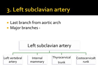
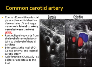
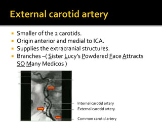
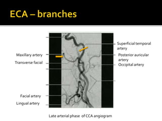
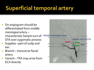
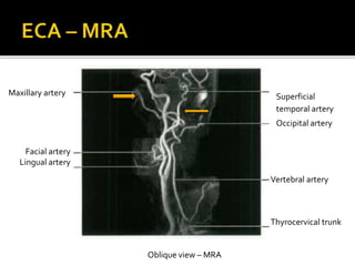
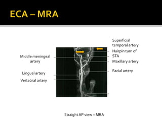
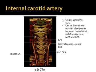
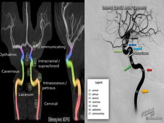
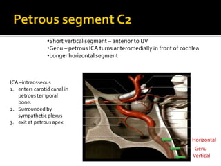
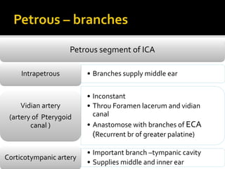
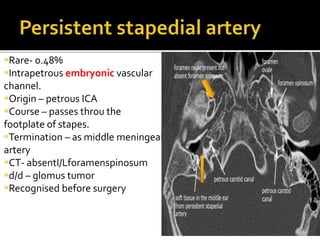
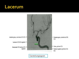
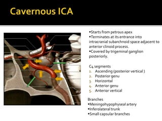
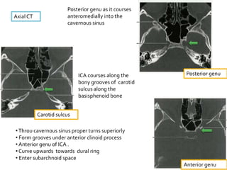
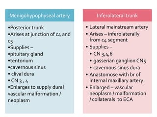
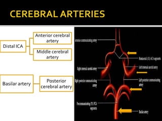
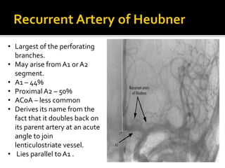
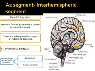
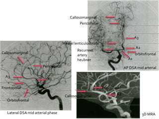
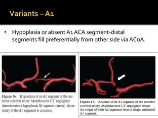
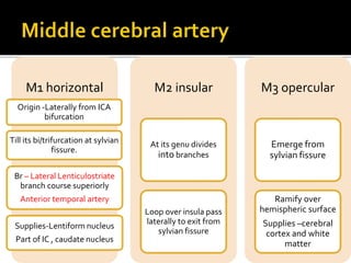
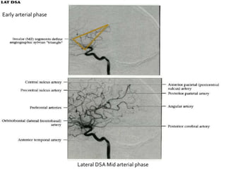
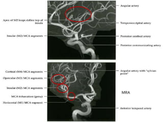
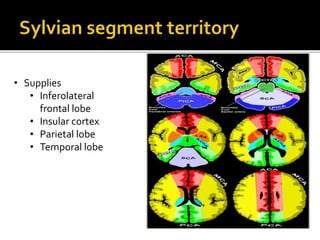
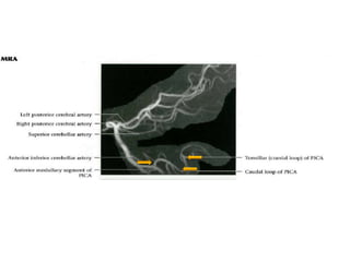
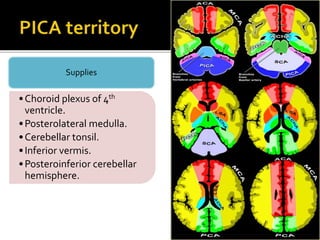
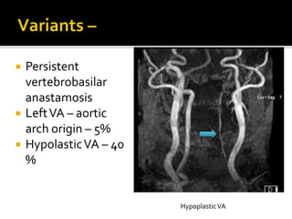
Presenter: Dr. Nikhil panpalia
Guide: Dr.K.R.Naik
 Understanding vascular anatomy is fundamental
to neuroimaging.
 About 18% of the total blood volume in the body circulates in
the brain, which accounts for about 2% of the body weight.
 The blood transports oxygen, nutrients, and other substances
necessary for proper functioning of the brain tissues and carries
away metabolites.
 Loss of consciousness occurs in less than 15 seconds after
blood flow to the brain has stopped, and irreparable damage to
the brain tissue occurs within 5 minutes.
 Cerebrovascular disease or stroke, occurs as a result of
vascular compromise or haemorrhage and is one of the most
frequent sources of neurologic disability.
 Part 1 –
 Aortic arch and great vessels
 Carotid arteries
 Circle ofWillis
 Part 2 –
 Cerebral arteries
 Posterior fossa arteries – vertebrobasilar system
1. Conventional intra-arterial angiography – DSA
system - techniques of image acquisition
 Standard radiographic projections
 carotid angio-
▪ Lateral projection – centered on pituitary fossa
▪ AP view – with petrous ridge projected over the roof of orbit
▪ I/L anterior oblique – for aneurysms in SAH
 Vertebral angio –
▪ lateral , half-axial (Towne’s) and AP – petrous ridge superimposed
on lower border of orbit
2. Computed tomography angiography
3. Magnetic resonance angiography
4. Doppler ultrasound
Starts from aortic arch :
Aortic arch
Innonimate or
brachiocephalic
artery
Left common
carotid
Left subclavian
Brachiocephalic
or innonimate
Rt common
carotid
Rt subclavian
3 . Innonimate artery
10. Left subclavian
artery
15. Left common
carotid artery
4. Right subclavian
artery
5. Right common
carotid artery
Right subclavian artery
Right
vertebral
artery
Internal
mammary artery
Thyrocervical
trunk
Costocervical
trunk
6. Right vertebral artery
9. Internal mammary artery
16.Thyrocervical trunk
16
 Common arch anomaly
 0.5-1% of all cases
 Here it is the last brachiocephalic
vessel arising from aortic arch -4th
branch
 Often asymptomatic – 10 % of
people can have dysphagia lusoria.
 Right common carotid arises
directly from arch – first branch
Barium studies – fixed narrowing of
esophagus at the level of arch
without mucosal deformity –
bayonet deformity
 1st Branch of right subclavian artery
 Right vertebral artery dominant -25%
 Anomalous origin – uncommon
 Arises from proximal brachicephalic
 Only cervical part as it arises caudally
 RCCA – directly from aortic arch ( when right
SCA is aberrant )
RCCA
RSCA
 2nd major branch from aortic arch
 Thoracic and cervical part –in thoracic it travels upwards throu superior
mediastinum to the level of left sternoclavicular joint and continues as
cervical
15.Left common
carotid
CCA bifurcates into ICA and ECA
at midcervical level C3-C6 level.
LCCA- common
origin with IA
 LCCA – hypoplastic
or absent – here the
ECA and ICA arise
directly from aortic
arch
 Last branch from aortic arch
 Major branches -
Left subclavian artery
Left vertebral
artery
Internal
mammary
Thyrocervical
trunk
Costocervicalt
runk
 First branch of left subclavian artery
 Dominant in 50-60%
 In 25% right and leftVA are equal in size
11.Left vertebral artery
14.Left internal mammary
 Left vertebral artery –directly from aortic arch -5%
( nondominant )
RSCA
LSCA
Innominate
artery
LCCA
RCCA
RVA
LVA
 Course - Runs within a fascial
plane – the carotid sheath –
also contains IJV and vagus
nerve( vein lateral to artery ,
nerve between the two)
(VNA)
 Runs obliquely upwards from
the level of sternoclavicular
joint to the level of thyroid
cartilage
 Bifurcates at the level of C3-
C5 into external and internal
carotid artery
 At bifurcation ICA usually lies
posterior and lateral to the
ECA
 Smaller of the 2 carotids.
 Origin anterior and medial to ICA.
 Supplies the extracranial structures.
 Branches –( Sister Lucy’s Powdered Face Attracts
SO Many Medicos )
Internal carotid artery
External carotid artery
Common carotid artery
External carotid artery
Anterior
Superior
thyroidal (sister)
Lingual (lucy)
Facial (face)
Posterior
Occipital
Posterior
auricular
(powdered)
Medial
Ascending
pharyngeal
(attracts)
Terminal
Maxillary (many)
Superficial
temporal (so)
ECA – branches
Superior thyroid artery
Lingual artery
Facial artery
Occipital artery
Posterior auricular
artery
Ascending pharyngeal
artery
Early arterial phase of CCA angiogram
Late arterial phase of CCA angiogram
Posterior auricular
artery
Occipital artery
Facial artery
Lingual artery
Superficial temporal
artery
Maxillary artery
Transverse facial
 Internal maxillary artery-
 Runs forward deep to the
mandible.
 Branches – inferior alveolar,
middle meningeal, deep
temporal , accessory
meningeal , sphenopalatine ,
infraorbital , descending
palatine, muscular branches.
 Middle meningeal artery –
runs superiorly crosses STA on
lateral projection thro
foramen spinosum.
 Supplies – dura and inner
table of skull.
 On angiogram should be
differentiated from middle
meningeal artery –
characteristic hairpin turn of
STA over zygomatic process
 Supplies –part of scalp and
ear.
 Branch – transverse facial
artery
 Variant –TFA may arise from
ECA directly
STA
Middle meningeal artery
hairpin turn of STA
Oblique view – MRA
Vertebral artery
Thyrocervical trunk
Facial artery
Lingual artery
Superficial
temporal artery
Occipital artery
Maxillary artery
Straight AP view – MRA
Superficial
temporal artery
Hairpin turn of
STA
Maxillary artery
Facial artery
Lingual artery
Vertebral artery
Middle meningeal
artery
Left CCA
Right CCA
Internal carotid- carotid
bulb
ECA
3-D CTA
• Origin -Lateral to
ECA.
• Can be divided into
number of segments
between the bulb and
its bifurcation into
MCA and ACA.
Cervical
Intraosseous /
petrous
Lacerum
Cavernous
Intracranial /
supraclinoid
Opthalmic
Communicating
ICA
Carotid bulb
Petrous
Cavernous
Supraclinoid
Cervical
Oblique DSALateral DSA
 Distal 2-4 cm of CCA
 Bulbous dilatation of ICA
origin
 Thinner media and thicker
adventitia containing many
receptor endings of
glossopharyngeal nerve
 No narrowing
 No dilatation
 No branches
 No tapering
Course – crosses
behind and
medial to ECA
ICA
ICA
ECA
 10%- ICA
originates medial
to ECA
 Anomalous ECA
branches arises
from cervical ICA
 Persistent
embryonic vesels
may anastomose
with
vertebrobasilar
system
ICA
ECA
Vertical
•Short vertical segment – anterior to IJV
•Genu – petrous ICA turns anteromedially in front of cochlea
•Longer horizontal segment
ICA –intraosseous
1. enters carotid canal in
petrous temporal
bone.
2. Surrounded by
sympathetic plexus
3. exit at petrous apex
Horizontal
Genu
Petrous segment of ICA
• Branches supply middle earIntrapetrous
• Inconstant
• Throu Foramen lacerum and vidian
canal
• Anastomose with branches of ECA
(Recurrent br of greater palatine)
Vidian artery
(artery of Pterygoid
canal )
• Important branch –tympanic cavity
• Supplies middle and inner ear
Corticotympanic artery
Axial NECT inferior to superior ( bone window )
Aberrant course
•Posterolateral course thro temporal bone
•Vertical segment of carotid canal absent
Normal course of ICA
•Anteromedial course thro temporal bone
•3 segments
Rare- 0.48%
Intrapetrous embryonic vascular
channel.
Origin – petrous ICA
Course – passes throu the
footplate of stapes.
Termination – as middle meningeal
artery
CT- absentI/Lforamenspinosum
d/d – glomus tumor
Recognised before surgery
Small segment that extends from petrous apex above foramen lacerum curving upwards
and then becomes the cavernous segment
Covered by trigeminal ganglion
No branches
Carotid angiogram
C4 segments
1. Ascending (posterior vertical )
2. Posterior genu
3. Horizontal
4. Anterior genu
5. Anterior vertical
Branches
Meningohypophyseal artery
Inferolateral trunk
Small capsular branches
Starts from petrous apex
Terminates at its entrance into
intracranial subarchnoid space adjacent to
anterior clinoid process.
Covered by trigeminal ganglion
posteriorly.
Carotid
angiogram
Axial CT
Posterior genu as it courses
anteromedially into the
cavernous sinus
ICA courses along the
bony grooves of carotid
sulcus along the
basisphenoid bone
•Throu cavernous sinus proper turns superiorly
• Form grooves under anterior clinoid process
• Anterior genu of ICA .
• Curve upwards towards dural ring
• Enter subarchnoid space
Posterior genu
Carotid sulcus
Anterior genu
Menigohypophyseal artery
•Posterior trunk
•Arises at junction of c4 and
c5
•Supplies –
•pituitary gland
•tentorium
•cavernous sinus
• clival dura
• CN 3 , 4
•Enlarges to supply dural
vascular malformation /
neoplasm
Inferolateral trunk
• Lateral mainstream artery
• Arises – inferolaterally
from c4 segment
• Supplies –
• CN 3,4,6
• gasserian ganglion CN5
• cavernous sinus dura
• Anastomose with br of
internal maxillary artery .
• Enlarged – vascular
neoplasm / malformation
/ collaterals to ECA
•Starts distal ly to cavernous sinus
•Ends as near anterior clinoid
process
•No important branches
Extends from superior
clinoid to just below
posterior
communicating artery
(PCoA) origin
Branches –
•Opthalmic artery
•Superior hypophyseal
artery
CECT
Anterior clinoid process C6
Origin –
• Intradural
•Antero-superior ICA
• Medial to anterior clinoid process
Course –
Anterior throu optic canal
Below optic nerve
Crosses superomedially over the nerve
Supply -globe
Gives off ocular , lacrimal , muscular branches
•Anastomose with ECA
Mid arterial phase DSA
Lateral view MRA
Lateral DSA
Arises from posteromedial aspect of
supraclinoid ICA
Course – across the ventral surface of
optic chaisma
Terminates- pituitary stalk and gland
Supplies – anterior pituitary ,
Infundibulum , optic nerve and chaisma
Anastomose - with hypophyseal branch
from the contralateral ICA forms plexus –
superior hypophyseal plexus
DSA – usually not visualized if not
enlarged
•Extends from below PCoA to
terminal ICA bifurcation.
•Passes between optic and
occulumotor nerve.
C7 segment branches
Posterior communicating artery Anterior choroidal artery
Lateral DSA
AChA
PCoA
3D CTA
•Arises – posterior aspect of
intradural ICA just below
anterior choroidal artery
•Course – posterolaterally
above the occulumotor nerve
to join posterior cerebral
artery
•Branches – anterior
thalamoperforating arteries
•Supplies – optic chiasma,
pituitary stalk , thalamus ,
hypothalamus.
Lateral late
arterial DSA
MRA
1. Hypoplasia – 1/3 rd cases
2. PCoA duplication/ fenestraion –
rare
PCoA fenestration
PCoA hypoplasia
• PCoM is larger than P1 segment of PCA and supplies the bulk of PCA . PCA therefore
is a part of anterior circulation
•Infundibular dilatation of PCoA
at origin from ICA- 5-15%
•Should be 2 mm or less
•Funnel shaped , conical
•PCoA arises from apex
Within suprasellar cistern under optic tract
Posteromedially around temporal lobe uncus
Cisternal Course :
Intraventricular course:
AChA angles sharply laterally
Enters choroidal fissure of temporal bone
Abrupt kink – plexal point
AChA-origin few mms above PCoA
Cisternal segment
Intraventricular segment
Supplies
Choroidal plexus of lateral ventricle (
temporal horn and atrium )
Optic tract and cerebral peduncle
Uncal and parahippocampal gyri of
temporal lobe .
Thalamus and posterior limb of internal
capsule.
Anastamoses – with AChA segments and
LPChA and MPChA
Variants – uncommon
Aplasia rare
Hypoplasia – 3 %
Hyperplasia – 2.3 %
AP mid arterial DSA
AP Late arterial DSAMRA lateral view
Choriodal
blush
Terminal ICA
Anterior cerebral artery Middle cerebral artery
3D CTA
MCA
ACA
ICA
3D CTAMid arterial phase DSA
Maxillary artery
• Middle meningeal
artery
• Foramen rotundum
artery
• Accessory meningeal
• Vidian artery
• Ant / mid deep
temporal
ICA
• Ethmoidal br of
opthalmic artery
• Inferlolateral trunk of
ICA
• Inferolateral trunk
• Intratemporal ICA
• Opthalmic artery
• Occipital
• Ascending pharyngeal
artery
• Ascending pharyngeal
artery
• Facial artery
• Posterior auricular
artery
• Vertebral
• Vertebral C3 level
• ICA (petrous and
cavernous )
• ICA (opthalmic artery)
• ICA (stylomastoid
artery)
2ICAs
Horizontal segment A1
of both ACAs
2 Posterior
communicating arteries
Anterior
communicating artery
Horizontal segment P1
of both PCA s
Basilar artery
Interconnected arterial
polygon
Location – surrounds
ventral surface of
diencephalon,
adjacent to optic nerve and
tracts, inferolateral to
hypothalamus
Anterior
circulation
2 B/L ICAs
2ACAs
UnpairedACoA
anteriorly
Posterior
circulation
Basilar bifurcation
from mergedVAs
2PCAs from BAs
B/L PCoAs
3DVRT CTA MRA
CT MRA
1. A1
2. P1
3. PCoA
4. ACoA
• Medial lenticulostriate arteries
• Recurrent artery of HeubnerACAs
• Perforating branches – hypothalamus , optic
chiasma , cingulate gyrus , corpus callosum , fornix
• Large vessel – median artery of corpus callosum
arises from ACoA
ACoA
• Anterior thalamoperforating arteriesPCoA
• Posterior thalamoperforating arteries
• Thalamogeniculate arteries
Basilar artery,
PCAs
Supplies-
1.Optic
chiasma and
tracts
2.Infundibulum
3.Hypothalam
us
4.Base of
brain
 Complete COW –only 20 –
25%
 Posterior circle anomalies –
50% anatomy specimens
Common variants
•Hypoplasia of 1 or both PCoA
– 34%
•Fetal origin of PCA from ICA
•Hypoplasia or absent A1 ACA
segment.
•Absent , duplicate or
multichannelACoA – 10-15%
 Represent persistent embryonic
circulatory patterns
 Channels between caudal carotid
artery and paired basilar and
vertebral arteries fail to regress.
1. Primitive persistent trigeminal artery
2. Primitive hypoglossal artery
3. Persistent otic artery
4. Proatlantal intersegmental artery
PCoA
PTA
Otic
Hypoglossal
Proatlantal
intersegmental
•Most common carotid vertebro basilar anastomoses - 0.1- 0.6%
•In utero – embryonic trigeminal artery supplies basilar artery before the PCoA and vertebral
artery develops
•As these vessels enlarge – PTA normally disappears
course – arise when ICA exists carotid canal and enters
cavernous sinus
Runs posterolaterally along trigeminal nerve 41%
Crosses over / throu dorsum sella before joining
basilar artery
Connects ICA to vertebrobasilar system trident shape on lateral DSA
 2nd most common- 0.027-
0.26%
 .
 Intracranial aneurysms
 If present – single artery that
supplies brain stem and
cerebellum
Courses thro hypoglossal canal
Parallel to CN 12
Connects cervical ICA with basilar
artery
Red – PHA Blue – sigmoid sinus Pink – coil mass with basilar tip
aneurysm
 Origin – petrous ICA
 Course – medially thro internal
auditory meatus and joins caudal
basilar artery
 VA – hypoplastic / absent – POA is the
sole arterial supply to basilar artery
Basilar artery
POA
 Cerebral arteries
 Vertebral artery
 Basilar artery
Distal ICA
Anterior cerebral
artery
Middle cerebral
artery
Basilar artery
Posterior
cerebral artery
A1 horizontal
segment
• From ACA origin to
ACoA junction.
• Inferior br – supply
superior surface of optic
nerve and chaisma.
• Superior br – anterior
hypothalamus , septum
pellucidum , anterior
commisure , fornix ,
anterior inferior portion
of corpus straitum.
Arise fromA1 segment-
perforating branches.
• Pass cephalad
thro anterior
perforated
substance.
• Supply head of
caudate nucleus
and anterior limb
of IC, putamen .
• Largest of the perforating
branches.
• May arise fromA1 or A2
segment.
• A1 – 44%
• Proximal A2 – 50%
• ACoA – less common
• Derives its name from the
fact that it doubles back on
its parent artery at an acute
angle to join
lenticulostriate vessel.
• Lies parallel to A1 .
From ACoA junction
Ascend in front of 3rd ventricle in cistern
of lamina terminalis
br –Orbitofrontal, frontopolar
Curves around corpus callosum genu
gives terminal branches
A2 terminal
branches-
Pericollasal
Collasomarginal
• Supply the anterior 2/3rds
of medial hemispheric
surface + small superior
area over the convexities.
• Callosomarginal a.– lies in
cingulate gyrus supplies
medial frontal lobe
• Pericallosal a.– course
along the posterior aspect
of corpus callosum and
supplies it and medial
parietal lobe
Lateral DSA mid arterial phase
A1
A2
A3
orbitofrontal
Callosomarginal
Pericollasal
Medial lenticulostriate
Recurrent
artery
heubner
Pericollasal
A2
Orbitofrontal
Frontopolar
A3
Callosomarginal
AP DSA mid arterial
3D MRA
A2
Pericollasal
Callosomarginal
 ACA – ACoA complex – normal 1/3rd anatomy
dissection
 Absent , duplicate or multichannelACoA –
10-15%
• Hypoplasia or absent A1 ACA segment-distal
segments fill preferentially from other side via ACoA.
 Fenestration /
duplication of
ACA
M1 horizontal
Origin -Laterally from ICA
bifurcation
Till its bi/trifurcation at sylvian
fissure.
Br – Lateral Lenticulostriate
branch course superiorly
Anterior temporal artery
Supplies-Lentiform nucleus
Part of IC , caudate nucleus
M2 insular
At its genu divides
into branches
Loop over insula pass
laterally to exit from
sylvian fissure
M3 opercular
Emerge from
sylvian fissure
Ramify over
hemispheric surface
Supplies –cerebral
cortex and white
matter
1. Orbitofrontal artery (lateral
frontobasal )
2. Prefrontal arteries
3. Precentral (prerolandic )
4. Central sulcus (rolandic)
5. Postcentral sulcus (anterior
parietal) artery
6. Posterior parietal artery
7. Angular artery
8. Posterior temporal
9. Temporooccipital artery
10. Medial temporal
AP DSA mid arterial phase
AP DSA early arterial phase
Early arterial phase
Lateral DSA Mid arterial phase
Lateral
•M1 horizontal
•MCA bifurcation
•M2 insular
•M3 opercular
CT
MRA
• Origin - M1
• Supplies –
• Part of head
and body of
caudate
• Globus pallidus
• Putamen
• Posterior limb
of internal
capsule
• Supplies
• Inferolateral
frontal lobe
• Insular cortex
• Parietal lobe
• Temporal lobe
Supplies –
• Lateral
cerebrum
• Insula
• Ant- lateral
temporal
lobe
 Less frequent
 Fenestration and duplication
 Single trunk
 Accessory arteries
All uncommon
≤5 %
• It is either hypertrophied RA heubner or medial ACA perforator.
• To be called accessory MCA it should have cortical branches.
PCA origin from bifurcation of basilar artery in interpeduncular cistern.
Lies above occulomotar nerve.
Circles midbrain above tentorium cerebelli.
P1 precommunicating /
peduncular
• Basilar bifurcation extends
laterally
• Junction with PCoA
• Br –
• Post thalamoperforating-
thalamus , midbrain
• Medial posterior choroidal
artery – anteromedially along
roof of 3rd ventricle –tectal
plate , midbrain , thalamus
posterior , pineal gland , tele
choroidae of 3rd ventricle.
P2 ambient / crural
• PCA- PCoA junction posterior
• Above trochlear nerve and
tentorial incisura
• Br –
• Thalamogeniculate arteries-
MGB , pulvinar , brachium
superior colliculus , crus
cerebri , LGB
• Lateral post choroidal artery –
over pulvinar of thalamus –
posterior thalamus , lateral
ventricular choroid plexus
P3 quadrigeminal
Behind midbrain in quadrigeminal
plate cistern
Reciprocal relationship with MCA
Inferior temporal artery
• Undersurface of temporal bone
• Anastamose -MCA
Parietooccipital artery
• Posterior 1/3rd interhemispheric
surface
• ACA
Calcarine artery( P4 )
• Visual cortex
• Occipital pole
Posterior pericollasal artery
(splenial)
• Splenium of corpus callosum
• ACA
AP DSA
AP DSA mid arterial
phase
Early arterial
phase
Lateral DSA Mid
arterial phase
MRA
CTA
• Supply –
• Medial +posterior
temporal lobe
• Medial parietal
lobe
• Occipital lobe
 Fetal origin of PCA from ICA instead of basilar – 15- 20 %
 Carotid basilar anastomosis – supply PCA via trigeminal artery or
other persistent channels
V1 Courses –Cephalad to enter
transverse foramina at C6
Ascend directly to C2 (V2)
Turns laterally and superiorly thro
C1 vertebral foramina
Looping posteriorly along atlasV3
extraspinal
EachVA passes superomedially
thro foramen magnum
In Posterior fossa
anterior to medulla (intradural )
VAs unite to form basilar artery
From subclavian arteries
LeftVA dominant 50%
Vertebral artery
Anterior spinal artery
Joins ASA from oppositeVA
along anteromedial sulcus of cervical
cord.
Medial medullary syndrome
Posterior inferior cerebellar
artery
Arises from distalVA
Lateral medullary syndrome
Lateral DSA
AP DSA
V1- extraosseous
V2 –foraminal
V3 – extraspinal
V4 – intradural
• Front of medullaAnterior medullary segment
• Along side of medulla caudally to level of CN 9-11Lateral medullary segment
• Around inferior half of cerebellar tonsilTonsilomedullary segment
• Cleft btw tela choridae and inferior medullary velum rostrally
and superior pole of tonsil caudallyTelovelotonsillar segment
Cortical / hemispheric
segment
Lateral DSA early
arterial
Lateral DSA
late arterial
Anterior medullary segment
Posterior medullary segment
Lateral medullary segment
•Choroid plexus of 4th
ventricle.
•Posterolateral medulla.
•Cerebellar tonsil.
•Inferior vermis.
•Posteroinferior cerebellar
hemisphere.
Supplies
 Persistent
vertebrobasilar
anastamosis
 LeftVA – aortic
arch origin – 5%
 HypolasticVA – 40
%
HypoplasticVA
VA terminates in PICA – 1%
Orange arrow –
duplicatedVA
Red – originalVA from
subclavian
VA duplication- ocassionally
VA fenestration –
occasionally
Extradural origin of PICA
PICA fromVA below foramen magnum
Right and leftVA s
unite – BA
Course cephalad in front of
pons
Pontine cistern in the
space delineated by
lateral margin of clivus
and dorsum sellae
Terminates in
interpeduncular cistern
Divides into PCAs
•Average length – 3 cm
•Width 1.5- 4 mm
•Diameter <4.5 mm
1. AICA – Anterior Inferior
CerebellarArtery
 1st major branch.
 Posterior laterally in
cerebellopontine angle cistern
toward the internal auditory canal.
Here typically anteroinferior to
facial and vestibulocochlear nerve.
 Few mms from origin AICA crossed
by abducens nerve.
 Supplies-
▪ Nerves
▪ Inferolateral pons
▪ Middle cerebellar peduncle
▪ Flocculus
▪ Anterolateral cerebelllar hemisphere
2. SCA- SuperiorCerebellar
Artery –
 Arises from BA apex.
 Posterolaterally around Pons
and mesencephalon below
tentorial incisura and CNS 3 n
4.
 Supplies –
▪ Superior surface of vermis n
cerebellar hemisphere.
▪ Deep cerebellar white matter.
▪ Dentate nucleus.
 Perforating branches – short
n long segment
 BA – terminates into PCA s
AP DSA
MRA
SCAs- can
arise from P1
segment
 Osborne radiology
 Gionni Boris bradac –Cerebral angiography
Normal cerebral angiogram final

Normal cerebral angiogram final

  • 1.
    Presenter: Dr. Nikhilpanpalia Guide: Dr.K.R.Naik
  • 2.
     Understanding vascularanatomy is fundamental to neuroimaging.  About 18% of the total blood volume in the body circulates in the brain, which accounts for about 2% of the body weight.  The blood transports oxygen, nutrients, and other substances necessary for proper functioning of the brain tissues and carries away metabolites.  Loss of consciousness occurs in less than 15 seconds after blood flow to the brain has stopped, and irreparable damage to the brain tissue occurs within 5 minutes.  Cerebrovascular disease or stroke, occurs as a result of vascular compromise or haemorrhage and is one of the most frequent sources of neurologic disability.
  • 3.
     Part 1–  Aortic arch and great vessels  Carotid arteries  Circle ofWillis  Part 2 –  Cerebral arteries  Posterior fossa arteries – vertebrobasilar system
  • 4.
    1. Conventional intra-arterialangiography – DSA system - techniques of image acquisition  Standard radiographic projections  carotid angio- ▪ Lateral projection – centered on pituitary fossa ▪ AP view – with petrous ridge projected over the roof of orbit ▪ I/L anterior oblique – for aneurysms in SAH  Vertebral angio – ▪ lateral , half-axial (Towne’s) and AP – petrous ridge superimposed on lower border of orbit 2. Computed tomography angiography 3. Magnetic resonance angiography 4. Doppler ultrasound
  • 5.
    Starts from aorticarch : Aortic arch Innonimate or brachiocephalic artery Left common carotid Left subclavian Brachiocephalic or innonimate Rt common carotid Rt subclavian
  • 6.
    3 . Innonimateartery 10. Left subclavian artery 15. Left common carotid artery
  • 7.
    4. Right subclavian artery 5.Right common carotid artery
  • 8.
    Right subclavian artery Right vertebral artery Internal mammaryartery Thyrocervical trunk Costocervical trunk
  • 9.
    6. Right vertebralartery 9. Internal mammary artery 16.Thyrocervical trunk 16
  • 10.
     Common archanomaly  0.5-1% of all cases  Here it is the last brachiocephalic vessel arising from aortic arch -4th branch  Often asymptomatic – 10 % of people can have dysphagia lusoria.  Right common carotid arises directly from arch – first branch
  • 11.
    Barium studies –fixed narrowing of esophagus at the level of arch without mucosal deformity – bayonet deformity
  • 12.
     1st Branchof right subclavian artery  Right vertebral artery dominant -25%  Anomalous origin – uncommon
  • 13.
     Arises fromproximal brachicephalic  Only cervical part as it arises caudally
  • 14.
     RCCA –directly from aortic arch ( when right SCA is aberrant ) RCCA RSCA
  • 15.
     2nd majorbranch from aortic arch  Thoracic and cervical part –in thoracic it travels upwards throu superior mediastinum to the level of left sternoclavicular joint and continues as cervical 15.Left common carotid CCA bifurcates into ICA and ECA at midcervical level C3-C6 level.
  • 16.
  • 17.
     LCCA –hypoplastic or absent – here the ECA and ICA arise directly from aortic arch
  • 18.
     Last branchfrom aortic arch  Major branches - Left subclavian artery Left vertebral artery Internal mammary Thyrocervical trunk Costocervicalt runk
  • 19.
     First branchof left subclavian artery  Dominant in 50-60%  In 25% right and leftVA are equal in size 11.Left vertebral artery 14.Left internal mammary
  • 20.
     Left vertebralartery –directly from aortic arch -5% ( nondominant )
  • 21.
  • 22.
     Course -Runs within a fascial plane – the carotid sheath – also contains IJV and vagus nerve( vein lateral to artery , nerve between the two) (VNA)  Runs obliquely upwards from the level of sternoclavicular joint to the level of thyroid cartilage  Bifurcates at the level of C3- C5 into external and internal carotid artery  At bifurcation ICA usually lies posterior and lateral to the ECA
  • 23.
     Smaller ofthe 2 carotids.  Origin anterior and medial to ICA.  Supplies the extracranial structures.  Branches –( Sister Lucy’s Powdered Face Attracts SO Many Medicos ) Internal carotid artery External carotid artery Common carotid artery
  • 24.
    External carotid artery Anterior Superior thyroidal(sister) Lingual (lucy) Facial (face) Posterior Occipital Posterior auricular (powdered) Medial Ascending pharyngeal (attracts) Terminal Maxillary (many) Superficial temporal (so)
  • 25.
  • 26.
    Superior thyroid artery Lingualartery Facial artery Occipital artery Posterior auricular artery Ascending pharyngeal artery Early arterial phase of CCA angiogram
  • 27.
    Late arterial phaseof CCA angiogram Posterior auricular artery Occipital artery Facial artery Lingual artery Superficial temporal artery Maxillary artery Transverse facial
  • 28.
     Internal maxillaryartery-  Runs forward deep to the mandible.  Branches – inferior alveolar, middle meningeal, deep temporal , accessory meningeal , sphenopalatine , infraorbital , descending palatine, muscular branches.  Middle meningeal artery – runs superiorly crosses STA on lateral projection thro foramen spinosum.  Supplies – dura and inner table of skull.
  • 29.
     On angiogramshould be differentiated from middle meningeal artery – characteristic hairpin turn of STA over zygomatic process  Supplies –part of scalp and ear.  Branch – transverse facial artery  Variant –TFA may arise from ECA directly STA Middle meningeal artery hairpin turn of STA
  • 30.
    Oblique view –MRA Vertebral artery Thyrocervical trunk Facial artery Lingual artery Superficial temporal artery Occipital artery Maxillary artery
  • 31.
    Straight AP view– MRA Superficial temporal artery Hairpin turn of STA Maxillary artery Facial artery Lingual artery Vertebral artery Middle meningeal artery
  • 32.
    Left CCA Right CCA Internalcarotid- carotid bulb ECA 3-D CTA • Origin -Lateral to ECA. • Can be divided into number of segments between the bulb and its bifurcation into MCA and ACA.
  • 33.
  • 34.
  • 35.
     Distal 2-4cm of CCA  Bulbous dilatation of ICA origin  Thinner media and thicker adventitia containing many receptor endings of glossopharyngeal nerve
  • 36.
     No narrowing No dilatation  No branches  No tapering Course – crosses behind and medial to ECA ICA ICA ECA
  • 37.
     10%- ICA originatesmedial to ECA  Anomalous ECA branches arises from cervical ICA  Persistent embryonic vesels may anastomose with vertebrobasilar system ICA ECA
  • 38.
    Vertical •Short vertical segment– anterior to IJV •Genu – petrous ICA turns anteromedially in front of cochlea •Longer horizontal segment ICA –intraosseous 1. enters carotid canal in petrous temporal bone. 2. Surrounded by sympathetic plexus 3. exit at petrous apex Horizontal Genu
  • 39.
    Petrous segment ofICA • Branches supply middle earIntrapetrous • Inconstant • Throu Foramen lacerum and vidian canal • Anastomose with branches of ECA (Recurrent br of greater palatine) Vidian artery (artery of Pterygoid canal ) • Important branch –tympanic cavity • Supplies middle and inner ear Corticotympanic artery
  • 40.
    Axial NECT inferiorto superior ( bone window )
  • 42.
    Aberrant course •Posterolateral coursethro temporal bone •Vertical segment of carotid canal absent Normal course of ICA •Anteromedial course thro temporal bone •3 segments
  • 43.
    Rare- 0.48% Intrapetrous embryonicvascular channel. Origin – petrous ICA Course – passes throu the footplate of stapes. Termination – as middle meningeal artery CT- absentI/Lforamenspinosum d/d – glomus tumor Recognised before surgery
  • 44.
    Small segment thatextends from petrous apex above foramen lacerum curving upwards and then becomes the cavernous segment Covered by trigeminal ganglion No branches
  • 45.
  • 46.
    C4 segments 1. Ascending(posterior vertical ) 2. Posterior genu 3. Horizontal 4. Anterior genu 5. Anterior vertical Branches Meningohypophyseal artery Inferolateral trunk Small capsular branches Starts from petrous apex Terminates at its entrance into intracranial subarchnoid space adjacent to anterior clinoid process. Covered by trigeminal ganglion posteriorly.
  • 47.
  • 48.
    Axial CT Posterior genuas it courses anteromedially into the cavernous sinus ICA courses along the bony grooves of carotid sulcus along the basisphenoid bone •Throu cavernous sinus proper turns superiorly • Form grooves under anterior clinoid process • Anterior genu of ICA . • Curve upwards towards dural ring • Enter subarchnoid space Posterior genu Carotid sulcus Anterior genu
  • 50.
    Menigohypophyseal artery •Posterior trunk •Arisesat junction of c4 and c5 •Supplies – •pituitary gland •tentorium •cavernous sinus • clival dura • CN 3 , 4 •Enlarges to supply dural vascular malformation / neoplasm Inferolateral trunk • Lateral mainstream artery • Arises – inferolaterally from c4 segment • Supplies – • CN 3,4,6 • gasserian ganglion CN5 • cavernous sinus dura • Anastomose with br of internal maxillary artery . • Enlarged – vascular neoplasm / malformation / collaterals to ECA
  • 51.
    •Starts distal lyto cavernous sinus •Ends as near anterior clinoid process •No important branches
  • 52.
    Extends from superior clinoidto just below posterior communicating artery (PCoA) origin Branches – •Opthalmic artery •Superior hypophyseal artery CECT Anterior clinoid process C6
  • 53.
    Origin – • Intradural •Antero-superiorICA • Medial to anterior clinoid process Course – Anterior throu optic canal Below optic nerve Crosses superomedially over the nerve Supply -globe Gives off ocular , lacrimal , muscular branches •Anastomose with ECA
  • 54.
    Mid arterial phaseDSA Lateral view MRA Lateral DSA
  • 55.
    Arises from posteromedialaspect of supraclinoid ICA Course – across the ventral surface of optic chaisma Terminates- pituitary stalk and gland Supplies – anterior pituitary , Infundibulum , optic nerve and chaisma Anastomose - with hypophyseal branch from the contralateral ICA forms plexus – superior hypophyseal plexus DSA – usually not visualized if not enlarged
  • 56.
    •Extends from belowPCoA to terminal ICA bifurcation. •Passes between optic and occulumotor nerve. C7 segment branches Posterior communicating artery Anterior choroidal artery Lateral DSA AChA PCoA 3D CTA
  • 57.
    •Arises – posterioraspect of intradural ICA just below anterior choroidal artery •Course – posterolaterally above the occulumotor nerve to join posterior cerebral artery •Branches – anterior thalamoperforating arteries •Supplies – optic chiasma, pituitary stalk , thalamus , hypothalamus. Lateral late arterial DSA MRA
  • 58.
    1. Hypoplasia –1/3 rd cases 2. PCoA duplication/ fenestraion – rare PCoA fenestration PCoA hypoplasia
  • 59.
    • PCoM islarger than P1 segment of PCA and supplies the bulk of PCA . PCA therefore is a part of anterior circulation
  • 60.
    •Infundibular dilatation ofPCoA at origin from ICA- 5-15% •Should be 2 mm or less •Funnel shaped , conical •PCoA arises from apex
  • 61.
    Within suprasellar cisternunder optic tract Posteromedially around temporal lobe uncus Cisternal Course : Intraventricular course: AChA angles sharply laterally Enters choroidal fissure of temporal bone Abrupt kink – plexal point AChA-origin few mms above PCoA Cisternal segment Intraventricular segment
  • 62.
    Supplies Choroidal plexus oflateral ventricle ( temporal horn and atrium ) Optic tract and cerebral peduncle Uncal and parahippocampal gyri of temporal lobe . Thalamus and posterior limb of internal capsule. Anastamoses – with AChA segments and LPChA and MPChA Variants – uncommon Aplasia rare Hypoplasia – 3 % Hyperplasia – 2.3 % AP mid arterial DSA AP Late arterial DSAMRA lateral view Choriodal blush
  • 63.
    Terminal ICA Anterior cerebralartery Middle cerebral artery 3D CTA MCA ACA ICA 3D CTAMid arterial phase DSA
  • 65.
    Maxillary artery • Middlemeningeal artery • Foramen rotundum artery • Accessory meningeal • Vidian artery • Ant / mid deep temporal ICA • Ethmoidal br of opthalmic artery • Inferlolateral trunk of ICA • Inferolateral trunk • Intratemporal ICA • Opthalmic artery
  • 66.
    • Occipital • Ascendingpharyngeal artery • Ascending pharyngeal artery • Facial artery • Posterior auricular artery • Vertebral • Vertebral C3 level • ICA (petrous and cavernous ) • ICA (opthalmic artery) • ICA (stylomastoid artery)
  • 67.
    2ICAs Horizontal segment A1 ofboth ACAs 2 Posterior communicating arteries Anterior communicating artery Horizontal segment P1 of both PCA s Basilar artery
  • 68.
    Interconnected arterial polygon Location –surrounds ventral surface of diencephalon, adjacent to optic nerve and tracts, inferolateral to hypothalamus Anterior circulation 2 B/L ICAs 2ACAs UnpairedACoA anteriorly Posterior circulation Basilar bifurcation from mergedVAs 2PCAs from BAs B/L PCoAs
  • 69.
    3DVRT CTA MRA CTMRA 1. A1 2. P1 3. PCoA 4. ACoA
  • 70.
    • Medial lenticulostriatearteries • Recurrent artery of HeubnerACAs • Perforating branches – hypothalamus , optic chiasma , cingulate gyrus , corpus callosum , fornix • Large vessel – median artery of corpus callosum arises from ACoA ACoA • Anterior thalamoperforating arteriesPCoA • Posterior thalamoperforating arteries • Thalamogeniculate arteries Basilar artery, PCAs Supplies- 1.Optic chiasma and tracts 2.Infundibulum 3.Hypothalam us 4.Base of brain
  • 71.
     Complete COW–only 20 – 25%  Posterior circle anomalies – 50% anatomy specimens Common variants •Hypoplasia of 1 or both PCoA – 34% •Fetal origin of PCA from ICA
  • 72.
    •Hypoplasia or absentA1 ACA segment. •Absent , duplicate or multichannelACoA – 10-15%
  • 73.
     Represent persistentembryonic circulatory patterns  Channels between caudal carotid artery and paired basilar and vertebral arteries fail to regress. 1. Primitive persistent trigeminal artery 2. Primitive hypoglossal artery 3. Persistent otic artery 4. Proatlantal intersegmental artery PCoA PTA Otic Hypoglossal Proatlantal intersegmental
  • 74.
    •Most common carotidvertebro basilar anastomoses - 0.1- 0.6% •In utero – embryonic trigeminal artery supplies basilar artery before the PCoA and vertebral artery develops •As these vessels enlarge – PTA normally disappears course – arise when ICA exists carotid canal and enters cavernous sinus Runs posterolaterally along trigeminal nerve 41% Crosses over / throu dorsum sella before joining basilar artery Connects ICA to vertebrobasilar system trident shape on lateral DSA
  • 75.
     2nd mostcommon- 0.027- 0.26%  .  Intracranial aneurysms  If present – single artery that supplies brain stem and cerebellum Courses thro hypoglossal canal Parallel to CN 12 Connects cervical ICA with basilar artery
  • 76.
    Red – PHABlue – sigmoid sinus Pink – coil mass with basilar tip aneurysm
  • 77.
     Origin –petrous ICA  Course – medially thro internal auditory meatus and joins caudal basilar artery  VA – hypoplastic / absent – POA is the sole arterial supply to basilar artery Basilar artery POA
  • 78.
     Cerebral arteries Vertebral artery  Basilar artery
  • 79.
    Distal ICA Anterior cerebral artery Middlecerebral artery Basilar artery Posterior cerebral artery
  • 80.
    A1 horizontal segment • FromACA origin to ACoA junction. • Inferior br – supply superior surface of optic nerve and chaisma. • Superior br – anterior hypothalamus , septum pellucidum , anterior commisure , fornix , anterior inferior portion of corpus straitum.
  • 81.
    Arise fromA1 segment- perforatingbranches. • Pass cephalad thro anterior perforated substance. • Supply head of caudate nucleus and anterior limb of IC, putamen .
  • 82.
    • Largest ofthe perforating branches. • May arise fromA1 or A2 segment. • A1 – 44% • Proximal A2 – 50% • ACoA – less common • Derives its name from the fact that it doubles back on its parent artery at an acute angle to join lenticulostriate vessel. • Lies parallel to A1 .
  • 83.
    From ACoA junction Ascendin front of 3rd ventricle in cistern of lamina terminalis br –Orbitofrontal, frontopolar Curves around corpus callosum genu gives terminal branches A2 terminal branches- Pericollasal Collasomarginal
  • 84.
    • Supply theanterior 2/3rds of medial hemispheric surface + small superior area over the convexities. • Callosomarginal a.– lies in cingulate gyrus supplies medial frontal lobe • Pericallosal a.– course along the posterior aspect of corpus callosum and supplies it and medial parietal lobe
  • 85.
    Lateral DSA midarterial phase A1 A2 A3 orbitofrontal Callosomarginal Pericollasal Medial lenticulostriate Recurrent artery heubner Pericollasal A2 Orbitofrontal Frontopolar A3 Callosomarginal AP DSA mid arterial 3D MRA A2 Pericollasal Callosomarginal
  • 86.
     ACA –ACoA complex – normal 1/3rd anatomy dissection  Absent , duplicate or multichannelACoA – 10-15%
  • 87.
    • Hypoplasia orabsent A1 ACA segment-distal segments fill preferentially from other side via ACoA.
  • 88.
  • 89.
    M1 horizontal Origin -Laterallyfrom ICA bifurcation Till its bi/trifurcation at sylvian fissure. Br – Lateral Lenticulostriate branch course superiorly Anterior temporal artery Supplies-Lentiform nucleus Part of IC , caudate nucleus M2 insular At its genu divides into branches Loop over insula pass laterally to exit from sylvian fissure M3 opercular Emerge from sylvian fissure Ramify over hemispheric surface Supplies –cerebral cortex and white matter
  • 92.
    1. Orbitofrontal artery(lateral frontobasal ) 2. Prefrontal arteries 3. Precentral (prerolandic ) 4. Central sulcus (rolandic) 5. Postcentral sulcus (anterior parietal) artery 6. Posterior parietal artery 7. Angular artery 8. Posterior temporal 9. Temporooccipital artery 10. Medial temporal
  • 93.
    AP DSA midarterial phase AP DSA early arterial phase
  • 94.
    Early arterial phase LateralDSA Mid arterial phase
  • 95.
  • 96.
  • 97.
    • Origin -M1 • Supplies – • Part of head and body of caudate • Globus pallidus • Putamen • Posterior limb of internal capsule
  • 98.
    • Supplies • Inferolateral frontallobe • Insular cortex • Parietal lobe • Temporal lobe
  • 99.
    Supplies – • Lateral cerebrum •Insula • Ant- lateral temporal lobe
  • 100.
     Less frequent Fenestration and duplication  Single trunk  Accessory arteries All uncommon ≤5 %
  • 102.
    • It iseither hypertrophied RA heubner or medial ACA perforator. • To be called accessory MCA it should have cortical branches.
  • 103.
    PCA origin frombifurcation of basilar artery in interpeduncular cistern. Lies above occulomotar nerve. Circles midbrain above tentorium cerebelli.
  • 104.
    P1 precommunicating / peduncular •Basilar bifurcation extends laterally • Junction with PCoA • Br – • Post thalamoperforating- thalamus , midbrain • Medial posterior choroidal artery – anteromedially along roof of 3rd ventricle –tectal plate , midbrain , thalamus posterior , pineal gland , tele choroidae of 3rd ventricle. P2 ambient / crural • PCA- PCoA junction posterior • Above trochlear nerve and tentorial incisura • Br – • Thalamogeniculate arteries- MGB , pulvinar , brachium superior colliculus , crus cerebri , LGB • Lateral post choroidal artery – over pulvinar of thalamus – posterior thalamus , lateral ventricular choroid plexus
  • 106.
    P3 quadrigeminal Behind midbrainin quadrigeminal plate cistern Reciprocal relationship with MCA Inferior temporal artery • Undersurface of temporal bone • Anastamose -MCA Parietooccipital artery • Posterior 1/3rd interhemispheric surface • ACA Calcarine artery( P4 ) • Visual cortex • Occipital pole Posterior pericollasal artery (splenial) • Splenium of corpus callosum • ACA
  • 107.
    AP DSA AP DSAmid arterial phase
  • 108.
  • 109.
  • 110.
    • Supply – •Medial +posterior temporal lobe • Medial parietal lobe • Occipital lobe
  • 111.
     Fetal originof PCA from ICA instead of basilar – 15- 20 %  Carotid basilar anastomosis – supply PCA via trigeminal artery or other persistent channels
  • 112.
    V1 Courses –Cephaladto enter transverse foramina at C6 Ascend directly to C2 (V2) Turns laterally and superiorly thro C1 vertebral foramina Looping posteriorly along atlasV3 extraspinal EachVA passes superomedially thro foramen magnum In Posterior fossa anterior to medulla (intradural ) VAs unite to form basilar artery From subclavian arteries LeftVA dominant 50%
  • 113.
    Vertebral artery Anterior spinalartery Joins ASA from oppositeVA along anteromedial sulcus of cervical cord. Medial medullary syndrome Posterior inferior cerebellar artery Arises from distalVA Lateral medullary syndrome
  • 115.
    Lateral DSA AP DSA V1-extraosseous V2 –foraminal V3 – extraspinal V4 – intradural
  • 116.
    • Front ofmedullaAnterior medullary segment • Along side of medulla caudally to level of CN 9-11Lateral medullary segment • Around inferior half of cerebellar tonsilTonsilomedullary segment • Cleft btw tela choridae and inferior medullary velum rostrally and superior pole of tonsil caudallyTelovelotonsillar segment Cortical / hemispheric segment
  • 117.
  • 119.
    Anterior medullary segment Posteriormedullary segment Lateral medullary segment
  • 120.
    •Choroid plexus of4th ventricle. •Posterolateral medulla. •Cerebellar tonsil. •Inferior vermis. •Posteroinferior cerebellar hemisphere. Supplies
  • 121.
     Persistent vertebrobasilar anastamosis  LeftVA– aortic arch origin – 5%  HypolasticVA – 40 % HypoplasticVA
  • 122.
    VA terminates inPICA – 1%
  • 123.
    Orange arrow – duplicatedVA Red– originalVA from subclavian VA duplication- ocassionally
  • 124.
  • 125.
    Extradural origin ofPICA PICA fromVA below foramen magnum
  • 126.
    Right and leftVAs unite – BA Course cephalad in front of pons Pontine cistern in the space delineated by lateral margin of clivus and dorsum sellae Terminates in interpeduncular cistern Divides into PCAs •Average length – 3 cm •Width 1.5- 4 mm •Diameter <4.5 mm
  • 127.
    1. AICA –Anterior Inferior CerebellarArtery  1st major branch.  Posterior laterally in cerebellopontine angle cistern toward the internal auditory canal. Here typically anteroinferior to facial and vestibulocochlear nerve.  Few mms from origin AICA crossed by abducens nerve.  Supplies- ▪ Nerves ▪ Inferolateral pons ▪ Middle cerebellar peduncle ▪ Flocculus ▪ Anterolateral cerebelllar hemisphere
  • 128.
    2. SCA- SuperiorCerebellar Artery–  Arises from BA apex.  Posterolaterally around Pons and mesencephalon below tentorial incisura and CNS 3 n 4.  Supplies – ▪ Superior surface of vermis n cerebellar hemisphere. ▪ Deep cerebellar white matter. ▪ Dentate nucleus.  Perforating branches – short n long segment  BA – terminates into PCA s
  • 129.
  • 131.
  • 135.
  • 136.
     Osborne radiology Gionni Boris bradac –Cerebral angiography

Editor's Notes

  • #12 Vascualr structurs sbranching from the right side of superior aortic arch – aberrant rt sca
  • #28 Transverse facial – branch of sta
  • #29 Pterygoid muscle divede into 3 parts
  • #90 Larger of two 2 terminal icas
  • #117 9 glosso 10 vagus 1 1 spinal accessory