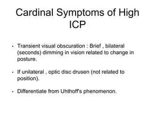
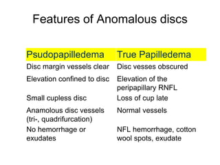
Is This Disc Normal ?
Raed Behbehani , MD FRCSC
Introduction
• Ophthalmologist are called to evaluate optic discs
frequently .
• R/O Papilledema , infiltrative processes,
compression or atrophy.
• Anomalous disc is not a diseases disc .
History
• Asymptomatic
• Loss of vision , eye pain .
• Cardinal symptoms of high intracranial pressure.
Cardinal Symptoms of High
ICP
• Headache : holocranial , tension-like band , worse
upon awakening and in supine position and is
reduced when they are active.
• Migraine : aura, unilateral , throbbing , light
sensitivity , phonophobia, nausea and vomiting ,
family history of migraine and car sickness.
Cardinal Symptoms of High
ICP
• Tinnitus : “swishing” sound , synchronous with pulse
and worse when supine. (transmission of high ICP
causes turbulent flow in venous sinuses).
• Tinnitus is very rare in other types of headaches .
Cardinal Symptoms of High
ICP
• Transient visual obscuration : Brief , bilateral
(seconds) dimming in vision related to change in
posture.
• If unilateral , optic disc drusen (not related to
position).
• Differentiate from Uhthoff's phenomenon.
Cardinal Symptoms of High
ICP
• Diplopia : Binocular horizontal (sixth nerve paresis).
Examination
• Optic nerve cup : usually small or absent in anomalous
optic disc. In papilledema it is present until late stage.
• Vessel branching : trifurcations , loops , increased
branching
• Blood vessels dilated , congested
• opacification of the RNFL —> blurring of vessels—
>hemorrhages and exudates
• Spontaneous venous pulsations : can be absent in normal.
Normal vs Anomalous
Early Papilledema
“as the disc swells lateralwards, it displaces the retina.- throwing it into a series of folds
which run concentric with the edge of the disc. This lateral bulging is due to the distension
of the most peripheral nerve fibers”
Paton and Holmes 1911
Papilledema ?
http://library.med.utah.edu/NOVEL/Hoyt
Papilledema ?
http://library.med.utah.edu/NOVEL/Hoyt
Features of Anomalous discs
Psudopapilledema True Papilledema
Disc margin vessels clear Disc vesses obscured
Elevation confined to disc Elevation of the
peripapillary RNFL
Small cupless disc Loss of cup late
Anamolous disc vessels
(tri-, quadrifurcation)
Normal vessels
No hemorrhage or
exudates
NFL hemorrhage, cotton
wool spots, exudate
Ancillary Testing
• Ultrasound : Hyper-reflective bodies
• OCT : Can be useful in differentiating true vs false
papilledema , buried drusen sometimes do not to exhibit a
significant difference in reflectivity from surrounding disc
tissue.
• Fluorecin Angiogroaphy : Red-free (drusen) , staining and
leakage with fluorescin (inflammation).
• Neuro-imaging : MRI, CT
• LP
Patton’s lines
Outer retinal folds (ORF)
Patton’s lines
Peripapillary Wrinkles (PPW)
Copyright © 2016 Journal of Neuro-Ophthalmology. Published by Lippincott Williams & Wilkins.
Deformation in Bruch’s Membrane
FIG. 6. A. Optic disc tissue in a patient with
papilledema, showing the upward angling and
displacement of Bruch's membrane (red
arrows) in the right and left eyes. B. There is a
change in displacement of Bruch's membrane
in the same eye before and after treatment of
raised intercranial pressure (23).
17
Optical Coherence Tomography in
Papilledema: What Am I Missing?
Kardon, Randy
Journal of Neuro-Ophthalmology. 34():S10-
S17, September 2014.
doi: 10.1097/WNO.0000000000000162
Copyright © 2016 Journal of Neuro-Ophthalmology. Published by Lippincott Williams & Wilkins.
OCT in Distinguishing Papilledema vs Pseudopapilledema
FIG. 7. A very large, coalescing druse imaged
in several SD-OCT modalities. A. Fundus
photo with 2 vertical markers placed on
either side of the druse (obtained with 3D
disc scan on Topcon 3D OCT 2000). B. Low-
resolution SD-OCT image, obtained on same
3D disc scan. C. High-resolution image,
obtained with 7-line Raster on Topcon 3D
OCT 2000. D. High-resolution (5-line Raster)
image, obtained with Zeiss Cirrus HD-OCT
(30). SD-OCT, spectral domain optical
coherence tomography.
18
Optical Coherence Tomography in
Papilledema: What Am I Missing?
Kardon, Randy
Journal of Neuro-Ophthalmology. 34():S10-
S17, September 2014.
doi: 10.1097/WNO.0000000000000162
Optic Nerve Drusen
• Most common cause for anomalous discs
• 2% population
• Can be superficial or buried.
• “Scalloped” disc margin.
Optic Nerve Drusen
Buried Drusen
SDOCT in Buried Drusen
Focal, round, hyper-reflective mass causing nasal optic disc elevation with an irregular contour
(arrows).
Disc Drusen
Tilted Disc
• Myopia
• Oblique insertion
(tilted) , margin
indistinct
Myelinated Nerve Fibers
• Bight-white , feathery
appearance over the retina.
Little red disc
Peri-papillary Atrophy
Case
 Failed a driving test.
 Started on Xalatan, two months later IOP was 19 OU.
 Has been driving for the last 10 years “with no
accidants”.Strabismic amblyopia OD , Strabismus
surgery.
 High Myopia ( -7 D OU ).
 Mother : Diabetes.
Segmental Optic Nerve
Hypoplasia
Segmental Optic Nerve
Hypoplasia
• Maternal diabetes in 1s trimester.
• No other systemic abnormalities.
• Can be found in families with no history of maternal
diabetes.
• Interference in gestational development of superior
retinal ganglion cells or their axons.
• Occasionally associated congenital lesions of the retina
, chiasm, and posterior visual pathway.
Segmental ONH
Homonymous Hemioptic Hypoplasia
140 By the kind permission of Professor WF Hoyt.
Morning Glory Disc Anomaly
• Funnel-shaped
excavation with
peripapillary pigment
changes.
• Usually unilateral .
• Central glial tuft.
• Vessels emanate from
periphery.
• MRI/MRA is warranted
Morning Glory Disc Anomaly
• Wide head , flat nose , hypertelorism , midline notch in
upper lip or palate.
• Agenesis of cropus callosum, achiasmia , posterior
dilatation of lateral ventricles.
• Panhypopitutarism.
• Ipsilateral intracranial vascular dysgensis (hypoplasia of
carotid and major vessels.
• PHACE syndrome (posterior fossa , facial hemangioma ,
arterial anamolies , cardiac and aorta and eye) anomalies.
PVL
Optic Nerve Coloboma
• Failure of closure of embryonic
fissure.
• Deep excavation more
prominent inferiorly.
• Unilateral or bilateral.
• Serous macular detachment.
• CHARGE , Aicardi syndrome,
Goldenhar.
Morning Glory Disc Coloboma
Symmetric defect Asymmetric (inferior)
central glial tuft No glial tuft
Peripapilalry pigmentary
changes
Minimal Peripapillary
pigmentary changes
Anomalous retinal vessles Normal vasculature
Aicardi Syndrome
• Infantile spasms , agenesis of
corpus callous, abnormal EEG.
• Anolmalous disc and
peripapillry chorioretinal
lacunae.
• Coloboma , ONH , and
pigmentation.
• Facial skeletal and vertebral
abnormalities.
Papillorenal syndrome
• No central vessels in
excavation (vacant disc)
• Progressive renal disease and
later renal failure.

Is This Disc Normal ?

  • 1.
    Is This DiscNormal ? Raed Behbehani , MD FRCSC
  • 2.
    Introduction • Ophthalmologist arecalled to evaluate optic discs frequently . • R/O Papilledema , infiltrative processes, compression or atrophy. • Anomalous disc is not a diseases disc .
  • 3.
    History • Asymptomatic • Lossof vision , eye pain . • Cardinal symptoms of high intracranial pressure.
  • 4.
    Cardinal Symptoms ofHigh ICP • Headache : holocranial , tension-like band , worse upon awakening and in supine position and is reduced when they are active. • Migraine : aura, unilateral , throbbing , light sensitivity , phonophobia, nausea and vomiting , family history of migraine and car sickness.
  • 5.
    Cardinal Symptoms ofHigh ICP • Tinnitus : “swishing” sound , synchronous with pulse and worse when supine. (transmission of high ICP causes turbulent flow in venous sinuses). • Tinnitus is very rare in other types of headaches .
  • 6.
    Cardinal Symptoms ofHigh ICP • Transient visual obscuration : Brief , bilateral (seconds) dimming in vision related to change in posture. • If unilateral , optic disc drusen (not related to position). • Differentiate from Uhthoff's phenomenon.
  • 7.
    Cardinal Symptoms ofHigh ICP • Diplopia : Binocular horizontal (sixth nerve paresis).
  • 8.
    Examination • Optic nervecup : usually small or absent in anomalous optic disc. In papilledema it is present until late stage. • Vessel branching : trifurcations , loops , increased branching • Blood vessels dilated , congested • opacification of the RNFL —> blurring of vessels— >hemorrhages and exudates • Spontaneous venous pulsations : can be absent in normal.
  • 9.
  • 10.
    Early Papilledema “as thedisc swells lateralwards, it displaces the retina.- throwing it into a series of folds which run concentric with the edge of the disc. This lateral bulging is due to the distension of the most peripheral nerve fibers” Paton and Holmes 1911
  • 11.
  • 12.
  • 13.
    Features of Anomalousdiscs Psudopapilledema True Papilledema Disc margin vessels clear Disc vesses obscured Elevation confined to disc Elevation of the peripapillary RNFL Small cupless disc Loss of cup late Anamolous disc vessels (tri-, quadrifurcation) Normal vessels No hemorrhage or exudates NFL hemorrhage, cotton wool spots, exudate
  • 14.
    Ancillary Testing • Ultrasound: Hyper-reflective bodies • OCT : Can be useful in differentiating true vs false papilledema , buried drusen sometimes do not to exhibit a significant difference in reflectivity from surrounding disc tissue. • Fluorecin Angiogroaphy : Red-free (drusen) , staining and leakage with fluorescin (inflammation). • Neuro-imaging : MRI, CT • LP
  • 15.
  • 16.
  • 17.
    Copyright © 2016Journal of Neuro-Ophthalmology. Published by Lippincott Williams & Wilkins. Deformation in Bruch’s Membrane FIG. 6. A. Optic disc tissue in a patient with papilledema, showing the upward angling and displacement of Bruch's membrane (red arrows) in the right and left eyes. B. There is a change in displacement of Bruch's membrane in the same eye before and after treatment of raised intercranial pressure (23). 17 Optical Coherence Tomography in Papilledema: What Am I Missing? Kardon, Randy Journal of Neuro-Ophthalmology. 34():S10- S17, September 2014. doi: 10.1097/WNO.0000000000000162
  • 18.
    Copyright © 2016Journal of Neuro-Ophthalmology. Published by Lippincott Williams & Wilkins. OCT in Distinguishing Papilledema vs Pseudopapilledema FIG. 7. A very large, coalescing druse imaged in several SD-OCT modalities. A. Fundus photo with 2 vertical markers placed on either side of the druse (obtained with 3D disc scan on Topcon 3D OCT 2000). B. Low- resolution SD-OCT image, obtained on same 3D disc scan. C. High-resolution image, obtained with 7-line Raster on Topcon 3D OCT 2000. D. High-resolution (5-line Raster) image, obtained with Zeiss Cirrus HD-OCT (30). SD-OCT, spectral domain optical coherence tomography. 18 Optical Coherence Tomography in Papilledema: What Am I Missing? Kardon, Randy Journal of Neuro-Ophthalmology. 34():S10- S17, September 2014. doi: 10.1097/WNO.0000000000000162
  • 19.
    Optic Nerve Drusen •Most common cause for anomalous discs • 2% population • Can be superficial or buried. • “Scalloped” disc margin.
  • 20.
  • 21.
  • 22.
    SDOCT in BuriedDrusen Focal, round, hyper-reflective mass causing nasal optic disc elevation with an irregular contour (arrows).
  • 23.
  • 24.
    Tilted Disc • Myopia •Oblique insertion (tilted) , margin indistinct
  • 25.
    Myelinated Nerve Fibers •Bight-white , feathery appearance over the retina.
  • 26.
  • 27.
  • 28.
    Case  Failed adriving test.  Started on Xalatan, two months later IOP was 19 OU.  Has been driving for the last 10 years “with no accidants”.Strabismic amblyopia OD , Strabismus surgery.  High Myopia ( -7 D OU ).  Mother : Diabetes.
  • 29.
  • 30.
    Segmental Optic Nerve Hypoplasia •Maternal diabetes in 1s trimester. • No other systemic abnormalities. • Can be found in families with no history of maternal diabetes. • Interference in gestational development of superior retinal ganglion cells or their axons. • Occasionally associated congenital lesions of the retina , chiasm, and posterior visual pathway.
  • 31.
  • 32.
    Homonymous Hemioptic Hypoplasia 140By the kind permission of Professor WF Hoyt.
  • 33.
    Morning Glory DiscAnomaly • Funnel-shaped excavation with peripapillary pigment changes. • Usually unilateral . • Central glial tuft. • Vessels emanate from periphery. • MRI/MRA is warranted
  • 34.
  • 35.
    Morning Glory DiscAnomaly • Wide head , flat nose , hypertelorism , midline notch in upper lip or palate. • Agenesis of cropus callosum, achiasmia , posterior dilatation of lateral ventricles. • Panhypopitutarism. • Ipsilateral intracranial vascular dysgensis (hypoplasia of carotid and major vessels. • PHACE syndrome (posterior fossa , facial hemangioma , arterial anamolies , cardiac and aorta and eye) anomalies.
  • 36.
  • 37.
    Optic Nerve Coloboma •Failure of closure of embryonic fissure. • Deep excavation more prominent inferiorly. • Unilateral or bilateral. • Serous macular detachment. • CHARGE , Aicardi syndrome, Goldenhar.
  • 38.
    Morning Glory DiscColoboma Symmetric defect Asymmetric (inferior) central glial tuft No glial tuft Peripapilalry pigmentary changes Minimal Peripapillary pigmentary changes Anomalous retinal vessles Normal vasculature
  • 39.
    Aicardi Syndrome • Infantilespasms , agenesis of corpus callous, abnormal EEG. • Anolmalous disc and peripapillry chorioretinal lacunae. • Coloboma , ONH , and pigmentation. • Facial skeletal and vertebral abnormalities.
  • 40.
    Papillorenal syndrome • Nocentral vessels in excavation (vacant disc) • Progressive renal disease and later renal failure.