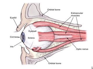
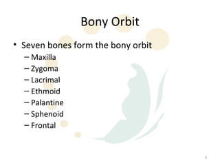
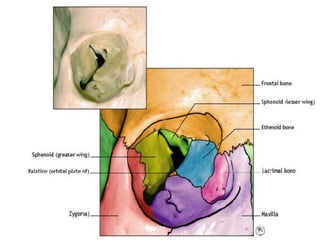
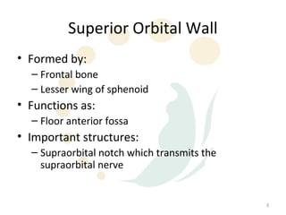
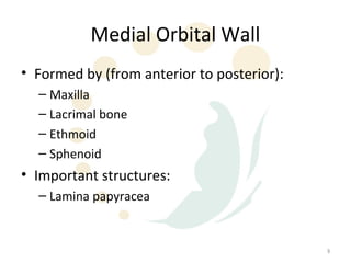
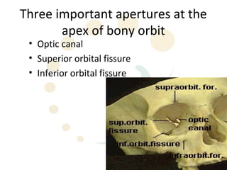
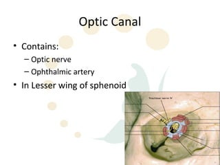
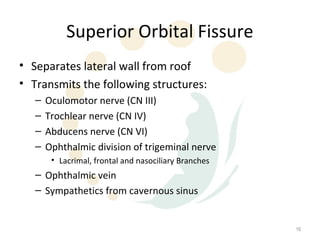
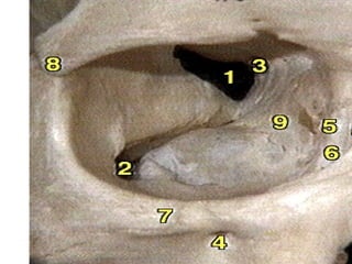
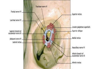
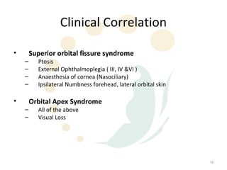
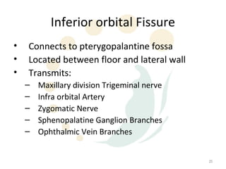
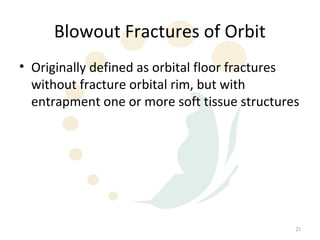
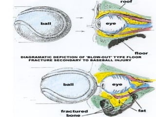
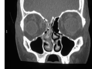
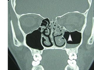
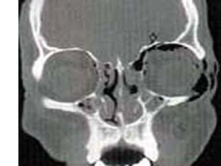
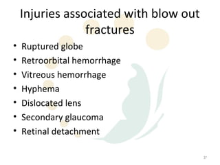
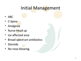
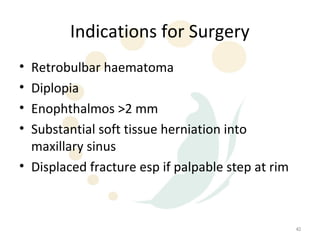
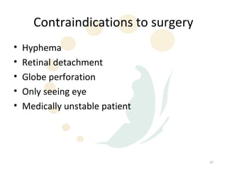
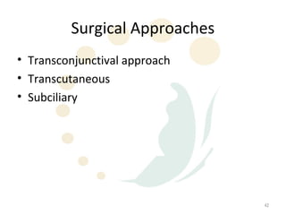
Orbital Fractures


                    2
Topics for Discussion
•   Orbital anatomy
•   Types of fractures
•   Signs and symptoms
•   Management


                                   3
Orbital Anatomy
• The bony orbit refers to the shell of bone
  which surrounds and protects the eye.
• The bony orbit is a pyramidal cavity with an
  elliptical base presenting anteriorly and the
  apex posteriorly


                                                  4
5
Bony Orbit
• Seven bones form the bony orbit
  – Maxilla
  – Zygoma
  – Lacrimal
  – Ethmoid
  – Palantine
  – Sphenoid
  – Frontal


                                    6
7
Superior Orbital Wall
• Formed by:
  – Frontal bone
  – Lesser wing of sphenoid
• Functions as:
  – Floor anterior fossa
• Important structures:
  – Supraorbital notch which transmits the
    supraorbital nerve


                                             8
Medial Orbital Wall
• Formed by (from anterior to posterior):
  – Maxilla
  – Lacrimal bone
  – Ethmoid
  – Sphenoid
• Important structures:
  – Lamina papyracea


                                            9
Lamina Papyracea
• Thin segment of the medial orbital wall
• Separates the orbit from the ethmoid air cells


                                                   10
Lateral Orbital Wall
• Formed by:
  – Zygomatic bone
  – Greater wing of sphenoid


                                 11
Orbital Floor
• Formed by:
  – Maxilla
  – Palatine
• Important structures:
  – Infraorbital groove
     • Transverses floor from lateral to medial
     • Location of infraorbital nerve which supplies sensation
       to check and ipsilateral upper alveolus and teeth


                                                                 12
Orbital Floor
• Forms roof of maxillary sinus
• Location of more blow out fractures due to
  inherent weakness of bone overlying maxillary
  sinus


                                              13
Three important apertures at the
       apex of bony orbit
 • Optic canal
 • Superior orbital fissure
 • Inferior orbital fissure


                                   14
Optic Canal
• Contains:
  – Optic nerve
  – Ophthalmic artery
• In Lesser wing of sphenoid


                               15
Superior Orbital Fissure
• Separates lateral wall from roof
• Transmits the following structures:
   –   Oculomotor nerve (CN III)
   –   Trochlear nerve (CN IV)
   –   Abducens nerve (CN VI)
   –   Ophthalmic division of trigeminal nerve
        • Lacrimal, frontal and nasociliary Branches
   – Ophthalmic vein
   – Sympathetics from cavernous sinus


                                                       16
17
18
Clinical Correlation

•       Superior orbital fissure syndrome
    –     Ptosis
    –     External Ophthalmoplegia ( III, IV &VI )
    –     Anaesthesia of cornea (Nasociliary)
    –     Ipsilateral Numbness forehead, lateral orbital skin

•       Orbital Apex Syndrome
    –     All of the above
    –     Visual Loss


                                                                19
Inferior orbital Fissure
•   Connects to pterygopalantine fossa
•   Located between floor and lateral wall
•   Transmits:
    –   Maxillary division Trigeminal nerve
    –   Infra orbital Artery
    –   Zygomatic Nerve
    –   Sphenopalatine Ganglion Branches
    –   Ophthalmic Vein Branches


                                              20
Blowout Fractures of Orbit
• Originally defined as orbital floor fractures
  without fracture orbital rim, but with
  entrapment one or more soft tissue structures


                                              21
Blowout Fractures
• Blowout fractures now refer to fractures of the:
   –   Orbital floor
   –   Medial wall
   –   Lateral wall
   –   Superior wall
• “pure” blowout fractures – trapdoor rotation to
  bone fragments involving central area of bone
• “impure” fracture – fracture line extends to orbital
  rim


                                                         22
Physiology of Blowout Fracture
• The bony defect is filled with soft tissue and
  fat from the orbit
• Alters support mechanisms for EOM
• EOM can become entrapped
• Direct muscle damage can result


                                                   23
Common causes of orbital
              fractures
•   Falling
•   Aggression
•   Sporting events
•   MVAs


                                   24
25
26
Initial Evaluation
• History
  – Time and mechanism of injury
  – Change in appearance of eye
  – State of vision immediately after injury
     • Immediate loss of vision – severe damage to retina
     • Loss of light perception - vascular occlusion or optic nerve
       compression
     • Initial good vision – compression optic neuropathy


                                                                      27
Initial Evaluation
• Physical Exam
  – Cranial nerve examination
       • EOM
       • Numbness check
  –   Palpation orbital rim
  –   Papillary function
  –   Visual acuity
  –   Fundus examine
  –   Ophthalmologic evaluation


                                     28
Visual Acuity
• Light perception
• Finger counting
• Visual acuity


                               29
Consultation


Do not hesitate to obtain an ophthalmologic
 consultation


                                              30
Common physical signs
•   Periorbital eccyhmosis
•   Impaired extraocular muscles
•   Hypoesthesia in V2 distribution
•   Intraorbital emphysema


                                      31
Common Symptoms
• Diplopia
• Pain with eye movement


                           32
Radiographic Evaluation
• CT scan of the orbits
• Plain films not useful due to a high rate of
  false negatives and non-diagnostic studies


                                                 33
34
35
36
Injuries associated with blow out
                 fractures
•   Ruptured globe
•   Retroorbital hemorrhage
•   Vitreous hemorrhage
•   Hyphema
•   Dislocated lens
•   Secondary glaucoma
•   Retinal detachment

                                        37
Treatment Options
• Nonsurgical
• Surgical


                              38
Initial Management
•   ABC
•   C-Spine
•   Analgesia
•   Nurse Head up
•   Ice affected area
•   Broad spectrum antibiotics
•   Steroids
•   No nose blowing

                                  39
Indications for Surgery
• Retrobulbar haematoma
• Diplopia
• Enophthalmos >2 mm
• Substantial soft tissue herniation into
  maxillary sinus
• Displaced fracture esp if palpable step at rim


                                                   40
Contraindications to surgery
•   Hyphema
•   Retinal detachment
•   Globe perforation
•   Only seeing eye
•   Medically unstable patient


                                     41
Surgical Approaches
• Transconjunctival approach
• Transcutaneous
• Subciliary


                               42
Factors to consider for surgery
•   Site
•   Location
•   Severity
•   What needs to be corrected


                                       43
Orbital Implants
• Use of implants based on degree of
  comminution and size of fracture
• Various implant material used
  – Autogenous bone and cartilage
  – Alloplastic material
     • Teflon
     • Marlex
     • PDS


                                       44
Complications of Surgery
•   Ectropion
•   Lid retraction
•   Persistent diplopia
•   Malposition of eye
•   Hypoaesthesia of V2
•   Extrusion of orbital floor implant
•   BLINDNESS

                                         45

Orbital fracturesw

  • 2.
  • 3.
    Topics for Discussion • Orbital anatomy • Types of fractures • Signs and symptoms • Management 3
  • 4.
    Orbital Anatomy • Thebony orbit refers to the shell of bone which surrounds and protects the eye. • The bony orbit is a pyramidal cavity with an elliptical base presenting anteriorly and the apex posteriorly 4
  • 5.
  • 6.
    Bony Orbit • Sevenbones form the bony orbit – Maxilla – Zygoma – Lacrimal – Ethmoid – Palantine – Sphenoid – Frontal 6
  • 7.
  • 8.
    Superior Orbital Wall •Formed by: – Frontal bone – Lesser wing of sphenoid • Functions as: – Floor anterior fossa • Important structures: – Supraorbital notch which transmits the supraorbital nerve 8
  • 9.
    Medial Orbital Wall •Formed by (from anterior to posterior): – Maxilla – Lacrimal bone – Ethmoid – Sphenoid • Important structures: – Lamina papyracea 9
  • 10.
    Lamina Papyracea • Thinsegment of the medial orbital wall • Separates the orbit from the ethmoid air cells 10
  • 11.
    Lateral Orbital Wall •Formed by: – Zygomatic bone – Greater wing of sphenoid 11
  • 12.
    Orbital Floor • Formedby: – Maxilla – Palatine • Important structures: – Infraorbital groove • Transverses floor from lateral to medial • Location of infraorbital nerve which supplies sensation to check and ipsilateral upper alveolus and teeth 12
  • 13.
    Orbital Floor • Formsroof of maxillary sinus • Location of more blow out fractures due to inherent weakness of bone overlying maxillary sinus 13
  • 14.
    Three important aperturesat the apex of bony orbit • Optic canal • Superior orbital fissure • Inferior orbital fissure 14
  • 15.
    Optic Canal • Contains: – Optic nerve – Ophthalmic artery • In Lesser wing of sphenoid 15
  • 16.
    Superior Orbital Fissure •Separates lateral wall from roof • Transmits the following structures: – Oculomotor nerve (CN III) – Trochlear nerve (CN IV) – Abducens nerve (CN VI) – Ophthalmic division of trigeminal nerve • Lacrimal, frontal and nasociliary Branches – Ophthalmic vein – Sympathetics from cavernous sinus 16
  • 17.
  • 18.
  • 19.
    Clinical Correlation • Superior orbital fissure syndrome – Ptosis – External Ophthalmoplegia ( III, IV &VI ) – Anaesthesia of cornea (Nasociliary) – Ipsilateral Numbness forehead, lateral orbital skin • Orbital Apex Syndrome – All of the above – Visual Loss 19
  • 20.
    Inferior orbital Fissure • Connects to pterygopalantine fossa • Located between floor and lateral wall • Transmits: – Maxillary division Trigeminal nerve – Infra orbital Artery – Zygomatic Nerve – Sphenopalatine Ganglion Branches – Ophthalmic Vein Branches 20
  • 21.
    Blowout Fractures ofOrbit • Originally defined as orbital floor fractures without fracture orbital rim, but with entrapment one or more soft tissue structures 21
  • 22.
    Blowout Fractures • Blowoutfractures now refer to fractures of the: – Orbital floor – Medial wall – Lateral wall – Superior wall • “pure” blowout fractures – trapdoor rotation to bone fragments involving central area of bone • “impure” fracture – fracture line extends to orbital rim 22
  • 23.
    Physiology of BlowoutFracture • The bony defect is filled with soft tissue and fat from the orbit • Alters support mechanisms for EOM • EOM can become entrapped • Direct muscle damage can result 23
  • 24.
    Common causes oforbital fractures • Falling • Aggression • Sporting events • MVAs 24
  • 25.
  • 26.
  • 27.
    Initial Evaluation • History – Time and mechanism of injury – Change in appearance of eye – State of vision immediately after injury • Immediate loss of vision – severe damage to retina • Loss of light perception - vascular occlusion or optic nerve compression • Initial good vision – compression optic neuropathy 27
  • 28.
    Initial Evaluation • PhysicalExam – Cranial nerve examination • EOM • Numbness check – Palpation orbital rim – Papillary function – Visual acuity – Fundus examine – Ophthalmologic evaluation 28
  • 29.
    Visual Acuity • Lightperception • Finger counting • Visual acuity 29
  • 30.
    Consultation Do not hesitateto obtain an ophthalmologic consultation 30
  • 31.
    Common physical signs • Periorbital eccyhmosis • Impaired extraocular muscles • Hypoesthesia in V2 distribution • Intraorbital emphysema 31
  • 32.
    Common Symptoms • Diplopia •Pain with eye movement 32
  • 33.
    Radiographic Evaluation • CTscan of the orbits • Plain films not useful due to a high rate of false negatives and non-diagnostic studies 33
  • 34.
  • 35.
  • 36.
  • 37.
    Injuries associated withblow out fractures • Ruptured globe • Retroorbital hemorrhage • Vitreous hemorrhage • Hyphema • Dislocated lens • Secondary glaucoma • Retinal detachment 37
  • 38.
  • 39.
    Initial Management • ABC • C-Spine • Analgesia • Nurse Head up • Ice affected area • Broad spectrum antibiotics • Steroids • No nose blowing 39
  • 40.
    Indications for Surgery •Retrobulbar haematoma • Diplopia • Enophthalmos >2 mm • Substantial soft tissue herniation into maxillary sinus • Displaced fracture esp if palpable step at rim 40
  • 41.
    Contraindications to surgery • Hyphema • Retinal detachment • Globe perforation • Only seeing eye • Medically unstable patient 41
  • 42.
    Surgical Approaches • Transconjunctivalapproach • Transcutaneous • Subciliary 42
  • 43.
    Factors to considerfor surgery • Site • Location • Severity • What needs to be corrected 43
  • 44.
    Orbital Implants • Useof implants based on degree of comminution and size of fracture • Various implant material used – Autogenous bone and cartilage – Alloplastic material • Teflon • Marlex • PDS 44
  • 45.
    Complications of Surgery • Ectropion • Lid retraction • Persistent diplopia • Malposition of eye • Hypoaesthesia of V2 • Extrusion of orbital floor implant • BLINDNESS 45

Editor's Notes

  • #46 Ectropion – turing outward eyelid