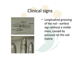
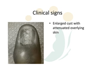
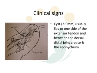
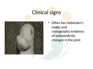
Mucous cysts of the DIPJ
Mucous cyst DIPJ
• Ganglion cyst of the DIPJ
• Usually occurs between the fifth and seventh
  decades
• Associated with osteophytes or spurring of
  the DIPJ
• Osteoarthritis in other joints
Ganglion/Mucous cyst
• Single or multiloculated cyst which appears smooth, white &
  translucent
• Wall is made up of compressed collagen fibres and is sparsely
  lined with flattened cells without evidence of an epithelial or
  synovial lining
• Mucin-filled “clefts” from the capsular attachment of the
  main cyst interconnect with the adjacent underlying joint via
  tortuous continuous ducts
• Stroma may show tightly packed collagen fibres or sparsely
  cellular areas with broken fibres and mucin-filled intercellular
  & extracellular lakes
• No inflammatory reaction or mitotic activity has been noted
Ganglion/Mucous cyst
• Contents of cyst characterized by a highly viscous, clear,
  sticky, jelly-like mucin made up of glucosamine, albumin,
  globulin, & high concentrations of hyaluronic acid
• Aetiology & pathogenesis remain obscure
• Most widely accepted theory - mucoid degeneration
  associated with degeneration of joint capsule or tendon
  sheath
• Injury & mechanical irritation may stimulate production of
  hyaluronic acid to form mucin, which may penetrate joint
  ligaments and capsules and then coalesce to form cyst
Clinical signs
       • Longitudinal grooving
         of the nail - earliest
         sign without a visible
         mass, caused by
         pressure on the nail
         matrix
Clinical signs
       • Enlarged cyst with
         attenuated overlying
         skin
Clinical signs
       • Cyst (3-5mm) usually
         lies to one side of the
         extensor tendon and
         between the dorsal
         distal joint crease &
         the eponychium
Clinical signs
       • Often has Heberden’s
         nodes and
         radiographic evidence
         of osteoarthritic
         changes in the joint
Treatment
• Primarily surgical
• Numerous alternative treatment reported in
  the past with moderate success:
  – Intralesional injection - eg. Sodium morrhuate,
    triamcinolone
  – Occlusive flurandrenolone tape
Surgical Management
• Excision of the cyst alone
• Wide excision of the cyst along with
  surrounding adjacent structures - eg.the
  overlying skin, osteophyte debridements
• Debridement of the DIPJ osteophytes only,
  without excision of the cyst itself or overlying
  skin
Operative technique
          • L-shaped / H-shaped /
            curved incision
          • Elliptical excision of
            attenuated or
            involved skin
Operative technique
          • Cyst mobilized, traced to
            the joint capsule &
            excised with the joint
            capsule
          • All tissue excised between
            the extensor tendon &
            the adjacent collateral
            ligaments
          • Insertion of the extensor
            tendon and the nail
            matrix must be protected
Operative technique
          • Excison of
            osteophytes
          • Skin closure may
            require rotation /
            advancement dorsal
            skin flap or a full-
            thickness graft
Alternative approach
          • Transverse incision
            centred over DIPJ
          • Base of mucous cyst
            identified & excised while
            leaving the distal &
            superficial portion of the
            cyst intact
          • Excision of osteophtyes &
            joint capsule with direct
            skin closure
          • Allow several weeks for
            involution of the
            remaining cyst
Complications
Residual nail deformities
Stiffness
Skin necrosis
Recurrence:
   - inadequate excision
   - ganglion extension to the other side of extensor
   tendon
   - persistent underlying arthritic process

Mucous cysts dip jw

  • 2.
  • 3.
    Mucous cyst DIPJ •Ganglion cyst of the DIPJ • Usually occurs between the fifth and seventh decades • Associated with osteophytes or spurring of the DIPJ • Osteoarthritis in other joints
  • 4.
    Ganglion/Mucous cyst • Singleor multiloculated cyst which appears smooth, white & translucent • Wall is made up of compressed collagen fibres and is sparsely lined with flattened cells without evidence of an epithelial or synovial lining • Mucin-filled “clefts” from the capsular attachment of the main cyst interconnect with the adjacent underlying joint via tortuous continuous ducts • Stroma may show tightly packed collagen fibres or sparsely cellular areas with broken fibres and mucin-filled intercellular & extracellular lakes • No inflammatory reaction or mitotic activity has been noted
  • 5.
    Ganglion/Mucous cyst • Contentsof cyst characterized by a highly viscous, clear, sticky, jelly-like mucin made up of glucosamine, albumin, globulin, & high concentrations of hyaluronic acid • Aetiology & pathogenesis remain obscure • Most widely accepted theory - mucoid degeneration associated with degeneration of joint capsule or tendon sheath • Injury & mechanical irritation may stimulate production of hyaluronic acid to form mucin, which may penetrate joint ligaments and capsules and then coalesce to form cyst
  • 6.
    Clinical signs • Longitudinal grooving of the nail - earliest sign without a visible mass, caused by pressure on the nail matrix
  • 7.
    Clinical signs • Enlarged cyst with attenuated overlying skin
  • 8.
    Clinical signs • Cyst (3-5mm) usually lies to one side of the extensor tendon and between the dorsal distal joint crease & the eponychium
  • 9.
    Clinical signs • Often has Heberden’s nodes and radiographic evidence of osteoarthritic changes in the joint
  • 10.
    Treatment • Primarily surgical •Numerous alternative treatment reported in the past with moderate success: – Intralesional injection - eg. Sodium morrhuate, triamcinolone – Occlusive flurandrenolone tape
  • 11.
    Surgical Management • Excisionof the cyst alone • Wide excision of the cyst along with surrounding adjacent structures - eg.the overlying skin, osteophyte debridements • Debridement of the DIPJ osteophytes only, without excision of the cyst itself or overlying skin
  • 12.
    Operative technique • L-shaped / H-shaped / curved incision • Elliptical excision of attenuated or involved skin
  • 13.
    Operative technique • Cyst mobilized, traced to the joint capsule & excised with the joint capsule • All tissue excised between the extensor tendon & the adjacent collateral ligaments • Insertion of the extensor tendon and the nail matrix must be protected
  • 14.
    Operative technique • Excison of osteophytes • Skin closure may require rotation / advancement dorsal skin flap or a full- thickness graft
  • 15.
    Alternative approach • Transverse incision centred over DIPJ • Base of mucous cyst identified & excised while leaving the distal & superficial portion of the cyst intact • Excision of osteophtyes & joint capsule with direct skin closure • Allow several weeks for involution of the remaining cyst
  • 16.
    Complications Residual nail deformities Stiffness Skinnecrosis Recurrence: - inadequate excision - ganglion extension to the other side of extensor tendon - persistent underlying arthritic process