
Recommended
More Related Content
What's hot
What's hot (20)
Similar to Blowout fractures
Similar to Blowout fractures (20)
More from Mohammed Sayed
Recently uploaded
Recently uploaded (20)
Blowout fractures
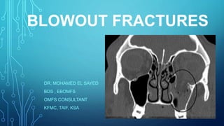
- 1. BLOWOUT FRACTURES DR. MOHAMED EL SAYED BDS , EBOMFS OMFS CONSULTANT KFMC, TAIF, KSA
- 2. ANATOMY • The orbit is the bony vault that houses the eyeball, or globe. • By age 5 years orbital growth is 85% complete, and it is finalized between 7 years of age and puberty. • Seven bones form the orbit: maxillary, zygomatic, frontal, ethmoidal, lacrimal, palatine, and sphenoid. Besides forming a protective socket for the globe, these bones also provide origins for the extraocular muscles, and foramina and fissures for cranial nerves and blood vessels. • The orbital floor and medial wall are most frequently fractured owing to their thinness and lack of support • The subsequent increased intraorbital pressure is most often relieved by traumatic expansion of the walls with herniation of orbital tissue into the maxillary sinus and/or ethmoid air cells adjacent to these walls. In essence, the paranasal sinuses and ethmoid air cells serve as air bags or shock absorbers to the globe and orbital contents.
- 4. MECHANISM • Orbital wall fractures can be divided into two sections, anterior and posterior. The anterior section is composed of the orbital rim. The posterior section is composed of the thinner roof, floor, and medial and lateral walls • There have been two major theories proposed regarding the mechanism of blowout fractures: 1. hydraulic mechanism whereby hydrostatic pressure within the globe or orbital contents is transmitted to the orbital walls. 2. impact against the orbital rim transmits force to the more fragile orbital walls, resulting in a blowout fracture
- 6. CLASSIFICATION • Isolated orbital wall fractures account for 4 to 16% of all facial fractures. If fractures that extend outside the orbit are included, such as those of the zygomatic complex (ZMC) and naso-orbitoethmoid (NOE), then this accounts for 30 to 55% of all facial fractures. • blowout injuries are further described as pure, for those that occur in the presence of an intact orbital rim, and impure, for those with a concomitant fracture of the orbital rim. Blowout fractures can occur on the floor, medial wall,
- 7. DIAGNOSIS • The clinical examination is also initially obscured by significant edema, which may mask visual observation of enophthalmos or vertical diplopia and palpation of bony step deformities. • Extraocular movements should be assessed by the evaluation of cardinal movements. If there is any question about muscle entrapment, a forced duction test of all four rectus muscles is indicated. • A blowout fracture should be suspected if paresthesia of the infraorbital nerve distribution is present following trauma, with limitation of normal ocular motion and no notable fracture of the rim. • Noncontrast CT scans with 1.5- or 2-mm axial, sagittal, and coronal cuts are the most appropriate for specific evaluation of the orbit. The indications for surgical intervention for an isolated, radiographically evident orbital blowout fracture is nonresolving diplopia within 2 to 3 weeks of injury or enophthalmos greater than 2 mm. • Other relative indications for repair include orbital floor defects larger than 1 cm2 or clinically notable hypoglobus.