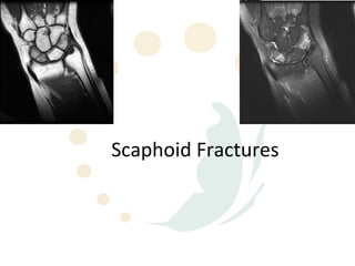
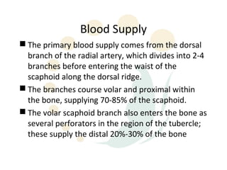
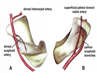
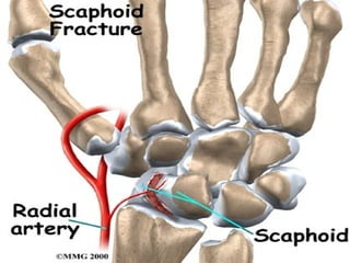
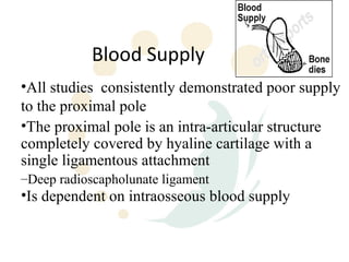
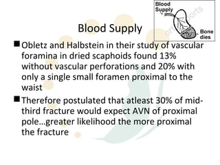
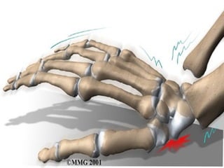
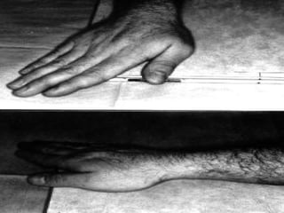
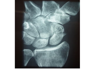
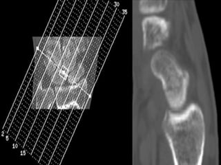
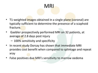
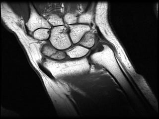
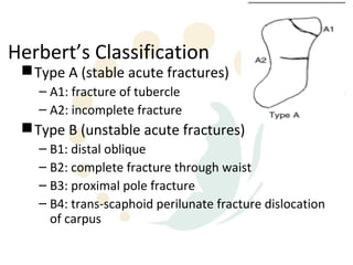
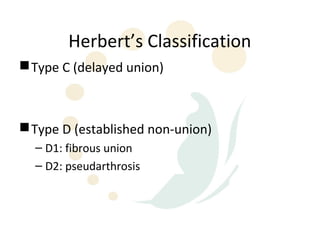
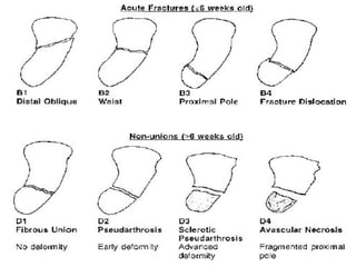
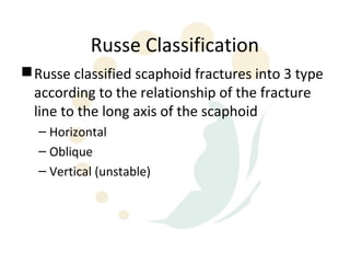
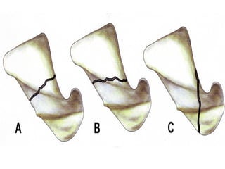
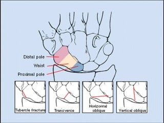
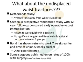
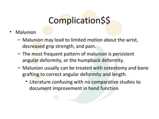
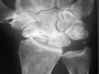
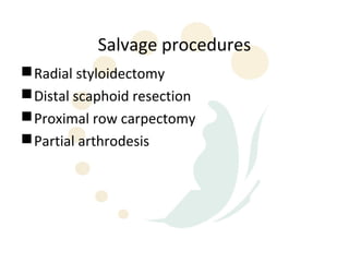
Scaphoid fractures are the most common carpal bone fractures, often occurring in young adults from falls on an outstretched hand. The scaphoid has a tenuous blood supply and is prone to non-union, especially for proximal pole fractures. Treatment depends on fracture type and stability, ranging from casting to operative fixation with screws. Complications include malunion, delayed union, non-union and avascular necrosis, requiring further procedures like bone grafting or carpal fusion.
elbow and wrist and hand fracture with managementkajalgoel8
describing anatomy of the wrist and hand ..
what is fracture
mechanism of injury of all the fracture
classification of fracture
clinical features
radiologicals exminations
management of the fracture
elbow and wrist and hand fracture with managementkajalgoel8
describing anatomy of the wrist and hand ..
what is fracture
mechanism of injury of all the fracture
classification of fracture
clinical features
radiologicals exminations
management of the fracture
paediatric injuries around the elbow
supracondylar elbow injuries
pulled elbow in paediatric age r
radiological signs around elbow in supracondylar fracture humerus
3. Scaphoid Fractures
• The scaphoid is the most frequently fractured
carpal bone, accounting for 71% of all carpal bone
fractures.
• Scaphoid fractures often occur in young and
middle-aged adults, typically those aged 15-60
years.
• About 5-12% of scaphoid fractures are associated
with other fractures
• 70-80% occur at the waist or mid-portion
• 10-20% proximal pole
4. Anatomy
• The scaphoid lies at the radial border of the
proximal carpal row, but its elongated shape and
position allow bridging between the 2 carpal rows
because it acts as a stabilizing rod.
• The scaphoid has 5 articulating surfaces:
– with the radius, lunate, capitate, trapezoid, and
trapezium.
• As a result, nearly the entire surface is covered by
hyaline cartilage.
5.
6. Blood Supply
• Vessels may enter only at the sites of
ligamentous attachment:
– the flexor retinaculum at the tubercle,
– the volar ligaments along the palmar surface,
– and the dorsal radiocarpal and radial collateral
ligaments along the dorsal ridge.
7. Blood Supply
Classically described as 3 principal arterial
groups, but in more recent investigations by
Gelberman and Menon described 2:
– Entering dorsally
– Volar side limited to tubercle
8. Blood Supply
The primary blood supply comes from the dorsal
branch of the radial artery, which divides into 2-4
branches before entering the waist of the
scaphoid along the dorsal ridge.
The branches course volar and proximal within
the bone, supplying 70-85% of the scaphoid.
The volar scaphoid branch also enters the bone as
several perforators in the region of the tubercle;
these supply the distal 20%-30% of the bone
9.
10.
11. Blood Supply
•All studies consistently demonstrated poor supply
to the proximal pole
•The proximal pole is an intra-articular structure
completely covered by hyaline cartilage with a
single ligamentous attachment
–Deep radioscapholunate ligament
•Is dependent on intraosseous blood supply
12. Blood Supply
Obletz and Halbstein in their study of vascular
foramina in dried scaphoids found 13%
without vascular perforations and 20% with
only a single small foramen proximal to the
waist
Therefore postulated that atleast 30% of mid-
third fracture would expect AVN of proximal
pole…greater likelihood the more proximal
the fracture
13.
14. Pathophysiology
The primary mechanism of injury to the scaphoid bone is a fall
on an outstretched hand.
A scaphoid fracture is part of a spectrum of injuries based on 4
factors:
– (1) the direction of 3-dimensional loading,
– (2) the magnitude and duration of the force,
– (3) the position of the hand and wrist at the time of injury,
and
– (4) the biomechanical properties of ligaments and bones.
These factors affect the end result of the fall: distal radius
fracture, ligamentous injury, scaphoid fracture, or a
combination of these.
15. Pathophysiology
Essentially fractures of scaphoid have been explained as a
failure of bone cause by compressive or tension load
Compression, as explained by Cobey and White, against
concave surface by head of capitate
Position of radial and ulnar deviation thought to
determine where it breaks
Fryman subjected cadaver wrists to loading and observed
that:
– extension of 35 degrees of less resulted in distal forearm
fractures
– >90degrees resulted in carpal fractures
Combination of radial deviation and wrist extension locks
scaphoid within the scaphoid fossa
16. Diagnosis
Suggested by:
– patient’s age,
– mechanism of injury and
– signs and symptoms
Imaging
– Xray
– CT Scan
– MRI
– Bone Scan
17. Radiography
The 4 essential views (ie, PA, lateral, supinated and
pronated obliques) identify majority of fractures.
The scaphoid view is a PA radiograph with the wrist
extended 30° and deviated ulnarly 20°. This view
helps to stretch out the scaphoid and is also used for
assessing the degree of scaphoid fracture angulation.
A clenched-fist radiograph has also been useful for
visualization of the scaphoid waist.
18.
19.
20. CT Scans
CT permits accurate anatomic assessment of the fracture.
Bone contusions are not evaluated with CT, but true fractures
can be excluded
21.
22. MRI
• T1-weighted images obtained in a single plane (coronal) are
typically sufficient to determine the presence of a scaphoid
fracture.
• Gaebler prospectively performed MRI on 32 patients, at
average of 2.8 days post injury
– 100% sensitivity and specificity
• In recent study Dorsay has shown that immediate MRI
provides cost benefit when compared to splintage and repeat
xray
• False positives due MRI’s sensitivity to marrow oedema
23.
24.
25. Nuclear Imaging
Radionuclide bone scanning typically is performed 3-7
days after the initial injury if the radiographic findings are
normal.
Best at 48hours, premature imaging may be obscured by
traumatic synovitis
Bone scan findings are considered positive for a fracture
when intense, focal tracer accumulation is identified.
Negative bone scan results virtually exclude scaphoid
fracture
Teil-van studied cost effectiveness and concluded that
initial xray followed by bone scan at 2 weeks if patient is
still symptomatic is most effective management option
Teil-van also suggested that more sensitive and less
expensive than MRI
27. Classification
Determining optimal treatment depends on
accurate diagnosis and fracture
classification
Herbert devised an alpha-numeric system
that combined fracture anatomy, stability
and chronicity of injury.
28. Herbert’s Classification
Type A (stable acute fractures)
– A1: fracture of tubercle
– A2: incomplete fracture
Type B (unstable acute fractures)
– B1: distal oblique
– B2: complete fracture through waist
– B3: proximal pole fracture
– B4: trans-scaphoid perilunate fracture dislocation
of carpus
31. Russe Classification
Russe classified scaphoid fractures into 3 type
according to the relationship of the fracture
line to the long axis of the scaphoid
– Horizontal
– Oblique
– Vertical (unstable)
35. Management
Proximal pole
– Depends on size and vascularity of fracture
– Growing sentiment that most should be
treated operatively because of high propensity
for non-union and increased duration of
immobilisation required for non-operative
management
– If large enough to accommodate a screw than
every attempt should be made
36. Management
DeMaagd and Engber showed 11 of 12 patients with proximal
pole fractures healed with Herbert screw
Retting and Raskin had 100% union in 17 cases with Herbert
screw
If fragment too small then K-wires can be used
37.
38.
39. Management
Distal Pole
– Are infrequent
– Usually extra-articular with good blood supply
– Best treated with short arm thumb spica for 3-6
weeks
40.
41. Management of waist fractures
Most common type of fracture
High rate of delayed and non-union
– With delays in treatment adversely affect results
Operative vs non-operative
– Controversial
42. Management of waist fractures
Most stable fractures can be treated with below
elbow thumb spica
Unstable fractures best treated with compression
screw fixation
– >1mm displacement
– Fragment angulation
– Abnormal carpal alignment
With advent of percutaneous techniques of
cannulated screws under flouroscopic control trend
towards operative management
43. What about the undisplaced
waist fractures???
Netherlands study:
– Average time away from work 4.5 months
Saeden in prospective randomised study with 12
year follow-up compared early operative vs cast
immobilisation
– Return to work quicker in operative
– No significant long term difference in functional
outcome between 2 groups
Bond has shown return to work 7 weeks earlier
and time of union 5 weeks quicker
– Other papers disagree
Some surgeons published union rates of 100%
with surgery(Green’s volume 1 page 721)
44.
45.
46.
47.
48. Complication$$
• Malunion
– Malunion may lead to limited motion about the wrist,
decreased grip strength, and pain.
– The most frequent pattern of malunion is persistent
angular deformity, or the humpback deformity.
– Malunion usually can be treated with osteotomy and bone
grafting to correct angular deformity and length.
• Literature confusing with no comparative studies to
document improvement in hand function
49. Complication$$
• Delayed union and non-union
– Delayed union is incomplete union after 4 months of cast
immobilization.
– Non-union is an unhealed fracture with smooth
fibrocartilage covering the fracture site.
– About 10-15% of all scaphoid fractures do not unite.
– Some degree of delayed union or non-union occurs in
nearly all proximal pole fractures and in 30% of scaphoid
waist fractures
50.
51. Complication$$
Delayed union is anticipated if fracture treatment is delayed
for several weeks.
The risk of non-union increases after a delay of 4 weeks.
These delays may be related to the patient's failure to seek
treatment for a presumed sprain, but they more frequently
are related to improper or incomplete immobilization or a
failure to diagnose and treat the acute fracture
52. Delayed union treatment
If the delayed union is stable and less than 6 months old
relative to the time of injury, prolonged cast
immobilization with or without electrical stimulation may
be used.
Treatment of choice for a symptomatic non-union is
placement of a bone graft and fixation.
– Russe corticocancellous iliac graft
– Fisk-Fernandez volar wedge graft
– Pronator pedicle graft
• Braun ‘83 reported 100% union in 8 pts
• Kawai, Kuhlmann, Papp reported 100% 37 pts
– Pechlaner reporrted 25 free vascularised iliac grafts
with 100%
Success rates for the treatment of non-union are as high
as 82%.
53. AVN
• Osteonecrosis occurs in 15-30% of all scaphoid fractures, and
most of these involve the proximal pole.
• Its incidence increases as the fracture line becomes more
proximal; this decreases the probability that the blood supply
to the proximal pole is preserved