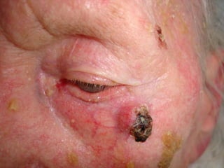
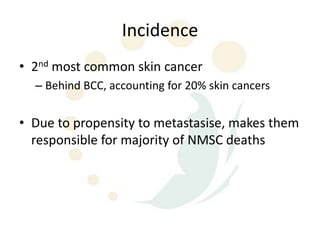
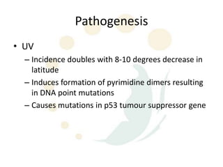
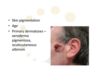
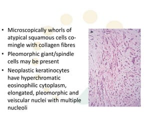
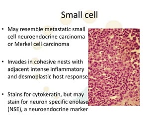
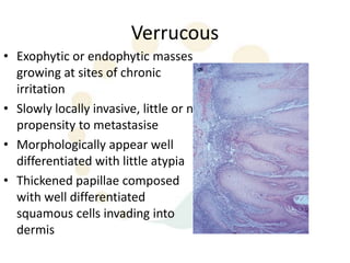
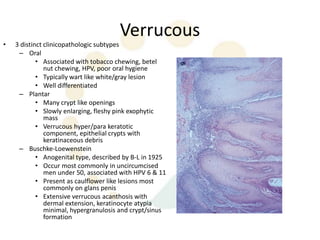
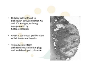
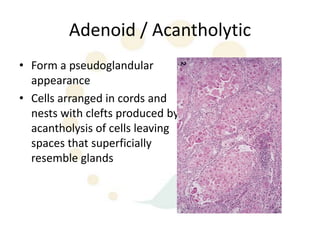
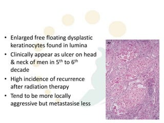
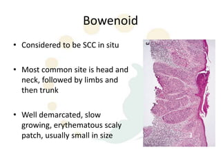
Squamous cell carcinoma (SCC) is the second most common skin cancer. It has a propensity to metastasize, making it responsible for most skin cancer deaths. Risk factors include UV exposure, age, immunosuppression, and primary dermatoses. Histological subtypes include pleomorphic, adenoid, small cell, verrucous, and keratoacanthoma. Tumor size greater than 2 cm and depth greater than 6 mm increase metastatic risk. Treatment involves surgical excision with adequate margins or other modalities like radiation for high risk cases.
guideline for long case presentation,include history,examination,,investigation,treatment option,surgical procedure of superficial parotidectomy,short discussion about plemorphic adenoma
We report a case of acquired anterior thoracic lung herniation in a 63-year-old female. This painful herniation developed four years after uncomplicated video-assisted thoracic surgery for lung cancer resection and adjuvant radiation for concomitant breast cancer. The herniation site was remote from all prior incisions, and demonstrated intercostal muscle denervation and radiation fibrosis. The 8 cm x 10 cm chest wall defect was reconstructed with inlay PROCEED mesh and reinforced with a pedicled latissimus dorsi flap. Five months postoperatively the patient had complete resolution of symptoms, no evidence of herniation, and a stable wound.
Skin cancers or cutaneous malignancies including Basal cell carcinoma, Squamous cell carcinoma and Melanoma and with a brief introduction of skin as an organ itself.
5. Incidence
• 2nd most common skin cancer
– Behind BCC, accounting for 20% skin cancers
• Due to propensity to metastasise, makes them
responsible for majority of NMSC deaths
6. Pathogenesis
• UV
– Incidence doubles with 8-10 degrees decrease in
latitude
– Induces formation of pyrimidine dimers resulting
in DNA point mutations
– Causes mutations in p53 tumour suppressor gene
8. • Immunosuppression – due to
immunosuppressive drugs, UVR, viral infection
esp HPV
– Reversed ratio of BCC:SCC, SCC being 3x more
common in transplant patients
– Higher rates – cumulative risk of SCC/ BCC in heart
transplant recipient is 3% at one year, 21% at 5
years, 35% at 10 years
10. Simplex
• Majority of SCCs
• Atypical keratinocytes
develop within epidermis and
invade the dermis
• Tumour cells are enlarged,
hyperchromatic, variably
pleomorphic nuclei,
prominent mitotic activity
• Keratin pearls
11. Actinic Keratosis
• Also SCC in situ, micro
invasive SCC, as there is
considerable overlap in the
histology
• Atypical keratinocytes that
have not breached the
dermal barrier
– SCCIS is typically full thickness
keratinocyte atypia
12. • Rate of malignant
transformation is 0.1%
per lesion per year
– About 16% will
eventually transform
– Can progress to other
skin cancers such as
sebaceous carcinoma
13. Pleomorphic
• AKA spindle / sarcomatoid, RARE
• Associated with previous trauma
or RTX
• Most commonly found on face
or sun exposed areas of elderly
• Commonly ulcerate, but may
present as an exophytic mass
14. • Microscopically whorls of
atypical squamous cells co-
mingle with collagen fibres
• Pleomorphic giant/spindle
cells may be present
• Neoplastic keratinocytes
have hyperchromatic
eosinophilic cytoplasm,
elongated, pleomorphic and
veiscular nuclei with multiple
nucleoli
15. Small cell
• May resemble metastatic small
cell neuroendocrine carcinoma
or Merkel cell carcinoma
• Invades in cohesive nests with
adjacent intense inflammatory
and desmoplastic host response
• Stains for cytokeratin, but may
stain for neuron specific enolase
(NSE), a neuroendocrine marker
16. Verrucous
• Exophytic or endophytic masses
growing at sites of chronic
irritation
• Slowly locally invasive, little or no
propensity to metastasise
• Morphologically appear well
differentiated with little atypia
• Thickened papillae composed
with well differentiated
squamous cells invading into
dermis
17. Verrucous
• 3 distinct clinicopathologic subtypes
– Oral
• Associated with tobacco chewing, betel
nut chewing, HPV, poor oral hygiene
• Typically wart like white/gray lesion
• Well differentiated
– Plantar
• Many crypt like openings
• Slowly enlarging, fleshy pink exophytic
mass
• Verrucous hyper/para keratotic
component, epithelial crypts with
keratinaceous debris
– Buschke-Loewenstein
• Anogenital type, described by B-L in 1925
• Occur most commonly in uncircumcised
men under 50, associated with HPV 6 & 11
• Present as caulflower like lesions most
commonly on glans penis
• Extensive verrucous acanthosis with
dermal extension, keratinocyte atypia
minimal, hypergranulosis and crypt/sinus
formation
18. Keratoacanthoma
• Period of rapid growth lasts 4-8
weeks
• Potential for spontaneous
involution usually within 4-6
months, sometimes with
considerable scarring
• Clinically tend to be rapid
growing smooth, firm nodule
with central keratin plug
19. • Histologically difficult to
distinguish between benign KA
and SCC KA type, so being
amalgamated by
histopathologists
• Atypical squamous proliferation
with intradermal invasion
• Typically crateriform
architecture with keratin plug
and well developed collarette
20. Adenoid / Acantholytic
• Form a pseudoglandular
appearance
• Cells arranged in cords and
nests with clefts produced by
acantholysis of cells leaving
spaces that superficially
resemble glands
21. • Enlarged free floating dysplastic
keratinocytes found in lumina
• Clinically appear as ulcer on head
& neck of men in 5th to 6th
decade
• High incidence of recurrence
after radiation therapy
• Tend to be more locally
aggressive but metastasise less
22. Bowenoid
• Considered to be SCC in situ
• Most common site is head and
neck, followed by limbs and
then trunk
• Well demarcated, slow
growing, erythematous scaly
patch, usually small in size
23. • Histologically shows
hyperkeratosis, acanthosis,
psoriasiform hyperplasia, full
thickness atypia, loss of polarity
reflecting cessation of
maturation
• When neoplastic keratinocytes
invade the dermis, this lesion is
termed Bowenoid SCC
• Especially associated with HPV
– HPV2 with extragenital
lesions, HPV16 with genital
lesions
24. Metastasis
• Overall risk is 2 – 6%, not 0.5%
• Recurrent SCC has metastatic rate of 30%, and
metastatic cases had a survival rate of 1/3
• Metastases tend to be to regional lymph nodes
• Most mets (and local recurrences) are found within
first 2 years, and 95% within first 5 years
25. Risk factors for metastasis and recurrence
• Recurrence rate doubled and tripled metastatic rate
– Size > 2cm
– Grade 3 & 4 vs. Grade 1 & 2 tumours
• Well differentiated has recurrence 7%, mod well diff 23%, poor
diff 28%
• Tumour thickness
– 3 year recurrence free survival is 98% for <3.5mm, 84%
for > 3.5mm (Breslow thickness)
• Rapid growth rate
26. • Sun exposed areas tend to metastasise and recur less than mucosal SCC
• Scar SCC are very aggressive
• Lip and ear SCC have higher metastatic rate than other head and neck
sites (16 & 10%)
– Probably due to decreased subcutaneous fat
– Nose and scalp, anogenital are intermediate risk
– Periungal SCC has high recurrence rate but almost never metastasises
• Previous treatment – recurrent cancers have a metastatic rate of 25%
– Location – ear 45%, lip 32% metastatic rate
27. • Histopathology
– Isolated strands, infiltrative pattern, haphazard growth vs.
broad pushing borders
– Perineural invasion (occurs in 2-14% SCC, most commonly
H&N in elderly men)
• Has been quoted as local recurrence 47%, regional mets 35%,
distant nodes 15%; so post op RTX commonly offered
– NO good evidence that any subtype has greater risk
recurrence or metastasis
31. Grades
Broder’s Grade Undifferentiated Ratio of
cells differentiated cells
I – Well < 25% 3:1
differentiated
II – Moderately 25 – 50% 1:1
well differentiated
III – Poorly 50 – 75% 1:3
differentiated
IV – Anaplastic or > 75% Nil
pleomorphic
32. Surgical Management
• Tumours < 2cm diameter are
successfully excised 95% of the
time with a margin of 4mm,
6mm for high risk cases
(Brodland & Zitelli)
• Tumours > 2cm diameter
require margin of 10mm for
equivalent local control rates
• Moh’s surgery
33. Other modalities
• Dessication and curettage
– Lesions less than 2cm diameter have cure rates of
97-98.8%
• Cryosurgery
– Well localised, superficial lesions on trunk or limbs
• 5FU & Imiquimod & Photodynamic therapy
– Useful for actinic keratoses
34. Radiation Therapy
– < 2cm tumours have a cure rate of 95%
– Adjunctive RTX must be given within 8 weeks for greatest efficiency
– (Late) changes include :
– atrophy, fibrosis, hypopigmentation, telangiectasia, ulceration
– “As late results of RTX can be poor, it is not recommended for patients
under 60 yo with uncomplicated primary SCC”
– May hasten natural history of KA