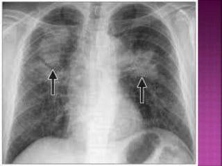
Downloaded 468 times

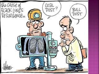
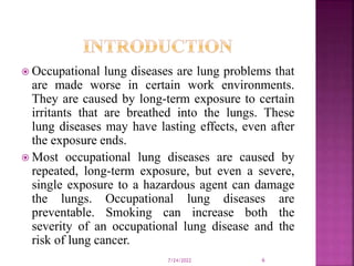
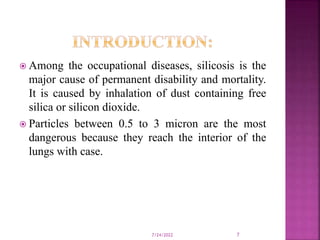


The document discusses occupational lung diseases, which are conditions exacerbated by prolonged exposure to harmful substances in the workplace, leading to permanent damage even after exposure ends. It covers various types of diseases such as silicosis and asbestosis, their causes, symptoms, diagnostic methods, and the importance of prevention. The document emphasizes the irreversibility of these conditions and the need for protective measures in workplaces to mitigate risks.