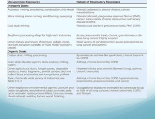
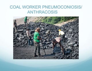
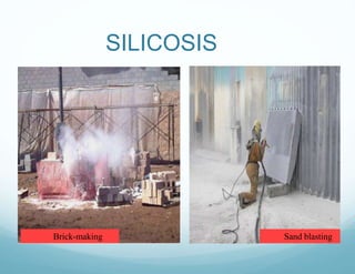
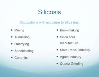
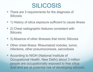
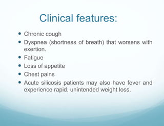
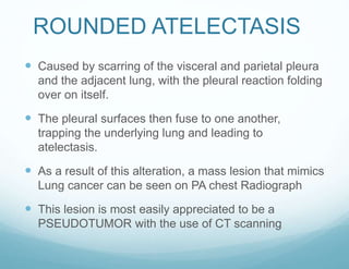
This document provides information on occupational lung diseases. It begins with a brief history of occupational lung diseases dating back to Roman times. It then defines occupational lung diseases and pneumoconiosis. The document classifies and describes various types of pneumoconiosis including anthracosis, silicosis, asbestosis, and berylliosis. For each type, it discusses associated occupations, pathogenesis, clinical features, diagnosis, and management. The document provides detailed information on the pathogenesis, clinical presentation, radiographic findings, and complications of major occupational lung diseases like silicosis and asbestosis.
Asbestos-related diseases include non-malignant disorders such as asbestosis, diffuse pleural thickening, pleural plaques, pleural effusion, rounded atelectasis and malignancies such as lung cancer and malignant mesothelioma.
Pneumoconiosis- dust with size and different types of occupational pneumoconic diseases, clinical features, diagnosis and prevention of pneumoconiosis.
Occupational lungs disease: It contains Asbestosis, Silicosis, Coal miner’s lung/coal worker’s, pneumoconiosis, Bagasosis, Bysinosis
and their nursing management and prevention.
Austin Journal of Lung Cancer Research is an open access, peer reviewed, scholarly journal committed to publication of unique contributions concerned with the causes, pathogenesis, diagnosis, prevention and management of lung cancer. Lung cancer occurs when the cells of lung divide uncontrollably and form tumor.
Austin Journal of Lung Cancer Research accepts original research articles, review articles, case reports, clinical images and rapid communication on all the aspects of this disease.
AIR POLLUTION CONTROL course material by Prof S S JAHAGIRDAR,NKOCET,SOLAPUR for BE (CIVIL ) students of Solapur university. Content will be also useful for SHIVAJI and PUNE university students
Restrictive lung diseases (interstitial lung diseases)
Histological Structure of Alveoli
The wall of the alveoli is formed by a thin sheet of tissue separating two neighbouring alveoli.
This sheet is formed by epithelial cells and intervening connective tissue.
Collagenous , reticular and elastic fibres are present.
Between the connective tissue fibres we find a dense, anastomosing network of pulmonary capillaries. The wall of the capillaries are in direct contact with the epithelial lining of the alveoli.
Neighbouring alveoli may be connected to each other by small alveolar pores (pores of Kohn).
The epithelium of the alveoli is formed by two cell types:
Alveolar type I cells (small alveolar cells or type I pneumocytes) are extremely flattened and form the bulk (95%) of the surface of the alveolar walls.
Alveolar type II cells (large alveolar cells or type II pneumocytes) are irregularly (sometimes cuboidal) shaped.
They form small bulges on the alveolar walls.
Type II alveolar cells contain are large number of granules called cytosomes (or multilamellar bodies), which consist of precursors to pulmonary surfactant (the mixture of phospholipids which keep surface tension in the alveoli low) .
Cilia are absent from the alveolar epithelium and cannot help to remove particulate matter which continuously enters the alveoli with the inspired air. Alveolar macrophages take care of this job. They migrate freely over the alveolar epithelium and ingest particulate matter.
FUNCTIONS OF PULMONARY CELLS
Type I pneumocytes
Permeable to Oxygen and CO2, do not divide
Type II pneumocytes
Reserve cells
secrete pulmonary surfactant
Serve as repair cells
Alveolar macrophages
Phagocytosis
Pores of Kohn (allow passage of Macrophages)
Complexities of occupational and environmental lung diseases, exploring their causes, symptoms, diagnosis, and prevention measures. For more information please contact us: 9779030507.
This lecture elucidates in detail the important occupational health diseases, the all-important concept of work absenteeism and, lastly, the prevention of occupational diseases.
MANAGEMENT OF ATRIOVENTRICULAR CONDUCTION BLOCK.pdfJim Jacob Roy
Cardiac conduction defects can occur due to various causes.
Atrioventricular conduction blocks ( AV blocks ) are classified into 3 types.
This document describes the acute management of AV block.
Title: Sense of Taste
Presenter: Dr. Faiza, Assistant Professor of Physiology
Qualifications:
MBBS (Best Graduate, AIMC Lahore)
FCPS Physiology
ICMT, CHPE, DHPE (STMU)
MPH (GC University, Faisalabad)
MBA (Virtual University of Pakistan)
Learning Objectives:
Describe the structure and function of taste buds.
Describe the relationship between the taste threshold and taste index of common substances.
Explain the chemical basis and signal transduction of taste perception for each type of primary taste sensation.
Recognize different abnormalities of taste perception and their causes.
Key Topics:
Significance of Taste Sensation:
Differentiation between pleasant and harmful food
Influence on behavior
Selection of food based on metabolic needs
Receptors of Taste:
Taste buds on the tongue
Influence of sense of smell, texture of food, and pain stimulation (e.g., by pepper)
Primary and Secondary Taste Sensations:
Primary taste sensations: Sweet, Sour, Salty, Bitter, Umami
Chemical basis and signal transduction mechanisms for each taste
Taste Threshold and Index:
Taste threshold values for Sweet (sucrose), Salty (NaCl), Sour (HCl), and Bitter (Quinine)
Taste index relationship: Inversely proportional to taste threshold
Taste Blindness:
Inability to taste certain substances, particularly thiourea compounds
Example: Phenylthiocarbamide
Structure and Function of Taste Buds:
Composition: Epithelial cells, Sustentacular/Supporting cells, Taste cells, Basal cells
Features: Taste pores, Taste hairs/microvilli, and Taste nerve fibers
Location of Taste Buds:
Found in papillae of the tongue (Fungiform, Circumvallate, Foliate)
Also present on the palate, tonsillar pillars, epiglottis, and proximal esophagus
Mechanism of Taste Stimulation:
Interaction of taste substances with receptors on microvilli
Signal transduction pathways for Umami, Sweet, Bitter, Sour, and Salty tastes
Taste Sensitivity and Adaptation:
Decrease in sensitivity with age
Rapid adaptation of taste sensation
Role of Saliva in Taste:
Dissolution of tastants to reach receptors
Washing away the stimulus
Taste Preferences and Aversions:
Mechanisms behind taste preference and aversion
Influence of receptors and neural pathways
Impact of Sensory Nerve Damage:
Degeneration of taste buds if the sensory nerve fiber is cut
Abnormalities of Taste Detection:
Conditions: Ageusia, Hypogeusia, Dysgeusia (parageusia)
Causes: Nerve damage, neurological disorders, infections, poor oral hygiene, adverse drug effects, deficiencies, aging, tobacco use, altered neurotransmitter levels
Neurotransmitters and Taste Threshold:
Effects of serotonin (5-HT) and norepinephrine (NE) on taste sensitivity
Supertasters:
25% of the population with heightened sensitivity to taste, especially bitterness
Increased number of fungiform papillae
- Video recording of this lecture in English language: https://youtu.be/lK81BzxMqdo
- Video recording of this lecture in Arabic language: https://youtu.be/Ve4P0COk9OI
- Link to download the book free: https://nephrotube.blogspot.com/p/nephrotube-nephrology-books.html
- Link to NephroTube website: www.NephroTube.com
- Link to NephroTube social media accounts: https://nephrotube.blogspot.com/p/join-nephrotube-on-social-media.html
New Directions in Targeted Therapeutic Approaches for Older Adults With Mantl...i3 Health
i3 Health is pleased to make the speaker slides from this activity available for use as a non-accredited self-study or teaching resource.
This slide deck presented by Dr. Kami Maddocks, Professor-Clinical in the Division of Hematology and
Associate Division Director for Ambulatory Operations
The Ohio State University Comprehensive Cancer Center, will provide insight into new directions in targeted therapeutic approaches for older adults with mantle cell lymphoma.
STATEMENT OF NEED
Mantle cell lymphoma (MCL) is a rare, aggressive B-cell non-Hodgkin lymphoma (NHL) accounting for 5% to 7% of all lymphomas. Its prognosis ranges from indolent disease that does not require treatment for years to very aggressive disease, which is associated with poor survival (Silkenstedt et al, 2021). Typically, MCL is diagnosed at advanced stage and in older patients who cannot tolerate intensive therapy (NCCN, 2022). Although recent advances have slightly increased remission rates, recurrence and relapse remain very common, leading to a median overall survival between 3 and 6 years (LLS, 2021). Though there are several effective options, progress is still needed towards establishing an accepted frontline approach for MCL (Castellino et al, 2022). Treatment selection and management of MCL are complicated by the heterogeneity of prognosis, advanced age and comorbidities of patients, and lack of an established standard approach for treatment, making it vital that clinicians be familiar with the latest research and advances in this area. In this activity chaired by Michael Wang, MD, Professor in the Department of Lymphoma & Myeloma at MD Anderson Cancer Center, expert faculty will discuss prognostic factors informing treatment, the promising results of recent trials in new therapeutic approaches, and the implications of treatment resistance in therapeutic selection for MCL.
Target Audience
Hematology/oncology fellows, attending faculty, and other health care professionals involved in the treatment of patients with mantle cell lymphoma (MCL).
Learning Objectives
1.) Identify clinical and biological prognostic factors that can guide treatment decision making for older adults with MCL
2.) Evaluate emerging data on targeted therapeutic approaches for treatment-naive and relapsed/refractory MCL and their applicability to older adults
3.) Assess mechanisms of resistance to targeted therapies for MCL and their implications for treatment selection
ARTIFICIAL INTELLIGENCE IN HEALTHCARE.pdfAnujkumaranit
Artificial intelligence (AI) refers to the simulation of human intelligence processes by machines, especially computer systems. It encompasses tasks such as learning, reasoning, problem-solving, perception, and language understanding. AI technologies are revolutionizing various fields, from healthcare to finance, by enabling machines to perform tasks that typically require human intelligence.
Lung Cancer: Artificial Intelligence, Synergetics, Complex System Analysis, S...Oleg Kshivets
RESULTS: Overall life span (LS) was 2252.1±1742.5 days and cumulative 5-year survival (5YS) reached 73.2%, 10 years – 64.8%, 20 years – 42.5%. 513 LCP lived more than 5 years (LS=3124.6±1525.6 days), 148 LCP – more than 10 years (LS=5054.4±1504.1 days).199 LCP died because of LC (LS=562.7±374.5 days). 5YS of LCP after bi/lobectomies was significantly superior in comparison with LCP after pneumonectomies (78.1% vs.63.7%, P=0.00001 by log-rank test). AT significantly improved 5YS (66.3% vs. 34.8%) (P=0.00000 by log-rank test) only for LCP with N1-2. Cox modeling displayed that 5YS of LCP significantly depended on: phase transition (PT) early-invasive LC in terms of synergetics, PT N0—N12, cell ratio factors (ratio between cancer cells- CC and blood cells subpopulations), G1-3, histology, glucose, AT, blood cell circuit, prothrombin index, heparin tolerance, recalcification time (P=0.000-0.038). Neural networks, genetic algorithm selection and bootstrap simulation revealed relationships between 5YS and PT early-invasive LC (rank=1), PT N0—N12 (rank=2), thrombocytes/CC (3), erythrocytes/CC (4), eosinophils/CC (5), healthy cells/CC (6), lymphocytes/CC (7), segmented neutrophils/CC (8), stick neutrophils/CC (9), monocytes/CC (10); leucocytes/CC (11). Correct prediction of 5YS was 100% by neural networks computing (area under ROC curve=1.0; error=0.0).
CONCLUSIONS: 5YS of LCP after radical procedures significantly depended on: 1) PT early-invasive cancer; 2) PT N0--N12; 3) cell ratio factors; 4) blood cell circuit; 5) biochemical factors; 6) hemostasis system; 7) AT; 8) LC characteristics; 9) LC cell dynamics; 10) surgery type: lobectomy/pneumonectomy; 11) anthropometric data. Optimal diagnosis and treatment strategies for LC are: 1) screening and early detection of LC; 2) availability of experienced thoracic surgeons because of complexity of radical procedures; 3) aggressive en block surgery and adequate lymph node dissection for completeness; 4) precise prediction; 5) adjuvant chemoimmunoradiotherapy for LCP with unfavorable prognosis.
micro teaching on communication m.sc nursing.pdfAnurag Sharma
Microteaching is a unique model of practice teaching. It is a viable instrument for the. desired change in the teaching behavior or the behavior potential which, in specified types of real. classroom situations, tends to facilitate the achievement of specified types of objectives.
The prostate is an exocrine gland of the male mammalian reproductive system
It is a walnut-sized gland that forms part of the male reproductive system and is located in front of the rectum and just below the urinary bladder
Function is to store and secrete a clear, slightly alkaline fluid that constitutes 10-30% of the volume of the seminal fluid that along with the spermatozoa, constitutes semen
A healthy human prostate measures (4cm-vertical, by 3cm-horizontal, 2cm ant-post ).
It surrounds the urethra just below the urinary bladder. It has anterior, median, posterior and two lateral lobes
It’s work is regulated by androgens which are responsible for male sex characteristics
Generalised disease of the prostate due to hormonal derangement which leads to non malignant enlargement of the gland (increase in the number of epithelial cells and stromal tissue)to cause compression of the urethra leading to symptoms (LUTS
These simplified slides by Dr. Sidra Arshad present an overview of the non-respiratory functions of the respiratory tract.
Learning objectives:
1. Enlist the non-respiratory functions of the respiratory tract
2. Briefly explain how these functions are carried out
3. Discuss the significance of dead space
4. Differentiate between minute ventilation and alveolar ventilation
5. Describe the cough and sneeze reflexes
Study Resources:
1. Chapter 39, Guyton and Hall Textbook of Medical Physiology, 14th edition
2. Chapter 34, Ganong’s Review of Medical Physiology, 26th edition
3. Chapter 17, Human Physiology by Lauralee Sherwood, 9th edition
4. Non-respiratory functions of the lungs https://academic.oup.com/bjaed/article/13/3/98/278874
Title: Sense of Smell
Presenter: Dr. Faiza, Assistant Professor of Physiology
Qualifications:
MBBS (Best Graduate, AIMC Lahore)
FCPS Physiology
ICMT, CHPE, DHPE (STMU)
MPH (GC University, Faisalabad)
MBA (Virtual University of Pakistan)
Learning Objectives:
Describe the primary categories of smells and the concept of odor blindness.
Explain the structure and location of the olfactory membrane and mucosa, including the types and roles of cells involved in olfaction.
Describe the pathway and mechanisms of olfactory signal transmission from the olfactory receptors to the brain.
Illustrate the biochemical cascade triggered by odorant binding to olfactory receptors, including the role of G-proteins and second messengers in generating an action potential.
Identify different types of olfactory disorders such as anosmia, hyposmia, hyperosmia, and dysosmia, including their potential causes.
Key Topics:
Olfactory Genes:
3% of the human genome accounts for olfactory genes.
400 genes for odorant receptors.
Olfactory Membrane:
Located in the superior part of the nasal cavity.
Medially: Folds downward along the superior septum.
Laterally: Folds over the superior turbinate and upper surface of the middle turbinate.
Total surface area: 5-10 square centimeters.
Olfactory Mucosa:
Olfactory Cells: Bipolar nerve cells derived from the CNS (100 million), with 4-25 olfactory cilia per cell.
Sustentacular Cells: Produce mucus and maintain ionic and molecular environment.
Basal Cells: Replace worn-out olfactory cells with an average lifespan of 1-2 months.
Bowman’s Gland: Secretes mucus.
Stimulation of Olfactory Cells:
Odorant dissolves in mucus and attaches to receptors on olfactory cilia.
Involves a cascade effect through G-proteins and second messengers, leading to depolarization and action potential generation in the olfactory nerve.
Quality of a Good Odorant:
Small (3-20 Carbon atoms), volatile, water-soluble, and lipid-soluble.
Facilitated by odorant-binding proteins in mucus.
Membrane Potential and Action Potential:
Resting membrane potential: -55mV.
Action potential frequency in the olfactory nerve increases with odorant strength.
Adaptation Towards the Sense of Smell:
Rapid adaptation within the first second, with further slow adaptation.
Psychological adaptation greater than receptor adaptation, involving feedback inhibition from the central nervous system.
Primary Sensations of Smell:
Camphoraceous, Musky, Floral, Pepperminty, Ethereal, Pungent, Putrid.
Odor Detection Threshold:
Examples: Hydrogen sulfide (0.0005 ppm), Methyl-mercaptan (0.002 ppm).
Some toxic substances are odorless at lethal concentrations.
Characteristics of Smell:
Odor blindness for single substances due to lack of appropriate receptor protein.
Behavioral and emotional influences of smell.
Transmission of Olfactory Signals:
From olfactory cells to glomeruli in the olfactory bulb, involving lateral inhibition.
Primitive, less old, and new olfactory systems with different path
3. HISTORY
It is likely that humans have
suffered from occupational lung
diseases since the change from
hunting to agriculture as a means of
providing food.
In Roman times it was recorded that
mining was a dangerous trade, fit
only for convicts and slaves.
The first recorded mention of
breathlessness among handlers of
grain was done by Ramazzini, the
father of occupational medicine, in
1713.
Bernardino Ramazzini
(1633 - 1714)
4. HISTORY
In the eighteenth and early nineteenth centuries, it was
thought that the symptoms from black lung disease were
asthma-related.
The term "black lung" was coined when medical
professionals discovered the blackening of miners' lungs
in post-mortem.
The first documented case of an asbestos-related death
was reported in 1906 when the autopsy of an asbestos
worker revealed lung fibrosis.
In the early twentieth century, it was observed that many
asbestos workers were dying unnaturally young.
5. HISTORY
In 1924, Nellie Kershaw, an English
textile worker was the first case of
asbestosis to be described in medical
literature.
Dr.William Edmund Cooke testified in
Kershaw's inquest that "mineral
particles in the lungs originated from
asbestos and were, beyond reasonable
doubt, the primary cause of the fibrosis
of the lungs and therefore of death"
Berylliosis was described first in
Germany in 1933 and in the USA in
1943.
Nellie Kershaw
(1891 –1924)
6. Definition
As per International Labour Organization (ILO)
The term “occupational disease” covers any disease
contracted as a result of an exposure to risk factors
arising from work activity.
Two main elements are present in the definition of an
occupational disease:
1) The causal relationship between exposure in a
specific working environment or work activity and a
specific disease; and
2) The fact that the disease occurs among a group of
exposed persons with a frequency above the average
morbidity of the rest of the population.
7. The term pneumoconiosis derives its meaning from
the Greek words: pneuma = air and konis = dust
The International Labour Organization defines
pneumoconiosis as “the accumulation of dust in the
lungs and the tissue reactions to its presence”.
Not included in the definition of pneumoconiosis are
conditions such as asthma, chronic obstructive
pulmonary disease (COPD), and hypersensitivity
pneumonitis, in which there is no requirement for dust
to accumulate in the lungs in the long term.
8. Determinants of inhalational exposure
Particles size of air contaminates
Particles > 10 -15 um diameter do not penetrate beyond the nose
and throat.
Particles are divided into three size fractions on the basis of their
size character and source .
1.Coarse-mode fraction –particles size of 2.5- 10 um contain
crustal elements such as silica, aluminum, and iron. Mostly deposit
relatively high in the tracheobronchial tree.
2.Fine mode fraction –particle size <2.5 um and carried to the
lower airways and get deposited .Fine particles are created by
burning of fossil fuels or high temperature industrial process,
gases ,fumes.
3. Ultra fine fraction - <0.1 um in size deposit in the lung and
they come in contact with the alveolar walls ,however particles of
this size range may penetrate into the circulation and be carried to
extra pulmonary sites.
9.
10. In other words
Pneumoconiosis can be defined as the non-neoplastic
reaction of lungs to inhaled minerals or organic dust
and the resultant alteration in their structure excluding
asthma, bronchitis and emphysema. – Textbook of
Pulmonary Medicine , D Behera
13. Types
Pneumoconiosis is usually divided into three groups:
Major pneumoconiosis
Minor pneumoconiosis
Benign pneumoconiosis
“ Fibrotic Pneumoconiosis”
14. Types
Major Pneumoconiosis: Inhalation of some dusts
results in “major fibrosis” of the lungs, which results
in interference of lung architecture or lung function
tests.
Examples are:
Silica silicosis
Asbestos asbestosis
Coal anthracosis
Healthy lung Silicotic lung
15. Types
Minor Pneumoconiosis: Inhalation of some dusts
results in “minor fibrosis” of the lungs
There is minimal fibrosis of the lungs without
interference of lung architecture or lung function tests.
These dusts include:
Mica pneumoconiosis
Koalin (china clay) pneumoconiosis
16. Types
Benign Pneumoconiosis: There isn't any reaction in
the lungs, but dust deposition casts a shadow in x-ray
of the lung. There is no fibrosis and no disturbance of
lung functions.
It can result from the inhalation of:
Iron dust siderosis
Tin dust stannosis
Calcium dust chalcosis
They are characterized by the presence of small
rounded dense opacities on a chest film due to
perivascular collections of dusts.
The deposits in the lung disappear when exposure is
discontinued.
17. PATHOGENESIS
For clinical pneumoconiosis to develop, 3 essential
factors are required:
Exposure to specific substance: coal, appear relatively
inert and may accumulate in considerable amounts with
minimal tissue response; while silica and asbestos,
have potent biologic effects.
Particles of appropriate size to be retained in lung (1-
5μm)
Exposure for a sufficient length of time (usually around
10 years)
21. ANTHRACOSIS
• Pathologic entity resulting from deposition of coal dust in
the lungs.
• Simple cwp – with prolonged exposure to coal dust for
15 to 20 years ,small rounded opacities develop usually
not associated with pulmonary impairment.
• Complicated cwp – appearance of nodules> 1cm in
diameter on chest radiography usually confined to upper
half of the lungs . And it progresses to PMF that is
accompanied by severe lung deficits causing chronic
bronchitis and COPD .
• When exposure is terminated the simple type will not
progress; PMF type will progress
22. CAPLANS SYNDROME
coal workers with rheumatoid disease may
develop nodules even after relatively low
exposures to dust.
The lesions are typically subpleural.
The lesions may grow rapidly, appear in crops(in
contrast to silicotic/CWP nodules that appears
over a period of time) cavitate and produce a
pneumothorax
23. CLINICAL FEATURES
SIMPLE CWP- disease state without symptoms or
physical signs.
Diagnosis is based on radiologic features
The symptoms of cough and sputum reported by most
coal miners- consequences of dust induced Chronic
Bronchitis
Breathlessness on effort- caused by associated
chronic airflow limitation/ by the development of PMF
Respiratory impairment and disability develop as PMF
progresses
26. Diagnosis and management
A history of occupational exposure to coal and a chest
radiograph are the fundamental elements in the
diagnosis of CWP. ( CT when chest x-ray is
insensitive)
Treatment of latent TB infection should be considered
for coal workers who are thought to have had
significant silica dust exposure or who have evidence
of Silicosis
Smoking cessation counselling
Management of Chronic Bronchitis and Emphysema
27. Anthracosis
In a study conducted by National Institute of
Occupational Health in collaboration with the
International Development Research Centre (IDRC),
Canada, (5777 underground coal miners and 1236
surface coal miners)
revealed that the prevalence of pneumoconiosis
(category 1/1 and more) in underground coal miners
was 2.84% and in the surface coal workers it was
2.10%.
The overall prevalence of functional abnormalities of
lung in underground coal miners and surface coal
workers was 45.4% and 42.2% respectively.
Source: www.nioh.org
29. Silicosis
Occupations with exposure to silica dust
Mining
Tunnelling
Quarrying
Sandblasting
Ceramics
Brick-making
Silica flour
manufacture
Slate Pencil Industry
Agate Industry
Quartz Grinding
30. SILICOSIS
There are 3 requirements for the diagnosis of
Silicosis:
1) History of silica exposure sufficient to cause illness
2) Chest radiographic features consistent with
Silicosis
3) Absence of other illnesses that mimic Silicosis
Other chest illness: Rheumatoid nodules, tumor,
infections, other pneumoconiosis, sarcoidosis
According to NIOH (National Institute of
Occupational Health, New Delhi) about 3 million
people are occupationally exposed to free silica
dust and are at potential risk of developing silicosis.
31. Clinical forms:
Acute silicosis- it develops with 10 months of silica
exposure
Clinical and pathologic features are similar to
pulmonary alveolar proteinosis .It is a quite severe
form and is progressive despite discontinuation of
exposure.
Xray - chest - profuse miliary infiltration or
consolidation,
No silicotic nodules
HRCT- characteristic pattern-Crazy paving [thickened
intra and interlobar septa producing polygonal shapes
].
Treatment- whole lung lavage provide symptomatic
relief
32. Clinical features:
Chronic cough
Dyspnea (shortness of breath) that worsens with
exertion.
Fatigue
Loss of appetite
Chest pains
Acute silicosis patients may also have fever and
experience rapid, unintended weight loss.
33. Simple silicosis-develops with long term (15-20
years) less intense exposure, without associated
impairment of lung function
Xray – chest –variably sized ,poorly defined small
nodules predominating in upper lobes .
Calcification of hilar nodes producing characteristic
feature “EGG shell” pattern.
Egg shell pattern
34. HRCT-numerous small nodules more pronounced
on upper lobes a number of sub pleural location
RCT shows numerous small nodules
35. Complicated silicosis-nodular form may progress in the
absence of further exposure with coalescence and
formation irregular masses > 1cm in diameter.
these masses can become quite large and when this
occurs progressive massive fibrosis term is applied.
Both restrictive and obstructive pattern may be associated
with PMF.
PMF
37. COMPLICATIONS
SILICOTUBERCULOSIS-silica is cytotoxic to alveolar
macrophages so patients are at increased risk of tuberculosis.
Also infection with atypical mycobacteria and fungus.
Autoimmune disorder- rheumatoid arthritis , scleroderma
malignancy
38. MANAGEMENT AND
CONTROL
Change in occupation to an environment free of silica
containing dust.
Lung lavage to remove silica from lung
Treatment of all forms of Silicosis should be directed towards
control of mycobacterial disease
All subjects with Silicosis should have a Tuberculin Skin Test
or IFN-gamma release assay
Smoking Cessation
Acute and accelerated silicosis- Lung Transplant
39. ASBESTOSIS
ASBESTOS is a fibrous hydrated Magnesium Silicate
with more than 3000 commercial uses due to its
indestructible nature, fire resistance etc.
Types:
1) Chrysolite (serpentine)- M.C. used
2) Amphibole( Amosite)- straight fibre
3) Crocidolite- needle shaped long fibre
40. Serpentine
(93% of commercial use)
Amphibole
(7% of commercial use)
Chrysolite
Actinolite, Amosite, Anthophyllite,
Crocidolite, Richterite, Tremolite
ASBESTOSIS
41. ASBESTOSIS
Significant occupational exposure to asbestos occurs
mainly in
Asbestos cement factories
Asbestos textile industry and
Asbestos mining and milling. Asbestos cement factories
Asbestos textile industry Asbestos mining
43. Asbestos-Related Pleural Abnormalities
Four types of abnormalities:
Pleural plaques
Benign asbestos pleural effusions
Diffuse pleural thickening
Pleural disease puts patient at risk for other asbestos related
diseases – 10% get interstitial fibrosis within 10 years
Mostly asymptomatic, though some can cause dyspnea or
cough
Latency periods: 10-30 years
(shorter latency is for pleural effusion)
No specific therapies
Pleurectomy in severe cases .
44. ASBESTOSIS
Symptoms
Average latency period is 20-30 years
Dyspnoea
Cough
Chest pain
In advanced cases, clubbing of fingers
At histopathologic analysis, asbestos bodies, which
may consist of a single asbestos fibre surrounded by
a segmented protein-iron coat, can be identified in
intra alveolar macrophages.
45. ASBESTOSIS
Translucent asbestos fiber (straight
arrow) surrounded by a protein-iron coat
and an alveolar macrophage (curved arrow)
Chest x-ray showing Small, irregular oval
opacities Interstitial fibrosis and “Shaggy
heart sign”
46. Chest Radiograph Findings: Asbestos-Related
Pleural Abnormalities
Pleural plaques
Areas of pleural thickening
Sometimes with calcification
Pleural effusions
Diffuse pleural thickening
Lobulated prominence of
pleura adjacent to thoracic margin
(over ¼ of chest wall)
Interlobar tissue thickening
Rounded atelectasis
Rounded pleural mass
Bands of lung tissue radiating outwards
47. ROUNDED ATELECTASIS
Caused by scarring of the visceral and parietal pleura
and the adjacent lung, with the pleural reaction folding
over on itself.
The pleural surfaces then fuse to one another,
trapping the underlying lung and leading to
atelectasis.
As a result of this alteration, a mass lesion that mimics
Lung cancer can be seen on PA chest Radiograph
This lesion is most easily appreciated to be a
PSEUDOTUMOR with the use of CT scanning
48. HRCT can non invasively demonstrate continuity to
areas of diffuse pleural thickening, evidence of volume
loss in the adjacent lung, or a characteristic comet tail
of vessels and bronchi sweeping into wedge shaped
mass.
HRCT scans localised most cases of rounded
atelectasis to the lower, posterior portion of lung(right)
49.
50. Parenchymal Asbestosis
Diffuse interstitial fibrosis with:
Associated more with crocidolite
Smokers more prone to disease
Radiographic changes: >10 years
Latency period: 20-40 years
51. Lung Carcinoma
Latency period: 20-30 years
Bronchogenic Ca: 5x higher incidence in non-smoking
asbestos workers, 90x higher in smoking asbestos workers.
Adeno Ca is most common.
Chrysotile highest risk bronchogenic Ca.
52. Malignant Pleural
Mesothelioma
Tumor arises from the thin pleural membrane
surrounding the lungs
Rapidly invasive
Rare, although incidences are increasing
Long latency period: Usually 30-40 years
54. • Beryllium – is a light weight metal
• Exposure –manufacture of alloys, ceramics, or
high technology electronics , nuclear reactors
• Beryllium may produce
- Acute pneumonitis,
-Chronic granulomatous inflammatory disesase
that is similar to sarcodisis.
- Lung cancer
• Pathogenesis is a result of delayed type
hypersensitivity reaction stimulating proliferation
of T-cells leading to inflammatory ,fibrosis and
granuloma formation.
BERYLLIOSIS
55. Clinical features- cough , chest pain, arthralgia's
,fatigue and weight loss
BeLPT[beryllium lymphocyte proliferation test ]-blood is
drawn and in the lab, the WBC are separated from the rest
of the blood cells and then mixed with beryllium solution. If
the immune system is sensitized to beryllium, the cells will
multiply, producing an abnormal BeLPT result.In normal
individuals cells will not multiply.
Fiberoptic bronchoscopy with transbronchial lung biopsy is
required to make diagnosis of CBD . Biopsy shows
noncaseating granulomas or monocytic infiltration in lung
tissue.
56. MANAGEMENT
The diagnosis of CBD- based on documented exposure to
Beryllium, evidence of lung disease compatible with the
diagnosis, and a positive BeLPT performed on blood or BAL
fluid.
Three categories of Be-associated disorders:
1) Be sensitisation-( +ve Blood or BAL BeLPT but negative
biopsy)
2) Subclinical Be-disease-( +ve BeLPT and biopsy but no
clinical or radiological features of
disease)
3) Chronic Berylliosis- (+ve BeLPT and Biopsy with clinical
and radiological evidence of
disease)
Most imp. – complete cessation of further exposure
57. BYSSINOSIS
In 1831 Kay described chest
tightness and fever that
commonly occurred on Monday
after workers had been off work
over the weekend. It was
beacause of this observation that
term MONDAY MORNING
FEVER was coined.
Certain jobs in the textile mills
are associated with a higher risk
for development of Bronchitis.
Ginning, opening or carding work
carry a higher degree of risk.
58. CLINICAL FEATURES AND RISK
FACTORS
SOB occurs often on the day back to the work at
textile mill after several days off as on a Monday after
being off over the weekend.
Workers with a higher grade of symptoms tend to
have a more rapid decline in Pulmonary Function.
Risk factors include:
1) Length of employment in a cotton mill
2) Level of dust exposure
Tobacco smoking has been shown to be synergistic
with exposure to cotton dust in producing Chronic
Bronchitis
59. Grading of
Byssinosis(SCHILLING)
GRADE 0 – No symptoms on day 1 of work
GRADE ½- Occasional chest tightness or irritation of
respiratory tract on the 1st workday of week.
GRADE 1- Chest tightness on every 1st day of
workweek
GRADE 2 - Chest tightness on 1st and other days of
work week
GRADE 3 - Chest tightness on 1st and other days of
work week and physiological evidence of permanent
disability.
60. PFT Abnormality
Characteristically Byssinosis is associated with a
reduction in FVC and FEV 1 on the day of return to
work after an absence.
The degree of reduction in these parameters
increases over the workday.
This change is generally more severe on the 1st day of
work after an absence then on subsequent days
61. PATHOGENESIS
The Histopath. of Byssinosis is similar to those of
bronchitis induced by tobacco smoke- with
hyperplasia of mucus glands and infiltration of bronchi
with PMNs.
Cotton dust also stimulate mast cells and
macrophages, to release molecules that attract
neutrophils.
New evidence points to a
Lipopolysaccharide(Endotoxin) produced by bacterial
contaminants of cotton as the causative agent of
Byssinosis.
Byssinosis has elevated serum Histamine levels.
62. TREATMENT AND PREVENTION
The most important treatment- removal of the
individual from offending work environment.
Screening PFTs at the workplace is important to
identify susceptible individuals who exhibit airflow
abnormalities.
Measures have been taken in developed countries to
control cotton dust levels in textile mills( ex. Steam
clean cotton while it is still in the bale)
66. Medical measures:
Pre-placement examination
Periodical examination
Medical and health care services
Notification
Maintenance and analysis of records
Health education and counselling
Practicing good personal hygiene
Preventive measures
67. Practicing good personal hygiene:
Washing hands and face before eating, drinking, going to
the toilet, smoking.
Do not eat, drink, smoke, or apply cosmetics in areas
where silica is being used.
Wear protective clothes and respiratory protection
(Respirators must fit tightly.)
Before leaving work, shower and change into clean
clothes. Leave dusty clothes at work.
Preventive measures
68. Engineering measures
Design of building
Conduct air monitoring to measure the workers’
exposure to crystalline silica.
Minimize exposures by controlling the creation of
airborne particles, for example, use wet drilling, local
exhaust ventilation.
Personal Protective Equipments: Provide workers with
protective clothes, respiratory protection, and facilities
for washing (showers) and changing.
Enclosure / isolation
Environmental monitoring
Preventive measures
72. Other measures:
Legal measures: Measures to minimize dust emissions
and exposure to dust.
Law compliance mechanisms, including effective
workplace inspection systems
Cooperation between management and workers and
their representatives
A mechanism for the collection and analysis of data on
occupational diseases
Collaboration with social security schemes covering
occupational injuries and diseases
Preventive measures
73. Preventive measures
Other measures:
Training of health professionals in occupational
diseases as majority of medical practitioners lack
training in occupational health and consequently lack
the skills to diagnose and prevent occupational
diseases.