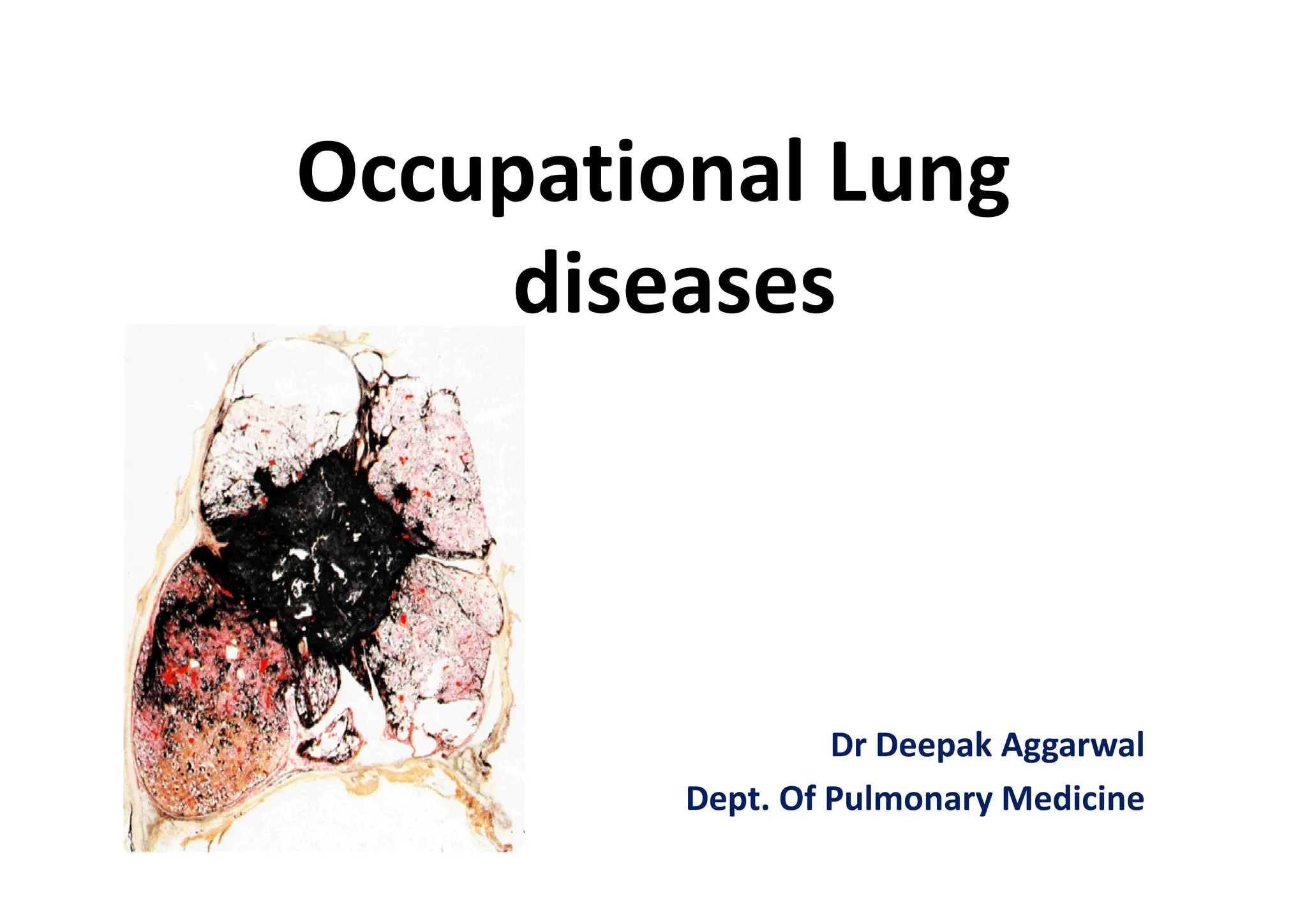
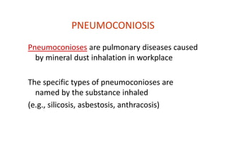
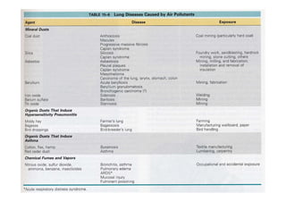
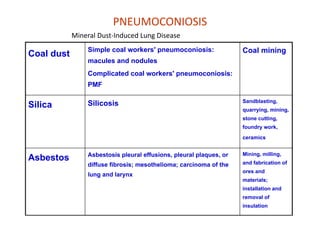
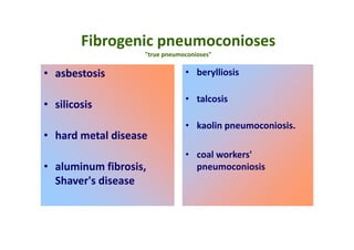
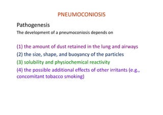
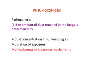
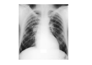
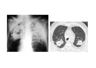
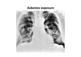
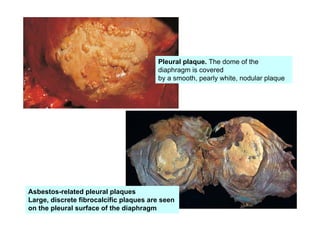
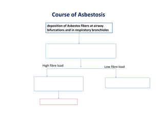
Occupational Lung
diseases
Dr Deepak Aggarwal
Dept. Of Pulmonary Medicine
To be discussed….
• Pneumoconiosis
• Hypersensitivity Pneumonitis
PNEUMOCONIOSIS
Pneumoconioses are pulmonary diseases caused
by mineral dust inhalation in workplace
The specific types of pneumoconioses are
named by the substance inhaled
(e.g., silicosis, asbestosis, anthracosis)
PNEUMOCONIOSIS
Mineral Dust‐Induced Lung Disease
Coal dust
Simple coal workers' pneumoconiosis:
macules and nodules
Complicated coal workers' pneumoconiosis:
PMF
Coal mining
Silica Silicosis Sandblasting,
quarrying, mining,
stone cutting,
foundry work,
ceramics
Asbestos Asbestosis pleural effusions, pleural plaques, or
diffuse fibrosis; mesothelioma; carcinoma of the
lung and larynx
Mining, milling,
and fabrication of
ores and
materials;
installation and
removal of
insulation
Fibrogenic pneumoconioses
"true pneumoconioses"
• asbestosis
• silicosis
• hard metal disease
• aluminum fibrosis,
Shaver's disease
• berylliosis
• talcosis
• kaolin pneumoconiosis.
• coal workers'
pneumoconiosis
Non‐fibrogenic pneumoconioses
benign pneumoconioses
Causes:
• antimony
• barium
• boric acid
• manganese
• iron
• tin
• titanium
• bismuth
PNEUMOCONIOSIS
Pathogenesis
The development of a pneumoconiosis depends on
(1) the amount of dust retained in the lung and airways
(2) the size, shape, and buoyancy of the particles
(3) solubility and physiochemical reactivity
(4) the possible additional effects of other irritants (e.g.,
concomitant tobacco smoking)
PNEUMOCONIOSIS
Pathogenesis
(1)The amount of dust retained in the lungs is
determined by
dust concentration in surrounding air
duration of exposure
effectiveness of clearance mechanisms
PNEUMOCONIOSIS
Pathogenesis
(2) the size, shape, and buoyancy of the particles
The most dangerous particles range from 1 to 5 μm in
diameter because they may reach the terminal small
airways and air sacs and settle in their linings
PNEUMOCONIOSIS
(3)The solubility and cytotoxicity of particles modify the
nature of the pulmonary response
Smaller particles tend to cause acute lung injury
Larger particles resist dissolution and so may persist within
the lung parenchyma for years ‐ tend to evoke fibrosing
collagenous pneumoconioses
PNEUMOCONIOSIS
Pathogenesis
The pulmonary alveolar macrophage is a key
cellular element in the initiation and perpetuation
of lung injury and fibrosis
The more reactive particles trigger the
macrophages to release a number of products that
mediate an inflammatory response and initiate
fibroblast proliferation and collagen deposition
PNEUMOCONIOSIS
Pathogenesis
(4) the possible additional effects of other
irritants (e.g., concomitant tobacco smoking)
tobacco smoking worsens the effects of all
inhaled mineral dusts
Coal worker pneumoconiosis
• Coal Workers’ Pneumoconiosis Is Due to
Inhalation of Carbon Particles
• Associated with coal mining industry
• Carbon + silica (anthracosilicosis)
Findings
The spectrum of lung findings in coal workers is wide,
varying from
(1) Asymptomatic anthracosis
(2) Simple CWP with little to no pulmonary
dysfunction
(3) Complicated CWP (progressive massive
fibrosis)
Anthracosis (urban dwellers)
morphology
• Carbon particles (anthrocotic pigment) in
alveolar and interstitial macrophages,in
connective tissue and lymphatics and lung
hilus.
• Generally asymptomatic
Simple CWP
Coal macules (1 to 2 mm in diameter, consists of peribronchiolar carbon‐
laden macrophages and dilated terminal bronchiole
larger coal nodules (contains small amounts of a delicate network of
collagen
located primarily adjacent to respiratory bronchioles
Microscopy:
•Carbon laden macrophages & delicate collagen fibres
adjacent to respiratory bronchioles initially (where dust
settles), later interstium & alveoli
• Dilatation of respiratory bronchioles –focal dust emphysema
Radiographic finding of bilateral small parenchymal nodules
• CXR typically shows upper lung field nodules
Complicated CWP
• Gross
• Multiple.,>2 cm ,v dark scars
• Microscopy:
• Dense collagen and carbon pigment.
• Central necrosis (+/‐)
• Progression does NOT correlate with amount of coal dust
deposition in lungs.
• Cigarette smoking increases rate of deterioration of
pulmonary function.
Caplans syndrome
• 1st described in coal workers, may be seen in
other pneumoconiosis
• ?? Immunopathologic mechanism
• Rheumatoid arthritis (RA) + Rheumatoid
nodules (Caplan nodules) in the lung
• Rheumatoid arthritis + pneumoconioses
• Caplans nodule = necrosis surrounded by
fibroblasts,monocytes and collagen
• s/s RA > lung symptoms
Clinical course
• Usually asymptomatic with little decrease of lung
function
• Progressive Dyspnoea, cough, expectoration
• PMF pulmonary dysfunction (restrictive)
• Pulmonary hypertension, cor pulmonale
• Progressive even if further exposure to dust is
prevented
• ↑ chronic bronchitis and emphysema
• No association with TB or carcinoma
Management
• Diagnosis by presence of clinical features
along with history of exposure to coal dust of
the magnitude that is sufficient to cause the
disease
• Imaging, PFT, sputum examination
• Treatment: no specific treatment, removal
from further exposure is important.
• Treat underlying airway disease
Silicosis
• Silica is silicon dioxide, the oxide of silicon,
chemical formula SiO2.
• SiO2 is the most abundant mineral on
earth
• Silicosis (also known as Grinder's disease and
Potter's rot) is a form of occupational lung
disease caused by inhalation of crystalline
silica dust, and is marked by inflammation and
scarring in forms of nodular lesions in the
upper lobes of the lungs
• It is found in sand, many rocks such as granite,
sandstone, flint and slate, and in some coal
and metallic ores.
• The cutting, breaking, crushing, drilling,
grinding, or abrasive blasting of these
materials may produce fine silica dust.
Silicosis – Foundry work
•
Silicosis ‐ Stone cutting
•
Silicosis ‐ Tunnel construction
Worst single incidence of silicosis in U.S. –
Hawk’s Nest Tunnel, Gauley Bridge, W. Va., 1930‐1931
•
Silicosis ‐ Sandblasting
• Compressed air at high pressure is used to blow fine sand or
other abrasive material through a hardened spray nozzle. The
abrasive particles quickly eat away whatever they are directed
at, leaving a clean, matte surface.
Diseases Associated with Exposure to
Silica Dust
• Silicosis
– Chronic silicosis
– Accelerated silicosis
– Acute silicosis (silicoproteinosis)(fine dust,
intense exposure , high silica)
– Progressive massive fibrosis
• Chronic Obstructive Pulmonary Disease
– Emphysema
– Chronic bronchitis
– Mineral dust‐induced small airway disease
Diseases Associated with Exposure to
Silica Dust
• Lung Cancer
• Mycobacterial Infection
• Immune‐Related Diseases
– Progressive systemic sclerosis
– Rheumatoid arthritis
– Chronic renal disease
– Systemic lupus erythematosus
Silicosis
• The most prevalent occupational disease in the
world.
• The induction period between initial silica exposure
and development of radiographically detectable nodular
silicosis is usually >10 years. Shorter induction periods
are associated with heavy exposures, and acute silicosis
may develop within 6 months to 2 years following
massive silica exposure
Three ‘types’ of Silicosis
• Simple chronic silicosis
• Most common form
• After long-term exposure (10-20 years) to low amounts
of silica dust.
• Nodules of chronic inflammation and scarring form in
the lungs and chest lymph nodes.
• Patients often asymptomatic, seen for other reasons.
• subdivided into:
simple
complicated silicosis(PMF)
Accelerated silicosis (= PMF, progressive
massive fibrosis)
• Occurs after exposure to larger amounts of silica
over a shorter period of time (5-10 years).
• Inflammation, scarring, and symptoms progress
faster in accelerated silicosis
• Patients have symptoms, especially shortness
of breath.
Acute silicosis
• From short-term exposure to very large
amounts of fine silica dust.
• The lungs become very inflamed, causing
severe shortness of breath and low blood
oxygen level.
• Killed hundreds of workers during Hawk’s
Nest Tunnel construction in early 1930s.
CLINICAL FEATURES
• The main symptom is breathlessness, first noted
during exertion and later at rest as the large
working reserve of the lung is diminished.
• a patient with chronic silicosis may present
without symptoms for assessment of an
abnormal chest radiograph
• Cough and sputum production are common
symptoms and usually relate to chronic bronchitis
• Clubbing is also not a feature of silicosis
• Patients with silicosis are particularly
susceptible to tuberculosis (TB) infection ‐
known as silicotuberculosis. The reason for
the increased risk ‐ 10‐30 fold increased
incidence ‐ is not well understood. It is
thought that silica damages pulmonary
macrophages, inhibiting their ability to kill
mycobacteria
Diagnosis of Silicosis
• In general, three key elements play a role in
the diagnosis of silicosis:
• A history of silica exposure sufficient to cause
the degree of illness and the appropriate
latency from the time of first exposure
• Chest imaging (usually a conventional chest
radiograph) that shows opacities consistent
with silicosis
• Absence of another diagnosis more likely to
be responsible for the observed abnormalities
• Pulmonary function tests are helpful to gauge
severity of impairment, but NOT for diagnosis.
• Lung biopsy rarely indicated (since no effective
treatment, biopsy is done only when other
diagnoses are being considered)
Silicosis can be misdiagnosed
• Silicosis can mimic:
– Sarcoidosis (benign inflammation of unknown cause)
– Idiopathic pulmonary fibrosis (lung scarring of unknown
cause)
– Lung cancer
– Several other lung conditions (chronic infection, collagen‐
vascular disease, etc.)
Can usually make right diagnosis with
detailed history (occupational & medical)
or, rarely, a lung biopsy.
• The three main radiographic presentations of
silicosis are:
• simple silicosis
• progressive massive fibrosis
• silicoproteinosis
• Simple silicosis refers to a profusion of small
(less than 10 mm in diameter) nodular
opacities (nodules). The nodules are generally
rounded but can be irregular, and are
distributed predominantly in the upper lung
zones
Eggshell calcification – almost
exclusively silicosis
• Progressive massive fibrosis (PMF, or
conglomerate silicosis) occurs when these
small opacities gradually enlarge and coalesce
to form larger, upper‐ or mid‐zone opacities
more than 10 mm in diameter
• The hila are retracted upward in association
with upper lobe fibrosis and lower lobe
hyperinflation
Silicoproteinosis
• Silicoproteinosis occurs following
overwhelming exposure to respirable
crystalline silica over a short time, and is the
radiographic hallmark of acute silicosis The
chest radiograph demonstrates a
characteristic basilar alveolar filling pattern,
without rounded opacities or lymph node
calcifications.
Treatment
• Silicosis is an irreversible condition with no cure.
Treatment options currently focus on alleviating
the symptoms and preventing complications
• The disease will generally progress even without
further exposure,but the rate of deterioration is
probably reduced
• Treatment of all forms of silicosis should be
directed toward control of mycobacterial disease.
• Lung lavage, transplantation
• Prevention is the key
Asbestosis
• The pulmonary parenchymal fibrosis develops
mostly in the bases.
• Generally occurs with >10 years exposure, but the
latency period can be >30 years.
• Smoking has a synergistic effect with asbestosis in
the development of lung cancer.
• Clinically is indistinguishable from IPF
• Associated lung CA: Squamous and
adenocarcinoma, NOT small or large cell.
• Asbestos is a fibrous hydrated magnesium silicate
with more than 3000 commercial uses due to its
indestructible nature, fire resistance, and
spinnability
• fireproof textiles, as insulation for boilers and
pipes, used in paper, paints, cloth, tape, filters,
and wire insulation. More recently, asbestos has
been used in cement pipes and in friction
materials, including brake linings, and roofing and
floor products.
Manifestations
• Pleural Plaque: most common
• They are focal, irregular, raised white lesions found on
the parietal and, rarely, the visceral pleura
• commonly they occur in the lateral and posterior
midlung zones, where they may follow rib contours and
the diaphragm.
• Histologically: paucity of cells, extensive collagen fibrils
arranged in a basket‐weave pattern, and a thin
covering of mesothelial cells
• Usually bilateral and remain stable over months
• No treatment; observation with periodic CXR
Asbestos exposure
Pleural plaque. The dome of the
diaphragm is covered
by a smooth, pearly white, nodular plaque
Asbestos-related pleural plaques
Large, discrete fibrocalcific plaques are seen
on the pleural surface of the diaphragm
• Diffuse Pleural thickening
• thick white peel that can encase significant pulmonary
structures.
• diffuse pleural thickening or fibrosis is a disease of the
visceral pleura
• Develops either due to confluence of pleural plaques,
due to extension of subpleural fibrosis or due to fibrotic
resoluation of benign pleural effusion.
• Assymptomatic or may cause s/s
• No specific therapy
• Rounded atelactasis
• Rare complication
• It is caused by
scarring of the
visceral and parietal
pleura and the
adjacent lung, with
the pleural reaction
folding over on itself.
Malignancies
• Mesothelioma: Malignant mesotheliomas are associated
(80%) with asbestos exposure, and latency period can be
as long as 40 years. Unlike in asbestosis, it is NOT
associated with smoking and tends to be rapidly fatal
• Lung cancer: besides smoking tobacco, asbestos
exposure has been linked to incraesed incidenceof lung
cancer.
• Adenocarcinoma: most common histological type
Asbestosis
• Interstitial pneumonitis and fibrosis caused
by exposure to asbestos fibers.
• Macrophage accumulation is a prominent
feature of this cellularity.
• The prevalence of parenchymal asbestosis
among asbestos workers increases as the
length of employment increases.
Course of Asbestosis
deposition of Asbestos fibers at airway
bifurcations and in respiratory bronchioles
Macrophages accumlate in and around the
bronchioles and alveolar ducts causing
alveolar macrophage alveolitis
High fibre load Low fibre load
Incomplete phagocytosis and
secretion of pro‐inflammatory
cytokines
Residual fibrosis ensues
Most fibres are cleared
leaving lung unscarred
• Depending on the duration and intensity of
exposure, the latent period for the
development of symptoms can vary from 1
decade to 2‐ 3 decades
• Dyspnoea, cough, rales (bilateral, late to
paninspiratory) heard best at posterior lung
bases
• Bilateral diffuse reticulo‐nodular pattern on
CXR
CT scan thorax
1. curvilinear subpleural Lines
2. increased intralobular
septa
3. dependent opacities,
4. parenchymal bands and
interlobular core structures
5. honeycombing.
Diagnosis
• Presence of symptoms along with history of
exposure to asbestos
• Duration, onset, type, intensity of exposure
• CXR /CT
• PFT
Bronchoscopy
• Bronchoscopy: biopsy/Bal may show the presence of coated
asbestos fibres which are called as asbestos bodies
• The presence of more than one coated fiber has been cited as a
necessary criterion for the pathological diagnosis of asbestosis
Treatment
• No established treatment available for the
disease
• Medical surveillance is recommended due to
risk of lung cancer and mesothelioma
Organic Dust (Byssinosis/Brown lung disease)
• Caused by inhalation of cotton, flax, or hemp dust.
• Not immune‐related, no sensitization is needed.
• Early stage: occasional chest tightness
• Late stage: regular chest tightness toward the end of
the 1st day of the workweek “Monday chest
tightness” and may slowly increase to include more
days.
• Tt: Early on may focus on reversing obstructive
disease with antihistamines and bronchodilators.
Removal of causative agent.
Hypersensitivity Pneumonitis
• An immune‐mediated
granulomatous
inflammatory reaction to
organic antigens in the
alveoli and in the respiratory
bronchioles
• Also called: extrinsic allergic
alveolitis
• Dx and etiology is often
in the history HRCT in Acute HP
Examples of EAA
• Farmer's lung
• Saw mill worker's lung
• Bird fancier's lung
• Mushroom worker´s lung
• Malt worker´s lung
• Humidifier lung
• Cheese washer's lung
• Suberosis
• Diisocyanate lung
• Hard metal worker's lung
Etiology
mouldy hay
mouldy wood dust
proteins in bird droppings
spores, moulds
mouldy malt
contaminated humidifier
water
Penicillium casei
cork dust mould
polyurethane hardeners
hard metal dust, cobalt
Symptoms
• flu‐like illness
• cough
• high fever, chills
• dyspnea, chest tightness
• malaise, myalgia
4‐8 hours after exposure
• Chronic disease: dyspnea in strain, sputum
production, fatigue, anorexia, weight loss
Acute: Hypersensitivity Pneumonitis
• may have febrile illness, tachypnea, cough and chest
tightness 3‐8 hours after exposure.
• Transient hypoxemia and leukocytosis may occur.
Hypoxemia may be severe if persons inhale large
quantities of antigen.
• CXR may show small noduar opacities or patchy
infiltrates.
• Symptoms typically peak –24 hours after onset and
resolve in 1‐3 days.
Chronic Hypersensitivity Pneumonitis
• Can consist of constitutional symptoms such as wt
loss, fever and fatigue.
• Radiographic findings more c/w with typical
interstitial fibrosis – dyspnea, bilateral crackles, cxr
with reticulonodular opacities and honeycombing,
poor response to steroids.
• Of note: eosinophilia is NOT characteristic of
hypersensitivity pneumonitis.
• Status
• Chest X-ray
HRCT
• lung function
• lab. tests
• BAL
EAA, clinical findings
dyspnea, cyanosis,
crepitant rales
digital glubbing (chronic form)
normal or small nodules/diffuse infiltrates/
ground glass appearance
chronic form: pulmonary fibrosis
normal or ground glass appearance
centrilobular micronodules
restriction, diffusing capacity decreases,
hypoxemia, obstruction, hyperreactivity
rise of sedimentation rate,
leukocytosis, neutrophilia
marked lymphocytosis,
T helper / T supressor cells decreased
EAA: HRCT, acute disease
EAA: HRCT, chronic disease
Diagnosis
Main criteria
1. Exposure to arganic dust (history, spesific IgG
antibodies, work place measurements).
2. Typical symptoms
3. Chest X-ray findings
Additional criteria
1. Decreased diffusion capacity
2. Hypoxia during rest or decreasing during excercise
3. Restriction in spirometric values
4. Lung biopsy with findings of allergic alveolitis
5. Provocation test (at work place) positive
All main criteria and two of the additional ones are
needed for diagnosis.
Treatment
• Treatment: Remove the patient from the
offending antigen.
• Short course Corticosteroids may be of help
in acute disease.

occupational lung diseases.pdf good presentation

  • 1.
    Occupational Lung diseases Dr DeepakAggarwal Dept. Of Pulmonary Medicine
  • 2.
    To be discussed…. •Pneumoconiosis • Hypersensitivity Pneumonitis
  • 3.
    PNEUMOCONIOSIS Pneumoconioses are pulmonarydiseases caused by mineral dust inhalation in workplace The specific types of pneumoconioses are named by the substance inhaled (e.g., silicosis, asbestosis, anthracosis)
  • 5.
    PNEUMOCONIOSIS Mineral Dust‐Induced LungDisease Coal dust Simple coal workers' pneumoconiosis: macules and nodules Complicated coal workers' pneumoconiosis: PMF Coal mining Silica Silicosis Sandblasting, quarrying, mining, stone cutting, foundry work, ceramics Asbestos Asbestosis pleural effusions, pleural plaques, or diffuse fibrosis; mesothelioma; carcinoma of the lung and larynx Mining, milling, and fabrication of ores and materials; installation and removal of insulation
  • 6.
    Fibrogenic pneumoconioses "true pneumoconioses" •asbestosis • silicosis • hard metal disease • aluminum fibrosis, Shaver's disease • berylliosis • talcosis • kaolin pneumoconiosis. • coal workers' pneumoconiosis
  • 7.
    Non‐fibrogenic pneumoconioses benign pneumoconioses Causes: •antimony • barium • boric acid • manganese • iron • tin • titanium • bismuth
  • 8.
    PNEUMOCONIOSIS Pathogenesis The development ofa pneumoconiosis depends on (1) the amount of dust retained in the lung and airways (2) the size, shape, and buoyancy of the particles (3) solubility and physiochemical reactivity (4) the possible additional effects of other irritants (e.g., concomitant tobacco smoking)
  • 9.
    PNEUMOCONIOSIS Pathogenesis (1)The amount ofdust retained in the lungs is determined by dust concentration in surrounding air duration of exposure effectiveness of clearance mechanisms
  • 10.
    PNEUMOCONIOSIS Pathogenesis (2) the size,shape, and buoyancy of the particles The most dangerous particles range from 1 to 5 μm in diameter because they may reach the terminal small airways and air sacs and settle in their linings
  • 11.
    PNEUMOCONIOSIS (3)The solubility andcytotoxicity of particles modify the nature of the pulmonary response Smaller particles tend to cause acute lung injury Larger particles resist dissolution and so may persist within the lung parenchyma for years ‐ tend to evoke fibrosing collagenous pneumoconioses
  • 12.
    PNEUMOCONIOSIS Pathogenesis The pulmonary alveolarmacrophage is a key cellular element in the initiation and perpetuation of lung injury and fibrosis The more reactive particles trigger the macrophages to release a number of products that mediate an inflammatory response and initiate fibroblast proliferation and collagen deposition
  • 13.
    PNEUMOCONIOSIS Pathogenesis (4) the possibleadditional effects of other irritants (e.g., concomitant tobacco smoking) tobacco smoking worsens the effects of all inhaled mineral dusts
  • 14.
    Coal worker pneumoconiosis •Coal Workers’ Pneumoconiosis Is Due to Inhalation of Carbon Particles • Associated with coal mining industry • Carbon + silica (anthracosilicosis)
  • 15.
    Findings The spectrum oflung findings in coal workers is wide, varying from (1) Asymptomatic anthracosis (2) Simple CWP with little to no pulmonary dysfunction (3) Complicated CWP (progressive massive fibrosis)
  • 16.
    Anthracosis (urban dwellers) morphology •Carbon particles (anthrocotic pigment) in alveolar and interstitial macrophages,in connective tissue and lymphatics and lung hilus. • Generally asymptomatic
  • 17.
    Simple CWP Coal macules(1 to 2 mm in diameter, consists of peribronchiolar carbon‐ laden macrophages and dilated terminal bronchiole larger coal nodules (contains small amounts of a delicate network of collagen located primarily adjacent to respiratory bronchioles Microscopy: •Carbon laden macrophages & delicate collagen fibres adjacent to respiratory bronchioles initially (where dust settles), later interstium & alveoli • Dilatation of respiratory bronchioles –focal dust emphysema Radiographic finding of bilateral small parenchymal nodules • CXR typically shows upper lung field nodules
  • 18.
    Complicated CWP • Gross •Multiple.,>2 cm ,v dark scars • Microscopy: • Dense collagen and carbon pigment. • Central necrosis (+/‐) • Progression does NOT correlate with amount of coal dust deposition in lungs. • Cigarette smoking increases rate of deterioration of pulmonary function.
  • 19.
    Caplans syndrome • 1stdescribed in coal workers, may be seen in other pneumoconiosis • ?? Immunopathologic mechanism • Rheumatoid arthritis (RA) + Rheumatoid nodules (Caplan nodules) in the lung • Rheumatoid arthritis + pneumoconioses • Caplans nodule = necrosis surrounded by fibroblasts,monocytes and collagen • s/s RA > lung symptoms
  • 21.
    Clinical course • Usuallyasymptomatic with little decrease of lung function • Progressive Dyspnoea, cough, expectoration • PMF pulmonary dysfunction (restrictive) • Pulmonary hypertension, cor pulmonale • Progressive even if further exposure to dust is prevented • ↑ chronic bronchitis and emphysema • No association with TB or carcinoma
  • 23.
    Management • Diagnosis bypresence of clinical features along with history of exposure to coal dust of the magnitude that is sufficient to cause the disease • Imaging, PFT, sputum examination • Treatment: no specific treatment, removal from further exposure is important. • Treat underlying airway disease
  • 24.
    Silicosis • Silica issilicon dioxide, the oxide of silicon, chemical formula SiO2. • SiO2 is the most abundant mineral on earth • Silicosis (also known as Grinder's disease and Potter's rot) is a form of occupational lung disease caused by inhalation of crystalline silica dust, and is marked by inflammation and scarring in forms of nodular lesions in the upper lobes of the lungs
  • 25.
    • It isfound in sand, many rocks such as granite, sandstone, flint and slate, and in some coal and metallic ores. • The cutting, breaking, crushing, drilling, grinding, or abrasive blasting of these materials may produce fine silica dust.
  • 26.
  • 27.
  • 28.
    Silicosis ‐ Tunnelconstruction Worst single incidence of silicosis in U.S. – Hawk’s Nest Tunnel, Gauley Bridge, W. Va., 1930‐1931 •
  • 29.
    Silicosis ‐ Sandblasting •Compressed air at high pressure is used to blow fine sand or other abrasive material through a hardened spray nozzle. The abrasive particles quickly eat away whatever they are directed at, leaving a clean, matte surface.
  • 30.
    Diseases Associated withExposure to Silica Dust • Silicosis – Chronic silicosis – Accelerated silicosis – Acute silicosis (silicoproteinosis)(fine dust, intense exposure , high silica) – Progressive massive fibrosis • Chronic Obstructive Pulmonary Disease – Emphysema – Chronic bronchitis – Mineral dust‐induced small airway disease
  • 31.
    Diseases Associated withExposure to Silica Dust • Lung Cancer • Mycobacterial Infection • Immune‐Related Diseases – Progressive systemic sclerosis – Rheumatoid arthritis – Chronic renal disease – Systemic lupus erythematosus
  • 32.
    Silicosis • The mostprevalent occupational disease in the world. • The induction period between initial silica exposure and development of radiographically detectable nodular silicosis is usually >10 years. Shorter induction periods are associated with heavy exposures, and acute silicosis may develop within 6 months to 2 years following massive silica exposure
  • 33.
    Three ‘types’ ofSilicosis • Simple chronic silicosis • Most common form • After long-term exposure (10-20 years) to low amounts of silica dust. • Nodules of chronic inflammation and scarring form in the lungs and chest lymph nodes. • Patients often asymptomatic, seen for other reasons. • subdivided into: simple complicated silicosis(PMF)
  • 34.
    Accelerated silicosis (=PMF, progressive massive fibrosis) • Occurs after exposure to larger amounts of silica over a shorter period of time (5-10 years). • Inflammation, scarring, and symptoms progress faster in accelerated silicosis • Patients have symptoms, especially shortness of breath.
  • 35.
    Acute silicosis • Fromshort-term exposure to very large amounts of fine silica dust. • The lungs become very inflamed, causing severe shortness of breath and low blood oxygen level. • Killed hundreds of workers during Hawk’s Nest Tunnel construction in early 1930s.
  • 36.
    CLINICAL FEATURES • Themain symptom is breathlessness, first noted during exertion and later at rest as the large working reserve of the lung is diminished. • a patient with chronic silicosis may present without symptoms for assessment of an abnormal chest radiograph • Cough and sputum production are common symptoms and usually relate to chronic bronchitis • Clubbing is also not a feature of silicosis
  • 37.
    • Patients withsilicosis are particularly susceptible to tuberculosis (TB) infection ‐ known as silicotuberculosis. The reason for the increased risk ‐ 10‐30 fold increased incidence ‐ is not well understood. It is thought that silica damages pulmonary macrophages, inhibiting their ability to kill mycobacteria
  • 38.
    Diagnosis of Silicosis •In general, three key elements play a role in the diagnosis of silicosis: • A history of silica exposure sufficient to cause the degree of illness and the appropriate latency from the time of first exposure • Chest imaging (usually a conventional chest radiograph) that shows opacities consistent with silicosis • Absence of another diagnosis more likely to be responsible for the observed abnormalities
  • 39.
    • Pulmonary functiontests are helpful to gauge severity of impairment, but NOT for diagnosis. • Lung biopsy rarely indicated (since no effective treatment, biopsy is done only when other diagnoses are being considered)
  • 40.
    Silicosis can bemisdiagnosed • Silicosis can mimic: – Sarcoidosis (benign inflammation of unknown cause) – Idiopathic pulmonary fibrosis (lung scarring of unknown cause) – Lung cancer – Several other lung conditions (chronic infection, collagen‐ vascular disease, etc.) Can usually make right diagnosis with detailed history (occupational & medical) or, rarely, a lung biopsy.
  • 41.
    • The threemain radiographic presentations of silicosis are: • simple silicosis • progressive massive fibrosis • silicoproteinosis
  • 42.
    • Simple silicosisrefers to a profusion of small (less than 10 mm in diameter) nodular opacities (nodules). The nodules are generally rounded but can be irregular, and are distributed predominantly in the upper lung zones
  • 44.
    Eggshell calcification –almost exclusively silicosis
  • 45.
    • Progressive massivefibrosis (PMF, or conglomerate silicosis) occurs when these small opacities gradually enlarge and coalesce to form larger, upper‐ or mid‐zone opacities more than 10 mm in diameter • The hila are retracted upward in association with upper lobe fibrosis and lower lobe hyperinflation
  • 48.
    Silicoproteinosis • Silicoproteinosis occursfollowing overwhelming exposure to respirable crystalline silica over a short time, and is the radiographic hallmark of acute silicosis The chest radiograph demonstrates a characteristic basilar alveolar filling pattern, without rounded opacities or lymph node calcifications.
  • 49.
    Treatment • Silicosis isan irreversible condition with no cure. Treatment options currently focus on alleviating the symptoms and preventing complications • The disease will generally progress even without further exposure,but the rate of deterioration is probably reduced • Treatment of all forms of silicosis should be directed toward control of mycobacterial disease. • Lung lavage, transplantation • Prevention is the key
  • 50.
    Asbestosis • The pulmonaryparenchymal fibrosis develops mostly in the bases. • Generally occurs with >10 years exposure, but the latency period can be >30 years. • Smoking has a synergistic effect with asbestosis in the development of lung cancer. • Clinically is indistinguishable from IPF • Associated lung CA: Squamous and adenocarcinoma, NOT small or large cell.
  • 51.
    • Asbestos isa fibrous hydrated magnesium silicate with more than 3000 commercial uses due to its indestructible nature, fire resistance, and spinnability • fireproof textiles, as insulation for boilers and pipes, used in paper, paints, cloth, tape, filters, and wire insulation. More recently, asbestos has been used in cement pipes and in friction materials, including brake linings, and roofing and floor products.
  • 52.
    Manifestations • Pleural Plaque:most common • They are focal, irregular, raised white lesions found on the parietal and, rarely, the visceral pleura • commonly they occur in the lateral and posterior midlung zones, where they may follow rib contours and the diaphragm. • Histologically: paucity of cells, extensive collagen fibrils arranged in a basket‐weave pattern, and a thin covering of mesothelial cells • Usually bilateral and remain stable over months • No treatment; observation with periodic CXR
  • 53.
  • 54.
    Pleural plaque. Thedome of the diaphragm is covered by a smooth, pearly white, nodular plaque Asbestos-related pleural plaques Large, discrete fibrocalcific plaques are seen on the pleural surface of the diaphragm
  • 55.
    • Diffuse Pleuralthickening • thick white peel that can encase significant pulmonary structures. • diffuse pleural thickening or fibrosis is a disease of the visceral pleura • Develops either due to confluence of pleural plaques, due to extension of subpleural fibrosis or due to fibrotic resoluation of benign pleural effusion. • Assymptomatic or may cause s/s • No specific therapy
  • 56.
    • Rounded atelactasis •Rare complication • It is caused by scarring of the visceral and parietal pleura and the adjacent lung, with the pleural reaction folding over on itself.
  • 57.
    Malignancies • Mesothelioma: Malignantmesotheliomas are associated (80%) with asbestos exposure, and latency period can be as long as 40 years. Unlike in asbestosis, it is NOT associated with smoking and tends to be rapidly fatal • Lung cancer: besides smoking tobacco, asbestos exposure has been linked to incraesed incidenceof lung cancer. • Adenocarcinoma: most common histological type
  • 58.
    Asbestosis • Interstitial pneumonitisand fibrosis caused by exposure to asbestos fibers. • Macrophage accumulation is a prominent feature of this cellularity. • The prevalence of parenchymal asbestosis among asbestos workers increases as the length of employment increases.
  • 59.
    Course of Asbestosis depositionof Asbestos fibers at airway bifurcations and in respiratory bronchioles Macrophages accumlate in and around the bronchioles and alveolar ducts causing alveolar macrophage alveolitis High fibre load Low fibre load Incomplete phagocytosis and secretion of pro‐inflammatory cytokines Residual fibrosis ensues Most fibres are cleared leaving lung unscarred
  • 60.
    • Depending onthe duration and intensity of exposure, the latent period for the development of symptoms can vary from 1 decade to 2‐ 3 decades • Dyspnoea, cough, rales (bilateral, late to paninspiratory) heard best at posterior lung bases • Bilateral diffuse reticulo‐nodular pattern on CXR
  • 61.
    CT scan thorax 1.curvilinear subpleural Lines 2. increased intralobular septa 3. dependent opacities, 4. parenchymal bands and interlobular core structures 5. honeycombing.
  • 62.
    Diagnosis • Presence ofsymptoms along with history of exposure to asbestos • Duration, onset, type, intensity of exposure • CXR /CT • PFT
  • 63.
    Bronchoscopy • Bronchoscopy: biopsy/Balmay show the presence of coated asbestos fibres which are called as asbestos bodies • The presence of more than one coated fiber has been cited as a necessary criterion for the pathological diagnosis of asbestosis
  • 64.
    Treatment • No establishedtreatment available for the disease • Medical surveillance is recommended due to risk of lung cancer and mesothelioma
  • 65.
    Organic Dust (Byssinosis/Brownlung disease) • Caused by inhalation of cotton, flax, or hemp dust. • Not immune‐related, no sensitization is needed. • Early stage: occasional chest tightness • Late stage: regular chest tightness toward the end of the 1st day of the workweek “Monday chest tightness” and may slowly increase to include more days. • Tt: Early on may focus on reversing obstructive disease with antihistamines and bronchodilators. Removal of causative agent.
  • 66.
    Hypersensitivity Pneumonitis • Animmune‐mediated granulomatous inflammatory reaction to organic antigens in the alveoli and in the respiratory bronchioles • Also called: extrinsic allergic alveolitis • Dx and etiology is often in the history HRCT in Acute HP
  • 67.
    Examples of EAA •Farmer's lung • Saw mill worker's lung • Bird fancier's lung • Mushroom worker´s lung • Malt worker´s lung • Humidifier lung • Cheese washer's lung • Suberosis • Diisocyanate lung • Hard metal worker's lung Etiology mouldy hay mouldy wood dust proteins in bird droppings spores, moulds mouldy malt contaminated humidifier water Penicillium casei cork dust mould polyurethane hardeners hard metal dust, cobalt
  • 68.
    Symptoms • flu‐like illness •cough • high fever, chills • dyspnea, chest tightness • malaise, myalgia 4‐8 hours after exposure • Chronic disease: dyspnea in strain, sputum production, fatigue, anorexia, weight loss
  • 69.
    Acute: Hypersensitivity Pneumonitis •may have febrile illness, tachypnea, cough and chest tightness 3‐8 hours after exposure. • Transient hypoxemia and leukocytosis may occur. Hypoxemia may be severe if persons inhale large quantities of antigen. • CXR may show small noduar opacities or patchy infiltrates. • Symptoms typically peak –24 hours after onset and resolve in 1‐3 days.
  • 70.
    Chronic Hypersensitivity Pneumonitis •Can consist of constitutional symptoms such as wt loss, fever and fatigue. • Radiographic findings more c/w with typical interstitial fibrosis – dyspnea, bilateral crackles, cxr with reticulonodular opacities and honeycombing, poor response to steroids. • Of note: eosinophilia is NOT characteristic of hypersensitivity pneumonitis.
  • 71.
    • Status • ChestX-ray HRCT • lung function • lab. tests • BAL EAA, clinical findings dyspnea, cyanosis, crepitant rales digital glubbing (chronic form) normal or small nodules/diffuse infiltrates/ ground glass appearance chronic form: pulmonary fibrosis normal or ground glass appearance centrilobular micronodules restriction, diffusing capacity decreases, hypoxemia, obstruction, hyperreactivity rise of sedimentation rate, leukocytosis, neutrophilia marked lymphocytosis, T helper / T supressor cells decreased
  • 72.
  • 73.
  • 74.
    Diagnosis Main criteria 1. Exposureto arganic dust (history, spesific IgG antibodies, work place measurements). 2. Typical symptoms 3. Chest X-ray findings Additional criteria 1. Decreased diffusion capacity 2. Hypoxia during rest or decreasing during excercise 3. Restriction in spirometric values 4. Lung biopsy with findings of allergic alveolitis 5. Provocation test (at work place) positive All main criteria and two of the additional ones are needed for diagnosis.
  • 75.
    Treatment • Treatment: Removethe patient from the offending antigen. • Short course Corticosteroids may be of help in acute disease.