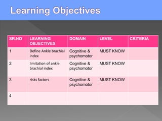
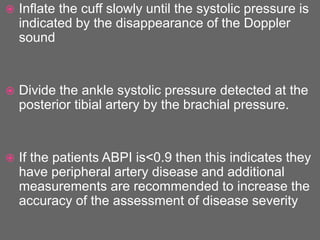
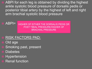
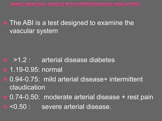
 At the end of the class the students will be able to
Define Ankle brachial index, limitation of ankle brachial
index, risks factors and clinical implications
SR.NO LEARNING
OBJECTIVES
DOMAIN LEVEL CRITERIA
1 Define Ankle brachial
index
Cognitive &
psychomotor
MUST KNOW
2 limitation of ankle
brachial index
Cognitive &
psychomotor
MUST KNOW
3 risks factors Cognitive &
psychomotor
MUST KNOW
4
 The Ankle brachial pressure index ABPI is the
ratio of the systolic blood pressure measured at
the ankle to that measured at the brachial artery.
 It is a noninvasive, Simple, Valid,reliable &
effective test which is used to detect lower
extremity Peripherial arterial disease PAD to
measure the severity of atherosclerosis in the
legs.
 It is not appropriate to detect PAD because of
possibility of false negative findings
 Lower extremity Peripherial arterial disease PAD
is a frequent, chronic ,progressive vascular
disease and associated with significant morbidity
& mortality
 Risk factors PAD are advanced >70 yrs,
smoking,past and present diabetes, family history
of cardivascular disease.
 The ABI is also used as a prognostic marker for
cardiovascular events in the absence of symptoms of
PAD
 INDICATIONS:
 Detection of PAD in all patients who present with
symptoms:
 Pain that restrict walking, ischemic pain at rest and
signs ( reduced or absent pedal pulses on palpation,
skin that is cool ,shiny, hairless or thick, thickening of
nails, abnormal capillary, pallor of distal extremity on
elevation, leg pain and tissue ulceration.
 Measuring ABI to detect peripheral artery disease
is a more sensitive and reliable test compared to
palpation of a pedal pulse, especially in patients
who are obese or who have significant edema.
 The ABI can provide also reliable information
about the severity of the disease
 It can exclude other causes of calf pain: spinal
stenosis, venous, raynaud phenomenon and
popliteal artery entrapment.
 Patients with PAD are frequently asymptomatic,
most commonly they have atypical leg pain and a
few present with typical claudication.
 ABI should be conducted on patients presenting
with risk factors to detect PAD so that therapeutic
interventions known to diminish their increased
risk of MI, stroke & death
 The ankle brachial pressure index ABPI is a non
invasive method for detecting or rulling out the
presence of peripheral artery disease.
 ABPI is a calculation of the ratio of the patients
systolic blood pressure at their ankle to the
systolic pressure in their arm.
 ABPI is generally between 1.0-1.4 in healthy
people i.e the systolic pressure at the ankle is
greater than the systolic pressure at the arm
 An abnormally low ABPI value i.e < 0.9 has a
sensitivity of 79-95% and specificity of
approximately 95% for peripheral artery disease.
 ABPI provides an indication of disease severity
and the urgency of referral.
 The presence of ischemic rest pain suggest
increased severity of peripheral artery disease
and an increased risk to the limb.
 Patients with ischemic rest pain often present with a
burning pain in the arch or distal foot that occurs
when their feet are elevated eg: in bed and resolves
when they place their feet on the floor
 An ABPI <0.4 indicates the patient has critical limb
ischemia
 This is a potentially life threatening condition
characterized by severly reduced circulation,
ischemia rest pain and tissue loss due to gangrene.
 The following equipment is recommended for
measuring the ankle brachial pressure index:
 A hand held portable Doppler device with a
frequency of 8-10 Hz although 5 Hz probes may
be better for patients with significant ankle edema.
 A sphygmomanometer
 Ultrasound transmission gel
 With the patient in a supine position:
 Place the blood pressure cuff approximately two
to three cm above the antecubital fossa for the
brachial pressure and approximately 5 cm above
the medial malleolus for ankle pressure
 The Doppler probe should detect a clear arterial
pulse before the cuff is inflated.
 Inflate the cuff slowly until the systolic pressure is
indicated by the disappearance of the Doppler
sound
 Divide the ankle systolic pressure detected at the
posterior tibial artery by the brachial pressure.
 If the patients ABPI is<0.9 then this indicates they
have peripheral artery disease and additional
measurements are recommended to increase the
accuracy of the assessment of disease severity
 Divide the highest ankle systolic pressure in each
of the posterior tibial and dorsalis pedis arteries in
both feet by the highest brachial systolic pressure
from each arm , the lowest resulting value is the
patients overall ABPI.
 The measurement may not be possible in all
patients as 12% of the general population has a
congenital absence of the dorsalis pedis pulse.
 ABPI for each leg is obtained by dividing the highest
ankle systolic blood pressure of dorsalis pedis or
posterior tibial artery by the highest of left and right
arm brachial systolic blood pressure
 ABPI=
 RISK FACTORS PAD:
 Old age
 Smoking past, present
 Diabetes
 Hypertension
 Renal function
HIGHER OF EITHER THE DORSALIS PEDIS OR
POST TIBIAL PRESSURE/HIGHER OF
BRACHIAL PRESSURE
 LIMITATION OF ABPI
 The Doppler device that is used in measurement
of ABPI indicates the velocity of blood flow and
this is related to blood volume
 The technique is unable to determine the exact
location of a patients arterial stenosis or occlusion
 ABPI can be falsely elevated in patients with
calcification of medial arteries eg in diabetes,
renal dysfunction ,RA
 Arterial insufficiency refers to a lack of adequate
blood flow to a region of the body
 PVD is a general term used to describe any
disorder that interfers with arterial or venous blood
flow of the extremities
 Factors that lead to PVD owing to arterial
insufficiency include smoking, cardiac disease
,Diabetes, hypertension ,renal disease and
elevated cholestrol, sedentry lifestyle are related
contributers in the cycle of disease and vessels
obstruction
 The damaged caused by these factors is reflected in
structual changes in the walls of arteries ,causing
abnormal blood flow
 Arteriosclerosis: thickening ,hardnening, loss of
elasticity of arterial walls
 Atherosclerosis the most common forms of
arteriosclerosis associated with damage to the
endothelial lining of the vessels and the formation of
lipid deposits, eventually leading to plaque formation.
 Thromboangitis obliterans (Buerger’s disease):
inflammation leads to arterial occlusion and tissue
ischemia especially in young men who smoke.
 Raynaud’s disease: a functional vasomotor
disease of small arteries and arterioles
 Ulceration : a peripheral sign of a long standing
disease process ,arterial ulcers are associated
with arterial insufficiency
 CLINICAL PRESENTATION:
 Most frequently located on the LE’S : Lateral
malleoli, dorsum of feet, toes
 When wounds are present on an ischemia limb,
atherosclerotic occlusion of the peripheral
vasculature is almost always present
 The majority of patients with arterial insufficiency
also have diabetes.
 Tropic changes are present and include abnormal
nail growth, decreased leg and foot hair , dry skin
 Skin is cool upon palpation
 Wounds are painful and patient may describe pain
in the leg and feet
 Wound base is necrotic and pale, lacking
granulation tissue.
 Skin around the wound may be black ,gangrenous
 Other signs of arterial insufficiency will be evident:
decreased pulses , pallor on elevation
 Painful cramping or aching of the LE during walking is
the most common complaint in patients with chronic
arterial occlusion of the LE’S.
 The pain is caused by intermittent claudication that
occurs when exercising muscle are not receiving
blood perfusion needed for normal function.
 Patients should be examined for other signs of
arterial insufficiency if intermittent claudication is
occuring
 Rest pain that develops at night awakens the
patient or requires analgesics for relief is
considered more severe than claudication.
 Diabetes will contribute to slower healing times
and difficulty fighting infection
 The ABI is a test designed to examine the
vascular system
 >1.2 : arterial disease diabetes
 1.19-0.95: normal
 0.94-0.75: mild arterial disease+ intermittent
claudication
 0.74-0.50: moderate arterial disease + rest pain
 <0.50 : severe arterial disease.
 What is ankle brachial Index?
 What are the limitation of Ankle brachial index?
 KISNER
 CASH
THANK YOU

Ankle brachial index

  • 2.
     At theend of the class the students will be able to Define Ankle brachial index, limitation of ankle brachial index, risks factors and clinical implications
  • 3.
    SR.NO LEARNING OBJECTIVES DOMAIN LEVELCRITERIA 1 Define Ankle brachial index Cognitive & psychomotor MUST KNOW 2 limitation of ankle brachial index Cognitive & psychomotor MUST KNOW 3 risks factors Cognitive & psychomotor MUST KNOW 4
  • 4.
     The Anklebrachial pressure index ABPI is the ratio of the systolic blood pressure measured at the ankle to that measured at the brachial artery.  It is a noninvasive, Simple, Valid,reliable & effective test which is used to detect lower extremity Peripherial arterial disease PAD to measure the severity of atherosclerosis in the legs.
  • 5.
     It isnot appropriate to detect PAD because of possibility of false negative findings  Lower extremity Peripherial arterial disease PAD is a frequent, chronic ,progressive vascular disease and associated with significant morbidity & mortality  Risk factors PAD are advanced >70 yrs, smoking,past and present diabetes, family history of cardivascular disease.
  • 6.
     The ABIis also used as a prognostic marker for cardiovascular events in the absence of symptoms of PAD  INDICATIONS:  Detection of PAD in all patients who present with symptoms:  Pain that restrict walking, ischemic pain at rest and signs ( reduced or absent pedal pulses on palpation, skin that is cool ,shiny, hairless or thick, thickening of nails, abnormal capillary, pallor of distal extremity on elevation, leg pain and tissue ulceration.
  • 7.
     Measuring ABIto detect peripheral artery disease is a more sensitive and reliable test compared to palpation of a pedal pulse, especially in patients who are obese or who have significant edema.  The ABI can provide also reliable information about the severity of the disease  It can exclude other causes of calf pain: spinal stenosis, venous, raynaud phenomenon and popliteal artery entrapment.
  • 8.
     Patients withPAD are frequently asymptomatic, most commonly they have atypical leg pain and a few present with typical claudication.  ABI should be conducted on patients presenting with risk factors to detect PAD so that therapeutic interventions known to diminish their increased risk of MI, stroke & death
  • 9.
     The anklebrachial pressure index ABPI is a non invasive method for detecting or rulling out the presence of peripheral artery disease.  ABPI is a calculation of the ratio of the patients systolic blood pressure at their ankle to the systolic pressure in their arm.  ABPI is generally between 1.0-1.4 in healthy people i.e the systolic pressure at the ankle is greater than the systolic pressure at the arm
  • 10.
     An abnormallylow ABPI value i.e < 0.9 has a sensitivity of 79-95% and specificity of approximately 95% for peripheral artery disease.  ABPI provides an indication of disease severity and the urgency of referral.  The presence of ischemic rest pain suggest increased severity of peripheral artery disease and an increased risk to the limb.
  • 11.
     Patients withischemic rest pain often present with a burning pain in the arch or distal foot that occurs when their feet are elevated eg: in bed and resolves when they place their feet on the floor  An ABPI <0.4 indicates the patient has critical limb ischemia  This is a potentially life threatening condition characterized by severly reduced circulation, ischemia rest pain and tissue loss due to gangrene.
  • 12.
     The followingequipment is recommended for measuring the ankle brachial pressure index:  A hand held portable Doppler device with a frequency of 8-10 Hz although 5 Hz probes may be better for patients with significant ankle edema.  A sphygmomanometer  Ultrasound transmission gel
  • 13.
     With thepatient in a supine position:  Place the blood pressure cuff approximately two to three cm above the antecubital fossa for the brachial pressure and approximately 5 cm above the medial malleolus for ankle pressure  The Doppler probe should detect a clear arterial pulse before the cuff is inflated.
  • 16.
     Inflate thecuff slowly until the systolic pressure is indicated by the disappearance of the Doppler sound  Divide the ankle systolic pressure detected at the posterior tibial artery by the brachial pressure.  If the patients ABPI is<0.9 then this indicates they have peripheral artery disease and additional measurements are recommended to increase the accuracy of the assessment of disease severity
  • 17.
     Divide thehighest ankle systolic pressure in each of the posterior tibial and dorsalis pedis arteries in both feet by the highest brachial systolic pressure from each arm , the lowest resulting value is the patients overall ABPI.  The measurement may not be possible in all patients as 12% of the general population has a congenital absence of the dorsalis pedis pulse.
  • 18.
     ABPI foreach leg is obtained by dividing the highest ankle systolic blood pressure of dorsalis pedis or posterior tibial artery by the highest of left and right arm brachial systolic blood pressure  ABPI=  RISK FACTORS PAD:  Old age  Smoking past, present  Diabetes  Hypertension  Renal function HIGHER OF EITHER THE DORSALIS PEDIS OR POST TIBIAL PRESSURE/HIGHER OF BRACHIAL PRESSURE
  • 19.
     LIMITATION OFABPI  The Doppler device that is used in measurement of ABPI indicates the velocity of blood flow and this is related to blood volume  The technique is unable to determine the exact location of a patients arterial stenosis or occlusion  ABPI can be falsely elevated in patients with calcification of medial arteries eg in diabetes, renal dysfunction ,RA
  • 20.
     Arterial insufficiencyrefers to a lack of adequate blood flow to a region of the body  PVD is a general term used to describe any disorder that interfers with arterial or venous blood flow of the extremities  Factors that lead to PVD owing to arterial insufficiency include smoking, cardiac disease ,Diabetes, hypertension ,renal disease and elevated cholestrol, sedentry lifestyle are related contributers in the cycle of disease and vessels obstruction
  • 21.
     The damagedcaused by these factors is reflected in structual changes in the walls of arteries ,causing abnormal blood flow  Arteriosclerosis: thickening ,hardnening, loss of elasticity of arterial walls  Atherosclerosis the most common forms of arteriosclerosis associated with damage to the endothelial lining of the vessels and the formation of lipid deposits, eventually leading to plaque formation.
  • 22.
     Thromboangitis obliterans(Buerger’s disease): inflammation leads to arterial occlusion and tissue ischemia especially in young men who smoke.  Raynaud’s disease: a functional vasomotor disease of small arteries and arterioles  Ulceration : a peripheral sign of a long standing disease process ,arterial ulcers are associated with arterial insufficiency
  • 23.
     CLINICAL PRESENTATION: Most frequently located on the LE’S : Lateral malleoli, dorsum of feet, toes  When wounds are present on an ischemia limb, atherosclerotic occlusion of the peripheral vasculature is almost always present  The majority of patients with arterial insufficiency also have diabetes.
  • 24.
     Tropic changesare present and include abnormal nail growth, decreased leg and foot hair , dry skin  Skin is cool upon palpation  Wounds are painful and patient may describe pain in the leg and feet
  • 25.
     Wound baseis necrotic and pale, lacking granulation tissue.  Skin around the wound may be black ,gangrenous  Other signs of arterial insufficiency will be evident: decreased pulses , pallor on elevation
  • 26.
     Painful crampingor aching of the LE during walking is the most common complaint in patients with chronic arterial occlusion of the LE’S.  The pain is caused by intermittent claudication that occurs when exercising muscle are not receiving blood perfusion needed for normal function.  Patients should be examined for other signs of arterial insufficiency if intermittent claudication is occuring
  • 27.
     Rest painthat develops at night awakens the patient or requires analgesics for relief is considered more severe than claudication.  Diabetes will contribute to slower healing times and difficulty fighting infection
  • 28.
     The ABIis a test designed to examine the vascular system  >1.2 : arterial disease diabetes  1.19-0.95: normal  0.94-0.75: mild arterial disease+ intermittent claudication  0.74-0.50: moderate arterial disease + rest pain  <0.50 : severe arterial disease.
  • 29.
     What isankle brachial Index?  What are the limitation of Ankle brachial index?
  • 30.
  • 31.