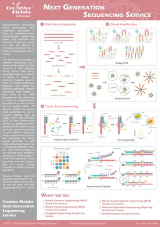
Natural killer (NK) cells are cytotoxic immune cells that play an important role in the defense against cancer.
https://mrna.creative-biolabs.com/custom-natual-killer-cell-reprogramming-by-mrna.htm
Natural killer (NK) cells are cytotoxic immune cells that play an important role in the defense against cancer.
https://mrna.creative-biolabs.com/custom-natual-killer-cell-reprogramming-by-mrna.htm
One mutational event is the integration of the viral genome into the host chromosome, The exogenous forces responsible for this event range from UV and ionizing radiation exposure to a defect in the virus itself.[a.3]
An additional event important in MCPyV-induced carcinogenesis is the mutation of the viral genome in a manner that renders the virus unable to replicate. The most common mutations occur in the carboxy terminus of LT, which generate a truncated LT that lacks the helicase domain required for replication yet preserve its oncogenic functions.[a.3]
Persistent expression of truncated LT and sT from the integrated MCPyV genome can inactivate pRb tumor suppressor function and promote and/or deregulate cap-dependent translation initiation, respectively. [a.3]
Ultimately, the cells in which integration of a replication-defective MCPyV is successful become addicted to viral oncogene expression and undergo clonal expansion and neoplastic progression to cause the development of MCC.[a.3]
A micro-array is a tool for analyzing gene expression that consists of a small membrane or glass slide containing samples of many genes arranged in a regular pattern.
This was made by me while I was in Masters. I have made few animations. I hope it makes understanding better.
The content is made by searching through internet and referencing books. I do not claim any content in whole presentation except the animations made on the subject.
The DNA microarray is a tool used to determine whether the DNA from a particular individual contains a mutation in genes like BRCA1 and BRCA2. The chip consists of a small glass plate encased in plastic. Some companies manufacture microarrays using methods similar to those used to make computer microchips.
A DNA microarray is a collection of microscopic DNA spots attached to a solid surface. Scientists use DNA microarrays to measure the expression levels of large numbers of genes simultaneously or to genotype multiple regions of a genome. Each DNA spot contains picomoles of a specific DNA sequence, known as probes.
This chapter provides an overview of DNA microarrays. Microarrays are a technology in which 1000’s of nucleic acids are bound to a surface and are used to measure the relative concentration of nucleic acid sequences in a mixture via hybridization and subsequent detection of the hybridization events. We first cover the history of microarrays and the antecedent technologies that led to their development. We then discuss the methods of manufacture of microarrays and the most common biological applications. The chapter ends with a brief discussion of the limitations of microarrays and discusses how microarrays are being rapidly replaced by DNA sequencing technologies.
The DNA microarray is a tool used to determine whether the DNA from a particular individual contains a mutation in genes like BRCA1 and BRCA2. The chip consists of a small glass plate encased in plastic. Some companies manufacture microarrays using methods similar to those used to make computer microchips.
Sequencing genes and genomes in biology. The most important technique available to the molecular biologist is DNA sequencing, by which the precise order of nucleotides in a piece of DNA can be determined
DNA Sequencing - DNA sequencing is like reading the instructions inside a cellAmitSamadhiya1
DNA sequencing is like reading the instructions inside a cell. It's figuring out the exact order of the building blocks that make up our DNA, represented by the letters A, T, C, and G. This order is like a code that tells our bodies how to function and grow.
By reading this code, scientists can understand genes, diagnose diseases, and even trace our ancestry. There are different ways to sequence DNA, kind of like having a few different ways to read a book. These techniques are constantly improving, making it faster and easier to unlock the secrets hidden in our DNA.
sequencing presentation. providing deep and insightful points about Sanger sequencing, Maxam-gilbert sequencing, Illumina sequencing, and single molecule sequencing.
Phage Display Screening for Single Domain Antibody (sdAb)ShawVivian
Phage display screening(https://www.creative-biolabs.com/sdab/phage-display-screening-for-single-domain-antibody-sdab.htm) is a well-established technique to identify antibody fragments and other binding molecules against any targets of interest. As each clone in phage display libraries represents a noncombinatorial functional domain of a naturally circulating antibody, phage display screening is a powerful method for sdAb discovery. Here, Creative Biolabs describes the phage display screening for sdAb generation.
Creative Biolabs offers a comprehensive range of quality recombinant antibodies for cell & organelle imaging. These antibodies are covering significant research areas such as cancer research, cell signaling, cell morphology and organelle detection.
Live cell imaging is the study of living cells using time-lapse microscopy. Scientists use it to better understand and validate the biological function through the study of cellular dynamics. Organelles are specialized and well-defined parts of the cell which are usually separately enclosed within their own lipid bilayers. Very often organelles have to be stained to follow cellular processes and the effects of changing parameters on these processes. Thus, there is a great significance and strong need for tools to stain and follow specific organelles in living cells.
https://www.antibody-creativebiolabs.com/category-introduction/cell-organelle-imaging-3.htm
Thanks to a longstanding presence in the market of sdAb development and our well-established screening platform, Creative Biolabs is the right partner to navigate the sdAb screening challenges. Focused on the field of sdAb discovery and development, we will use our skills to offer innovative and sophisticated sdAb screening services tailored to our customer's exact needs.
Secondary antibodies bind to the primary antibody to assist in detection, classification and isolation of interest antigen. To ensure detection, the secondary antibody must be specific for the isotype and antibody species of the primary antibody in the application. Secondary antibodies are typically polyclonal.
Secondary antibody conjugates are important tools in biochemical and cell-based analysis. Enzyme-labeled secondary antibodies have high catalytic turnover rates and are routinely used in various assays for their robust and rapid signal generation. Secondary antibodies conjugated to fluorescent dyes offer brighter signals and multiplexing capabilities in cell analysis and protein analysis applications including immunofluorescence microscopy and cell imaging. For both enzyme-labeled and dye-labeled secondary antibodies, high sensitivity and signal amplification are two desirable qualities when detecting target antigens of low abundance.
Antibody pair is two different antibodies developed against the same target that with different binding regions or epitopes. Differential epitope recognition renders simultaneous binding of both antibodies to the same target, to form sandwich ELISA format. Antibody pair can be used to detect and quantify interaction molecules in situ, ensure localization of sub-cellular events, reduce the risk of missing weak or transient interactions and improve detection specificity and sensitivity. Creative Biolabs has years of expertise in developing antibody pairs to provide a monoclonal antibody pair, a polyclonal antibody pair, or a combination for your specific requirement. Our high-quality products are suitable for used to quantify target analyte from complex samples across various fields including cancer, autoimmune, metabolic disease, endocrine, infectious disease, etc.
https://www.antibody-creativebiolabs.com/category/antibody-pairs-129.htm
Antibody arrays have emerged as an important tool, which enable multiplexed protein expression profiling such as cytokines, chemokines and growth factors, which may be involved in diseases. To design high-performing antibody arrays, it is necessary to develop an integrated technology platform, where the interplay between the surfaces and the antibody is anticipated to play an important role.
https://www.antibody-creativebiolabs.com/category/glass-slide-antibody-arrays-126.htm
Antibody arrays are valuable for the parallel analysis of multiple proteins in small sample volumes. The earliest and most widely used application of antibody arrays has been to measure multiple protein abundances, using sandwich assays and label-based assays, for biomarker discovery and biological studies. Modifications to these assays have led to studies profiling specific protein post-translational modifications, such as protein phosphorylation that is a mechanism of regulation that is extremely important in most cellular processes.
https://www.antibody-creativebiolabs.com/category-introduction/antibody-array-analysis-132.htm
Creative Biolabs provides a comprehensive range of recombinant antibodies of the key tumor for research processes and diagnostic assay developments for our clients. here is PIK3CA antibody for breast cancer.
https://www.antibody-creativebiolabs.com/category-introduction/tumor-marker-detection-1.htm
Single domain antibodies (sdAbs, or VHH) are the smallest antigen-binding units of antibodies, comprising either one variable domain or one engineered constant domain that solely facilitates target binding. They are popular as a novel class of proprietary therapeutic proteins containing unique structural and functional properties. At Creative Biolabs, we offer one-stop solutions for sdAb development and characterization to promote the identification of novel therapeutic sdAbs to meet your specific project demands.
https://www.creative-biolabs.com/sdab/sdab-functional-identification.htm
Next-generation sequencing (NGS) plays an important role in cancer diagnostics. Creative Biolabs offers SuPrecision™ Platform covering a full range of NGS-based services. This tool greatly improves our understanding of the multifaceted disease and achieves accurate, sensitive, high-throughput and cost effective cancer detection.
The genome diversity within an ecosystem of single cell species is far greater than currently accurate measurement. In the era of precision medicine, single cell genomics omics technology is highly promising owing to its potential in diagnosis, therapeutics, and prognosis. As a skillful expert in the single cell analysis, Creative Biolabs is committed to advancing clients' significant programs and provides end-to-end solutions for any challenging single cell genomics projects through our advanced platforms and bioinformatics profiling.
https://singlecell.creative-biolabs.com/single-cell-genomics-platform.htm
An ongoing outbreak of a novel coronavirus (SARS-CoV-2) has raised global concerns. It is identified as the cause of pneumonia with unknown etiology. Since the early outbreak in Wuhan, China, it has subsequently spread to all provinces of China and many other countries. The urgent epidemic situation has spurred the development of antiviral drugs and vaccines. As a leading service provider in the field of biological research and drug discovery, Creative Biolabs provides fast & elaborate therapeutic antibody discovery, drug candidates screening and vaccine development services to help combat this outbreak.
https://sars-cov-2.creative-biolabs.com/novel-coronavirus-sars-cov2.htm
Creative Biolabs is skilled in NGS-based cancer research. We support one-stop cancer research services based on a variety of sequencing technologies. Our whole gene sequencing (WGS) and whole exome sequencing (WES) platforms enable us to comprehensively analyze and identify known and unknown cancer gene mutations. Besides, we also provide high-quality target sequencing services, allowing us to analyze several or even hundreds of target gene mutations economically and effectively. Our services will facilitate our customers’ cancer research including cancer diagnosis and treatment.
https://www.creative-biolabs.com/suprecision/genetic-testing-for-cancer.htm
Creative Biolabs’ technical experts have successfully established the proprietary complement therapeutics system. We are looking forward to designing a strategy to carry on the therapeutic antibodies development services for our clients in complement therapeutic.
a full range of complement therapeutic services and complement based drug discovery services for customers at https://www.creative-biolabs.com/complement-therapeutics/
diversified species genetic information and a wide
range of antibodies information and one-stop customized service at https://modelorg-ab.creative-biolabs.com/
Lung Cancer: Artificial Intelligence, Synergetics, Complex System Analysis, S...Oleg Kshivets
RESULTS: Overall life span (LS) was 2252.1±1742.5 days and cumulative 5-year survival (5YS) reached 73.2%, 10 years – 64.8%, 20 years – 42.5%. 513 LCP lived more than 5 years (LS=3124.6±1525.6 days), 148 LCP – more than 10 years (LS=5054.4±1504.1 days).199 LCP died because of LC (LS=562.7±374.5 days). 5YS of LCP after bi/lobectomies was significantly superior in comparison with LCP after pneumonectomies (78.1% vs.63.7%, P=0.00001 by log-rank test). AT significantly improved 5YS (66.3% vs. 34.8%) (P=0.00000 by log-rank test) only for LCP with N1-2. Cox modeling displayed that 5YS of LCP significantly depended on: phase transition (PT) early-invasive LC in terms of synergetics, PT N0—N12, cell ratio factors (ratio between cancer cells- CC and blood cells subpopulations), G1-3, histology, glucose, AT, blood cell circuit, prothrombin index, heparin tolerance, recalcification time (P=0.000-0.038). Neural networks, genetic algorithm selection and bootstrap simulation revealed relationships between 5YS and PT early-invasive LC (rank=1), PT N0—N12 (rank=2), thrombocytes/CC (3), erythrocytes/CC (4), eosinophils/CC (5), healthy cells/CC (6), lymphocytes/CC (7), segmented neutrophils/CC (8), stick neutrophils/CC (9), monocytes/CC (10); leucocytes/CC (11). Correct prediction of 5YS was 100% by neural networks computing (area under ROC curve=1.0; error=0.0).
CONCLUSIONS: 5YS of LCP after radical procedures significantly depended on: 1) PT early-invasive cancer; 2) PT N0--N12; 3) cell ratio factors; 4) blood cell circuit; 5) biochemical factors; 6) hemostasis system; 7) AT; 8) LC characteristics; 9) LC cell dynamics; 10) surgery type: lobectomy/pneumonectomy; 11) anthropometric data. Optimal diagnosis and treatment strategies for LC are: 1) screening and early detection of LC; 2) availability of experienced thoracic surgeons because of complexity of radical procedures; 3) aggressive en block surgery and adequate lymph node dissection for completeness; 4) precise prediction; 5) adjuvant chemoimmunoradiotherapy for LCP with unfavorable prognosis.
Ozempic: Preoperative Management of Patients on GLP-1 Receptor Agonists Saeid Safari
Preoperative Management of Patients on GLP-1 Receptor Agonists like Ozempic and Semiglutide
ASA GUIDELINE
NYSORA Guideline
2 Case Reports of Gastric Ultrasound
TEST BANK for Operations Management, 14th Edition by William J. Stevenson, Ve...kevinkariuki227
TEST BANK for Operations Management, 14th Edition by William J. Stevenson, Verified Chapters 1 - 19, Complete Newest Version.pdf
Flu Vaccine Alert in Bangalore Karnatakaaddon Scans
As flu season approaches, health officials in Bangalore, Karnataka, are urging residents to get their flu vaccinations. The seasonal flu, while common, can lead to severe health complications, particularly for vulnerable populations such as young children, the elderly, and those with underlying health conditions.
Dr. Vidisha Kumari, a leading epidemiologist in Bangalore, emphasizes the importance of getting vaccinated. "The flu vaccine is our best defense against the influenza virus. It not only protects individuals but also helps prevent the spread of the virus in our communities," he says.
This year, the flu season is expected to coincide with a potential increase in other respiratory illnesses. The Karnataka Health Department has launched an awareness campaign highlighting the significance of flu vaccinations. They have set up multiple vaccination centers across Bangalore, making it convenient for residents to receive their shots.
To encourage widespread vaccination, the government is also collaborating with local schools, workplaces, and community centers to facilitate vaccination drives. Special attention is being given to ensuring that the vaccine is accessible to all, including marginalized communities who may have limited access to healthcare.
Residents are reminded that the flu vaccine is safe and effective. Common side effects are mild and may include soreness at the injection site, mild fever, or muscle aches. These side effects are generally short-lived and far less severe than the flu itself.
Healthcare providers are also stressing the importance of continuing COVID-19 precautions. Wearing masks, practicing good hand hygiene, and maintaining social distancing are still crucial, especially in crowded places.
Protect yourself and your loved ones by getting vaccinated. Together, we can help keep Bangalore healthy and safe this flu season. For more information on vaccination centers and schedules, residents can visit the Karnataka Health Department’s official website or follow their social media pages.
Stay informed, stay safe, and get your flu shot today!
Title: Sense of Taste
Presenter: Dr. Faiza, Assistant Professor of Physiology
Qualifications:
MBBS (Best Graduate, AIMC Lahore)
FCPS Physiology
ICMT, CHPE, DHPE (STMU)
MPH (GC University, Faisalabad)
MBA (Virtual University of Pakistan)
Learning Objectives:
Describe the structure and function of taste buds.
Describe the relationship between the taste threshold and taste index of common substances.
Explain the chemical basis and signal transduction of taste perception for each type of primary taste sensation.
Recognize different abnormalities of taste perception and their causes.
Key Topics:
Significance of Taste Sensation:
Differentiation between pleasant and harmful food
Influence on behavior
Selection of food based on metabolic needs
Receptors of Taste:
Taste buds on the tongue
Influence of sense of smell, texture of food, and pain stimulation (e.g., by pepper)
Primary and Secondary Taste Sensations:
Primary taste sensations: Sweet, Sour, Salty, Bitter, Umami
Chemical basis and signal transduction mechanisms for each taste
Taste Threshold and Index:
Taste threshold values for Sweet (sucrose), Salty (NaCl), Sour (HCl), and Bitter (Quinine)
Taste index relationship: Inversely proportional to taste threshold
Taste Blindness:
Inability to taste certain substances, particularly thiourea compounds
Example: Phenylthiocarbamide
Structure and Function of Taste Buds:
Composition: Epithelial cells, Sustentacular/Supporting cells, Taste cells, Basal cells
Features: Taste pores, Taste hairs/microvilli, and Taste nerve fibers
Location of Taste Buds:
Found in papillae of the tongue (Fungiform, Circumvallate, Foliate)
Also present on the palate, tonsillar pillars, epiglottis, and proximal esophagus
Mechanism of Taste Stimulation:
Interaction of taste substances with receptors on microvilli
Signal transduction pathways for Umami, Sweet, Bitter, Sour, and Salty tastes
Taste Sensitivity and Adaptation:
Decrease in sensitivity with age
Rapid adaptation of taste sensation
Role of Saliva in Taste:
Dissolution of tastants to reach receptors
Washing away the stimulus
Taste Preferences and Aversions:
Mechanisms behind taste preference and aversion
Influence of receptors and neural pathways
Impact of Sensory Nerve Damage:
Degeneration of taste buds if the sensory nerve fiber is cut
Abnormalities of Taste Detection:
Conditions: Ageusia, Hypogeusia, Dysgeusia (parageusia)
Causes: Nerve damage, neurological disorders, infections, poor oral hygiene, adverse drug effects, deficiencies, aging, tobacco use, altered neurotransmitter levels
Neurotransmitters and Taste Threshold:
Effects of serotonin (5-HT) and norepinephrine (NE) on taste sensitivity
Supertasters:
25% of the population with heightened sensitivity to taste, especially bitterness
Increased number of fungiform papillae
New Directions in Targeted Therapeutic Approaches for Older Adults With Mantl...i3 Health
i3 Health is pleased to make the speaker slides from this activity available for use as a non-accredited self-study or teaching resource.
This slide deck presented by Dr. Kami Maddocks, Professor-Clinical in the Division of Hematology and
Associate Division Director for Ambulatory Operations
The Ohio State University Comprehensive Cancer Center, will provide insight into new directions in targeted therapeutic approaches for older adults with mantle cell lymphoma.
STATEMENT OF NEED
Mantle cell lymphoma (MCL) is a rare, aggressive B-cell non-Hodgkin lymphoma (NHL) accounting for 5% to 7% of all lymphomas. Its prognosis ranges from indolent disease that does not require treatment for years to very aggressive disease, which is associated with poor survival (Silkenstedt et al, 2021). Typically, MCL is diagnosed at advanced stage and in older patients who cannot tolerate intensive therapy (NCCN, 2022). Although recent advances have slightly increased remission rates, recurrence and relapse remain very common, leading to a median overall survival between 3 and 6 years (LLS, 2021). Though there are several effective options, progress is still needed towards establishing an accepted frontline approach for MCL (Castellino et al, 2022). Treatment selection and management of MCL are complicated by the heterogeneity of prognosis, advanced age and comorbidities of patients, and lack of an established standard approach for treatment, making it vital that clinicians be familiar with the latest research and advances in this area. In this activity chaired by Michael Wang, MD, Professor in the Department of Lymphoma & Myeloma at MD Anderson Cancer Center, expert faculty will discuss prognostic factors informing treatment, the promising results of recent trials in new therapeutic approaches, and the implications of treatment resistance in therapeutic selection for MCL.
Target Audience
Hematology/oncology fellows, attending faculty, and other health care professionals involved in the treatment of patients with mantle cell lymphoma (MCL).
Learning Objectives
1.) Identify clinical and biological prognostic factors that can guide treatment decision making for older adults with MCL
2.) Evaluate emerging data on targeted therapeutic approaches for treatment-naive and relapsed/refractory MCL and their applicability to older adults
3.) Assess mechanisms of resistance to targeted therapies for MCL and their implications for treatment selection
The prostate is an exocrine gland of the male mammalian reproductive system
It is a walnut-sized gland that forms part of the male reproductive system and is located in front of the rectum and just below the urinary bladder
Function is to store and secrete a clear, slightly alkaline fluid that constitutes 10-30% of the volume of the seminal fluid that along with the spermatozoa, constitutes semen
A healthy human prostate measures (4cm-vertical, by 3cm-horizontal, 2cm ant-post ).
It surrounds the urethra just below the urinary bladder. It has anterior, median, posterior and two lateral lobes
It’s work is regulated by androgens which are responsible for male sex characteristics
Generalised disease of the prostate due to hormonal derangement which leads to non malignant enlargement of the gland (increase in the number of epithelial cells and stromal tissue)to cause compression of the urethra leading to symptoms (LUTS
2. DNA Library Preparation
1 Clonal Amplification
2
Cyclic Array Sequencing
3
T
5'
5'
3'
3'
T
5'
3'
T
C
A
A
A n
A n z
n z
z
C G T
T
5'
3'
A
A
T
5'
3'
A
G
A
A
A
A
A
T
5'
3'
A
A
A
A
3. 1
4 5
2 3
A
C
G
T
5'
3'
A n
G n z
n z
z
5'
5'
3'
A n
T n z
n z
z
3'
3'
T
n
C
A
G
n z
n
z
z
n
A
C n z
n z
5'
3'
A n
C n z
n z
z