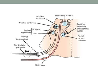
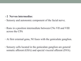
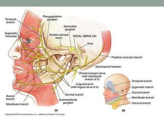
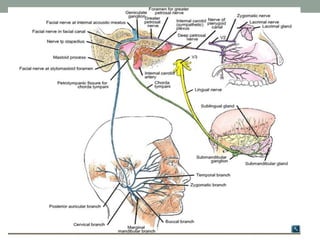
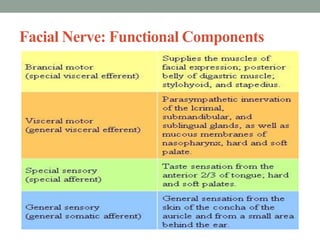
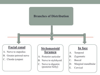
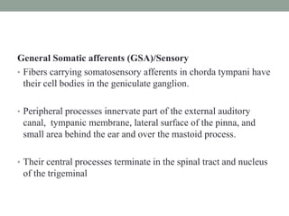
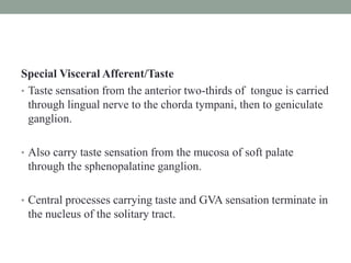
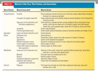
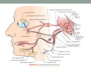
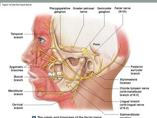
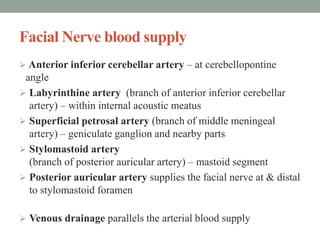
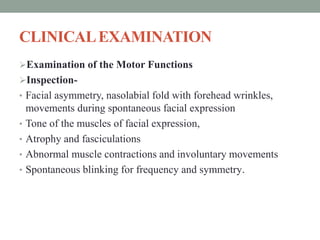
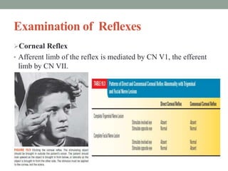
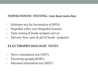
FACIAL NERVE
DR. SUMIT KAMBLE
SENIOR RESIDENT
DEPT. OF NEUROLOGY
GMC, KOTA
INTRODUCTION
• Facial nerve is seventh cranial nerve
• Mixed nerve with motor and sensory roots.
• Emerges from the brain stem between the pons and medulla,
controls muscles of facial expression, and muscles of the scalp
and ear, as well as buccinator, platysma, stapedius, stylohyoid,
and posterior belly of the digastric.
• Functions in conveyance of taste sensations from the anterior
two thirds of the tongue and oral cavity
• Carries parasympathetic secretory fibers to submandibular and
sublingual salivary glands, lacrimal gland and to mucous
membranes of oral and nasal cavities.
• Conveys exteroceptive sensation from eardrum and external
auditory canal, proprioceptive sensation from muscles it
supplies, and general visceral sensation from salivary glands
and mucosa of the nose and pharynx.
Facial nerve is formed mainly of two parts:
1 Facial nerve proper (motor):
• Arising from facial motor nucleus in pons.
• Supranuclear innervation to the muscles of facial expression
arises from the lower third of contralateral precentral gyrus in
facial area of motor homunculus.
• Portion of nucleus that innervates the lower half to two thirds
of the face has predominantly contralateral supranuclear
control;
• Portion that innervates upper third to half has bilateral control.
• Facial nucleus is special visceral efferent, or branchiomotor;
• It innervates the muscles of the second branchial arch
• Facial motor nucleus has lateral, medial, and dorsal subnuclei,
arranged in columns.
• Exits the pons laterally at the pontomedullar junction, just
caudal to the roots of CN V between the olive and the inferior
cerebellar peduncle
• Has two components, motor root, which makes up about 70% of
the fibers, and sensory root, which accounts for 30%.
2 Nervus intermedius:
• Sensory and autonomic component of the facial nerve.
• Runs in a position intermediate between CNs VII and VIII
across the CPA
• At first external genu, NI fuses with the geniculate ganglion.
• Sensory cells located in the geniculate ganglion are general
somatic afferent (GSA) and special visceral afferent (SVA).
• GSA fibers carry exteroceptive impulses from region of the
external auditory canal and tympanic membrane.
• SVA fibers convey taste from the anterior two-thirds of the
tongue.
• Autonomic component of the NI consists of preganglionic
general visceral efferent parasympathetic fibers from superior
salivatory and lacrimal nuclei
Course and relations:
• Intracranial (intrapetrosal) course
• Extracranial course
Facial Nerve: Functional Components
Branches of Distribution
Facial canal
A. Nerve to stapedius
B. Greater petrosal nerve
C. Chorda tympani
In face
A. Temporal
B. Zygomatic
C. Buccal
D. Marginal mandibular
E. Cervical
Stylomastoid
foramen
A. Posterior auricular
B. Nerve to stylohyoid
C. Nerve to digastric
(posterior belly)
Within the facial canal:
 1- Greater superficial petrosal nerve (GSPN) : carries
preganglionic parasympathetic fibers
• These fibers are conveyed by the NI to geniculate ganglion.
• Pass through the ganglion without synapsing into the greater
petrosal nerve, which goes forward through the hiatus of the
facial canal to join deep petrosal nerve from the carotid
sympathetic plexus to form the vidian nerve, or the nerve of the
pterygoid canal, which runs to the sphenopalatine ganglion,
from where postganglionic fibers proceed to the lacrimal gland.
 2-Nerve to stapedius: supplies the stapedius muscle.
3- Chorda tympani nerve:
• It arises from the facial nerve 6 mm above the stylomastoid
foramen and runs upwards to perforate the posterior bony wall
of the tympanic cavity.
• It carries taste and general visceral afferent (GVA) fibers as well
as preganglionic parasympathetics.
General Somatic afferents (GSA)/Sensory
• Fibers carrying somatosensory afferents in chorda tympani have
their cell bodies in the geniculate ganglion.
• Peripheral processes innervate part of the external auditory
canal, tympanic membrane, lateral surface of the pinna, and
small area behind the ear and over the mastoid process.
• Their central processes terminate in the spinal tract and nucleus
of the trigeminal
Special Visceral Afferent/Taste
• Taste sensation from the anterior two-thirds of tongue is carried
through lingual nerve to the chorda tympani, then to geniculate
ganglion.
• Also carry taste sensation from the mucosa of soft palate
through the sphenopalatine ganglion.
• Central processes carrying taste and GVA sensation terminate in
the nucleus of the solitary tract.
• Solitary tract sends communications to the superior and inferior
salivatory nuclei, which send parasympathetics to salivary
glands.
• Other fibers synapse in reticular formation; next order neurons
form a component of the reticulospinal tract bilaterally to
synapse with sympathetic neurons in the intermediolateral gray
column of the upper thoracic spinal cord.
• These send sympathetic innervation via the superior cervical
ganglion to the salivary glands.
General Visceral Efferent/Parasympathetic
• Chorda tympani also carries Preganglionic parasympathetic
fibers to the submandibular ganglion.
• Postganglionic fibers convey secretory and vasodilator impulses
to the submandibular and sublingual salivary glands and
mucous membranes of mouth and tongue.
• These glands also receive sympathetic innervation through
superior cervical ganglion and the carotid plexus.
• Parasympathetic fibers cause vasodilation and a copious, thin,
watery secretion high in enzymes;
• Sympathetic fibers cause vasoconstriction and a scant, thick,
mucoid secretion low in enzyme content.
II- At exit from the stylomastoid foramen
• 1- Posterior auricular nerve:
Supplies the occipitalis , posterior auricular, and transverse and
oblique auricular muscles.
• 2- Digastric branch:
Posterior belly of digastric muscle
• 3- Stylohyoid branch:
Stylohyoid muscle
GANGLIAASSOCIATED WITH FACIALNERVE
• Geniculate ganglion
• Submandibular ganglion
• Pterygopalatine ganglion
Geniculate Ganglion
• The geniculate ganglion (from Latin genu, for "knee") is an L-
shaped collection of fibers and sensory neurons of the facial
nerve located in the facial canal of the head.
• It receives fibers from the motor, sensory, and parasympathetic
components of the facial nerve and sends fibers that will
innervate the lacrimal glands, submandibular glands, sublingual
glands, tongue, palate, pharynx, external auditory meatus,
stapedius, posterior belly of the digastric muscle, stylohyoid
muscle, and muscles of facial expression.
Submandibular Ganglion
 The submandibular ganglion is small and fusiform in shape. It
is situated above the deep portion of the submandibular gland,
on the hyoglossus muscle, near the posterior border of the
mylohyoid muscle.
 The ganglion 'hangs' by two nerve filaments from the lower
border of the lingual nerve (itself a branch of the mandibular
nerve, CN V3). It is suspended from the lingual nerve by two
filaments, one anterior and one posterior. Through the
posterior of these it receives a branch from the chorda tympani
nerve which runs in the sheath of the lingual nerve.
Pterygopalatine Ganglion
• The pterygopalatine ganglion (meckel's ganglion, nasal
ganglion or sphenopalatine ganglion) is a parasympathetic
ganglion found in the pterygopalatine fossa.
• It's largely innervated by the greater petrosal nerve (a branch of
the facial nerve); and its axons project to the lacrimal glands
and nasal mucosa
Facial Nerve blood supply
 Anterior inferior cerebellar artery – at cerebellopontine
angle
 Labyrinthine artery (branch of anterior inferior cerebellar
artery) – within internal acoustic meatus
 Superficial petrosal artery (branch of middle meningeal
artery) – geniculate ganglion and nearby parts
 Stylomastoid artery
(branch of posterior auricular artery) – mastoid segment
 Posterior auricular artery supplies the facial nerve at & distal
to stylomastoid foramen
 Venous drainage parallels the arterial blood supply
CLINICALEXAMINATION
Examination of the Motor Functions
Inspection-
• Facial asymmetry, nasolabial fold with forehead wrinkles,
movements during spontaneous facial expression
• Tone of the muscles of facial expression,
• Atrophy and fasciculations
• Abnormal muscle contractions and involuntary movements
• Spontaneous blinking for frequency and symmetry.
Testing of Facial Nerve Branches
 Testing the temporal branches of the facial nerve –
patient is asked to frown and wrinkle his or her forehead.
 Testing the Zygomatic branches of the facial nerve patient
is asked to close their eyes tightly
 Testing the buccal branches of the facial nerve
• Puff up cheeks (buccinator)
• Smile and show teeth (orbicularis oris)
• Tap with finger over each cheek to detect ease of air expulsion
on the affected side
Examination of Reflexes
Corneal Reflex
• Afferent limb of the reflex is mediated by CN V1, the efferent
limb by CN VII.
Stapedius reflex
• Nerve to stapedius muscle test
• Impedence audiometry can record the presence or absence of
stapedius muscle contraction to sound stimuli 70 to 100 db
above hearing threshold
• Absence reflex or a reflex less than half the amplitude is due to
a lesion proximal to stapedius nerve
Examination of Sensory Functions
Hypesthesia of posterior wall of the external auditory meatus in
proximal facial nerve lesions.
Taste on anterior two-thirds of the tongue-use four substances
for testing:
• Sucrose (sweet), sodium chloride (salty), quinine (bitter), and
citric acid (sour).
• Patient with a peripheral pattern of facial weakness has
impaired taste, the lesion is proximal to the junction with the
chorda tympani.
Examination of Secretory Functions
• Tear production may be quantitated with the Schirmer test.
• Lacrimal reflex is tearing, usually bilateral, caused by
stimulating the cornea.
• Nasolacrimal reflex is elicited by mechanical stimulation of the
nasal mucosa, or by chemical stimulation using irritating
substances such as ammonia.
• Abnormalities of salivation are usually suggested by the history.
• TOPOGNOSTIC TESTING- tear-hear-taste-face
1. Schirmer test for lacrimation (GSPN)
2. Stapedial reflex test (Stapedial branch)
3. Taste testing (Chorda tympani nerve)
4. Salivary flow rates & pH (Chorda tympani)
• ELECTROPHYSIOLOGIC TESTS
1. Nerve stimulation test (NST)
2. Electromyography(EMG)
3. Maximal stimulation test (MST)
Facial Weakness
Two types of neurogenic facial nerve weakness:
• Peripheral or lower motor neuron - result from a lesion
anywhere from the CN VII nucleus in the pons to the terminal
branches in the face.
• Central facial palsy (CFP) - due to a lesion involving the
supranuclear pathways before they synapse on the facial
nucleus.
Peripheral Facial Palsy
• There is flaccid weakness of all the muscles of facial expression
on the involved side, both upper and lower face, and the
paralysis is usually complete
• Palpebral fissure is open wider than normal, and there may be
inability to close the eye (lagophthalmos).
• Very mild PFP may produce only slower and less complete
blink on the involved side.
• Bell’s phenomenon- Attempting to close involved eye causes a
reflex upturning of the eyeball
• Levator sign of Dutemps and Céstan- Patient look down, then
close the eyes slowly; because the function of levator palpebrae
superioris is no longer counteracted by orbicularis oculi, upper
lid on the paralyzed side moves upward slightly.
• Negro’s sign- eyeball on the paralyzed side deviates outward
and elevates more than the normal one when the patient raises
her eyes.
• Bergara-Wartenberg sign- loss of the fine vibrations palpable
with the thumbs or fingertips resting lightly on the lids as the
patient tries to close the eyes as tightly as possible.
• Platysma sign of Babinski- asymmetric contraction of the
platysma, less on the involved side, when the mouth is opened
House-Brackmann grading system
Grade I - Normal
Grade II - Mild dysfunction, slight weakness on close inspection,
normal symmetry at rest
Grade III - Moderate dysfunction, obvious but not disfiguring
difference between sides, eye can be completely closed with effort
Grade IV - Moderately severe, normal tone at rest, obvious weakness
or asymmetry with movement, incomplete closure of eye
Grade V - Severe dysfunction, only barely perceptible motion,
asymmetry at rest
Grade VI - No movement
Bell’s Palsy
• Most common form of lower motor neuron facial palsy.
• Incidence is 23/1,00,000
• 1 in 6o life time occurrence of single episode
• Affects men and women equally , all ages ,all times of the year.
• Increased occurrence in the elderly diabetics, hypertensives
than in the common people.
• Increased incidence in women during the third trimester of
pregnancy 2 weeks preceding delivery ,first two weeks
postpartum.
Etiology:
• Idiopathic
• Herpes simplex virus 1
• Herpes zoster is probably second most common viral infection
associated with PFP.
• Other viruses implicated include cytomegalovirus, Epstein-Barr
virus, human herpes virus 6, and coxsackie.
• Inactivated intra nasal influenza vaccine
Clinical features
• Onset of bell’s palsy is acute.
• ½ of the cases attain maximum paralysis in 48 hours.
• All cases are clinically prominent by 5 days.
• Pain behind the ear may precede the paralysis by a day or two .
• Impairement of taste is present to some degree in all cases –
rarely beyond second week of paralysis.
• Hyperacusis or distortion of sound in ipsilateral ear ---paralysis
of stapedius muscle.
• Paralysis is partial in 30%,complete in 70%cases.
• About 1% of cases are bilateral
• Enhancement of the facial nerve on gadolinium enhanced MRI
• Increased lymphocytes ,mononuclear cells in CSF.
Prognosis
• 80% patients recover within a few weeks.2-12 weeks.
• 10%--permanent long term sequelae.
• 8%--recurrence
Treatment
• Symptomatic
• Protection of eye during the sleep patch
• Massage of the weakened muscles
• Lubricating eye drops
• Prednisolone 60-80 mg/day in divided doses intial 4-5 days,then
taper over next 7-10 days.
• Acyclovir alone is not useful.
• Acyclovir 400mg 5 times a day –10 days
• Valacyclovir 1000mg /day 5-7 days.
• No evidence that surgical decompression of facial nerve is
effective ---may be harmful
•
Localization of Peripheral Facial
Nerve Palsy
Facial Weakness of Central Origin
• Weakness of the lower face, with relative sparing of upper face
• Upper face is not necessarily completely spared, but it is always
involved to a lesser degree than the lower face.
• Lesion involving the corticobulbar fibers anywhere prior to
their synapse on the facial nerve nucleus will cause a CFP
• Lesions are most often in the cortex or internal capsule.
• There are two variations of CFP:
(a) Volitional, or voluntary- weakness more marked on
voluntary contraction, when patient is asked to smile or bare
her teeth.
• Result from a lesion involving either the cortical center in the
lower third of the pre-central gyrus that controls facial
movements, or the corticobulbar tract.
(b) Emotional, or mimetic. –Facial asymmetry more apparent
with spontaneous expression, as when laughing.
• Most commonly results from thalamic or striatocapsular lesions,
usually infarction, rarely with brainstem lesions
THANK YOU
REFERENCES
• DeJong’s The Neurological examination, 7th edition
• Grays Anatomy : 39th Edition
• UptoDate. Com

Facial nerve

  • 1.
    FACIAL NERVE DR. SUMITKAMBLE SENIOR RESIDENT DEPT. OF NEUROLOGY GMC, KOTA
  • 2.
    INTRODUCTION • Facial nerveis seventh cranial nerve • Mixed nerve with motor and sensory roots. • Emerges from the brain stem between the pons and medulla, controls muscles of facial expression, and muscles of the scalp and ear, as well as buccinator, platysma, stapedius, stylohyoid, and posterior belly of the digastric. • Functions in conveyance of taste sensations from the anterior two thirds of the tongue and oral cavity
  • 3.
    • Carries parasympatheticsecretory fibers to submandibular and sublingual salivary glands, lacrimal gland and to mucous membranes of oral and nasal cavities. • Conveys exteroceptive sensation from eardrum and external auditory canal, proprioceptive sensation from muscles it supplies, and general visceral sensation from salivary glands and mucosa of the nose and pharynx.
  • 4.
    Facial nerve isformed mainly of two parts: 1 Facial nerve proper (motor): • Arising from facial motor nucleus in pons. • Supranuclear innervation to the muscles of facial expression arises from the lower third of contralateral precentral gyrus in facial area of motor homunculus. • Portion of nucleus that innervates the lower half to two thirds of the face has predominantly contralateral supranuclear control; • Portion that innervates upper third to half has bilateral control.
  • 5.
    • Facial nucleusis special visceral efferent, or branchiomotor; • It innervates the muscles of the second branchial arch • Facial motor nucleus has lateral, medial, and dorsal subnuclei, arranged in columns. • Exits the pons laterally at the pontomedullar junction, just caudal to the roots of CN V between the olive and the inferior cerebellar peduncle • Has two components, motor root, which makes up about 70% of the fibers, and sensory root, which accounts for 30%.
  • 7.
    2 Nervus intermedius: •Sensory and autonomic component of the facial nerve. • Runs in a position intermediate between CNs VII and VIII across the CPA • At first external genu, NI fuses with the geniculate ganglion. • Sensory cells located in the geniculate ganglion are general somatic afferent (GSA) and special visceral afferent (SVA).
  • 8.
    • GSA fiberscarry exteroceptive impulses from region of the external auditory canal and tympanic membrane. • SVA fibers convey taste from the anterior two-thirds of the tongue. • Autonomic component of the NI consists of preganglionic general visceral efferent parasympathetic fibers from superior salivatory and lacrimal nuclei
  • 9.
    Course and relations: •Intracranial (intrapetrosal) course • Extracranial course
  • 12.
  • 13.
    Branches of Distribution Facialcanal A. Nerve to stapedius B. Greater petrosal nerve C. Chorda tympani In face A. Temporal B. Zygomatic C. Buccal D. Marginal mandibular E. Cervical Stylomastoid foramen A. Posterior auricular B. Nerve to stylohyoid C. Nerve to digastric (posterior belly)
  • 14.
    Within the facialcanal:  1- Greater superficial petrosal nerve (GSPN) : carries preganglionic parasympathetic fibers • These fibers are conveyed by the NI to geniculate ganglion. • Pass through the ganglion without synapsing into the greater petrosal nerve, which goes forward through the hiatus of the facial canal to join deep petrosal nerve from the carotid sympathetic plexus to form the vidian nerve, or the nerve of the pterygoid canal, which runs to the sphenopalatine ganglion, from where postganglionic fibers proceed to the lacrimal gland.  2-Nerve to stapedius: supplies the stapedius muscle.
  • 15.
    3- Chorda tympaninerve: • It arises from the facial nerve 6 mm above the stylomastoid foramen and runs upwards to perforate the posterior bony wall of the tympanic cavity. • It carries taste and general visceral afferent (GVA) fibers as well as preganglionic parasympathetics.
  • 16.
    General Somatic afferents(GSA)/Sensory • Fibers carrying somatosensory afferents in chorda tympani have their cell bodies in the geniculate ganglion. • Peripheral processes innervate part of the external auditory canal, tympanic membrane, lateral surface of the pinna, and small area behind the ear and over the mastoid process. • Their central processes terminate in the spinal tract and nucleus of the trigeminal
  • 17.
    Special Visceral Afferent/Taste •Taste sensation from the anterior two-thirds of tongue is carried through lingual nerve to the chorda tympani, then to geniculate ganglion. • Also carry taste sensation from the mucosa of soft palate through the sphenopalatine ganglion. • Central processes carrying taste and GVA sensation terminate in the nucleus of the solitary tract.
  • 18.
    • Solitary tractsends communications to the superior and inferior salivatory nuclei, which send parasympathetics to salivary glands. • Other fibers synapse in reticular formation; next order neurons form a component of the reticulospinal tract bilaterally to synapse with sympathetic neurons in the intermediolateral gray column of the upper thoracic spinal cord. • These send sympathetic innervation via the superior cervical ganglion to the salivary glands.
  • 19.
    General Visceral Efferent/Parasympathetic •Chorda tympani also carries Preganglionic parasympathetic fibers to the submandibular ganglion. • Postganglionic fibers convey secretory and vasodilator impulses to the submandibular and sublingual salivary glands and mucous membranes of mouth and tongue. • These glands also receive sympathetic innervation through superior cervical ganglion and the carotid plexus. • Parasympathetic fibers cause vasodilation and a copious, thin, watery secretion high in enzymes; • Sympathetic fibers cause vasoconstriction and a scant, thick, mucoid secretion low in enzyme content.
  • 20.
    II- At exitfrom the stylomastoid foramen • 1- Posterior auricular nerve: Supplies the occipitalis , posterior auricular, and transverse and oblique auricular muscles. • 2- Digastric branch: Posterior belly of digastric muscle • 3- Stylohyoid branch: Stylohyoid muscle
  • 23.
    GANGLIAASSOCIATED WITH FACIALNERVE •Geniculate ganglion • Submandibular ganglion • Pterygopalatine ganglion
  • 24.
    Geniculate Ganglion • Thegeniculate ganglion (from Latin genu, for "knee") is an L- shaped collection of fibers and sensory neurons of the facial nerve located in the facial canal of the head. • It receives fibers from the motor, sensory, and parasympathetic components of the facial nerve and sends fibers that will innervate the lacrimal glands, submandibular glands, sublingual glands, tongue, palate, pharynx, external auditory meatus, stapedius, posterior belly of the digastric muscle, stylohyoid muscle, and muscles of facial expression.
  • 25.
    Submandibular Ganglion  Thesubmandibular ganglion is small and fusiform in shape. It is situated above the deep portion of the submandibular gland, on the hyoglossus muscle, near the posterior border of the mylohyoid muscle.  The ganglion 'hangs' by two nerve filaments from the lower border of the lingual nerve (itself a branch of the mandibular nerve, CN V3). It is suspended from the lingual nerve by two filaments, one anterior and one posterior. Through the posterior of these it receives a branch from the chorda tympani nerve which runs in the sheath of the lingual nerve.
  • 26.
    Pterygopalatine Ganglion • Thepterygopalatine ganglion (meckel's ganglion, nasal ganglion or sphenopalatine ganglion) is a parasympathetic ganglion found in the pterygopalatine fossa. • It's largely innervated by the greater petrosal nerve (a branch of the facial nerve); and its axons project to the lacrimal glands and nasal mucosa
  • 28.
    Facial Nerve bloodsupply  Anterior inferior cerebellar artery – at cerebellopontine angle  Labyrinthine artery (branch of anterior inferior cerebellar artery) – within internal acoustic meatus  Superficial petrosal artery (branch of middle meningeal artery) – geniculate ganglion and nearby parts  Stylomastoid artery (branch of posterior auricular artery) – mastoid segment  Posterior auricular artery supplies the facial nerve at & distal to stylomastoid foramen  Venous drainage parallels the arterial blood supply
  • 29.
    CLINICALEXAMINATION Examination of theMotor Functions Inspection- • Facial asymmetry, nasolabial fold with forehead wrinkles, movements during spontaneous facial expression • Tone of the muscles of facial expression, • Atrophy and fasciculations • Abnormal muscle contractions and involuntary movements • Spontaneous blinking for frequency and symmetry.
  • 30.
    Testing of FacialNerve Branches  Testing the temporal branches of the facial nerve – patient is asked to frown and wrinkle his or her forehead.  Testing the Zygomatic branches of the facial nerve patient is asked to close their eyes tightly  Testing the buccal branches of the facial nerve • Puff up cheeks (buccinator) • Smile and show teeth (orbicularis oris) • Tap with finger over each cheek to detect ease of air expulsion on the affected side
  • 31.
    Examination of Reflexes CornealReflex • Afferent limb of the reflex is mediated by CN V1, the efferent limb by CN VII.
  • 32.
    Stapedius reflex • Nerveto stapedius muscle test • Impedence audiometry can record the presence or absence of stapedius muscle contraction to sound stimuli 70 to 100 db above hearing threshold • Absence reflex or a reflex less than half the amplitude is due to a lesion proximal to stapedius nerve
  • 34.
    Examination of SensoryFunctions Hypesthesia of posterior wall of the external auditory meatus in proximal facial nerve lesions. Taste on anterior two-thirds of the tongue-use four substances for testing: • Sucrose (sweet), sodium chloride (salty), quinine (bitter), and citric acid (sour). • Patient with a peripheral pattern of facial weakness has impaired taste, the lesion is proximal to the junction with the chorda tympani.
  • 35.
    Examination of SecretoryFunctions • Tear production may be quantitated with the Schirmer test. • Lacrimal reflex is tearing, usually bilateral, caused by stimulating the cornea. • Nasolacrimal reflex is elicited by mechanical stimulation of the nasal mucosa, or by chemical stimulation using irritating substances such as ammonia. • Abnormalities of salivation are usually suggested by the history.
  • 36.
    • TOPOGNOSTIC TESTING-tear-hear-taste-face 1. Schirmer test for lacrimation (GSPN) 2. Stapedial reflex test (Stapedial branch) 3. Taste testing (Chorda tympani nerve) 4. Salivary flow rates & pH (Chorda tympani) • ELECTROPHYSIOLOGIC TESTS 1. Nerve stimulation test (NST) 2. Electromyography(EMG) 3. Maximal stimulation test (MST)
  • 37.
    Facial Weakness Two typesof neurogenic facial nerve weakness: • Peripheral or lower motor neuron - result from a lesion anywhere from the CN VII nucleus in the pons to the terminal branches in the face. • Central facial palsy (CFP) - due to a lesion involving the supranuclear pathways before they synapse on the facial nucleus.
  • 38.
    Peripheral Facial Palsy •There is flaccid weakness of all the muscles of facial expression on the involved side, both upper and lower face, and the paralysis is usually complete
  • 39.
    • Palpebral fissureis open wider than normal, and there may be inability to close the eye (lagophthalmos). • Very mild PFP may produce only slower and less complete blink on the involved side. • Bell’s phenomenon- Attempting to close involved eye causes a reflex upturning of the eyeball • Levator sign of Dutemps and Céstan- Patient look down, then close the eyes slowly; because the function of levator palpebrae superioris is no longer counteracted by orbicularis oculi, upper lid on the paralyzed side moves upward slightly.
  • 40.
    • Negro’s sign-eyeball on the paralyzed side deviates outward and elevates more than the normal one when the patient raises her eyes. • Bergara-Wartenberg sign- loss of the fine vibrations palpable with the thumbs or fingertips resting lightly on the lids as the patient tries to close the eyes as tightly as possible. • Platysma sign of Babinski- asymmetric contraction of the platysma, less on the involved side, when the mouth is opened
  • 41.
    House-Brackmann grading system GradeI - Normal Grade II - Mild dysfunction, slight weakness on close inspection, normal symmetry at rest Grade III - Moderate dysfunction, obvious but not disfiguring difference between sides, eye can be completely closed with effort Grade IV - Moderately severe, normal tone at rest, obvious weakness or asymmetry with movement, incomplete closure of eye Grade V - Severe dysfunction, only barely perceptible motion, asymmetry at rest Grade VI - No movement
  • 42.
    Bell’s Palsy • Mostcommon form of lower motor neuron facial palsy. • Incidence is 23/1,00,000 • 1 in 6o life time occurrence of single episode • Affects men and women equally , all ages ,all times of the year. • Increased occurrence in the elderly diabetics, hypertensives than in the common people. • Increased incidence in women during the third trimester of pregnancy 2 weeks preceding delivery ,first two weeks postpartum.
  • 43.
    Etiology: • Idiopathic • Herpessimplex virus 1 • Herpes zoster is probably second most common viral infection associated with PFP. • Other viruses implicated include cytomegalovirus, Epstein-Barr virus, human herpes virus 6, and coxsackie. • Inactivated intra nasal influenza vaccine
  • 44.
    Clinical features • Onsetof bell’s palsy is acute. • ½ of the cases attain maximum paralysis in 48 hours. • All cases are clinically prominent by 5 days. • Pain behind the ear may precede the paralysis by a day or two . • Impairement of taste is present to some degree in all cases – rarely beyond second week of paralysis. • Hyperacusis or distortion of sound in ipsilateral ear ---paralysis of stapedius muscle. • Paralysis is partial in 30%,complete in 70%cases. • About 1% of cases are bilateral
  • 45.
    • Enhancement ofthe facial nerve on gadolinium enhanced MRI • Increased lymphocytes ,mononuclear cells in CSF. Prognosis • 80% patients recover within a few weeks.2-12 weeks. • 10%--permanent long term sequelae. • 8%--recurrence
  • 46.
    Treatment • Symptomatic • Protectionof eye during the sleep patch • Massage of the weakened muscles • Lubricating eye drops • Prednisolone 60-80 mg/day in divided doses intial 4-5 days,then taper over next 7-10 days. • Acyclovir alone is not useful. • Acyclovir 400mg 5 times a day –10 days • Valacyclovir 1000mg /day 5-7 days. • No evidence that surgical decompression of facial nerve is effective ---may be harmful •
  • 47.
    Localization of PeripheralFacial Nerve Palsy
  • 49.
    Facial Weakness ofCentral Origin • Weakness of the lower face, with relative sparing of upper face • Upper face is not necessarily completely spared, but it is always involved to a lesser degree than the lower face. • Lesion involving the corticobulbar fibers anywhere prior to their synapse on the facial nerve nucleus will cause a CFP • Lesions are most often in the cortex or internal capsule.
  • 50.
    • There aretwo variations of CFP: (a) Volitional, or voluntary- weakness more marked on voluntary contraction, when patient is asked to smile or bare her teeth. • Result from a lesion involving either the cortical center in the lower third of the pre-central gyrus that controls facial movements, or the corticobulbar tract. (b) Emotional, or mimetic. –Facial asymmetry more apparent with spontaneous expression, as when laughing. • Most commonly results from thalamic or striatocapsular lesions, usually infarction, rarely with brainstem lesions
  • 52.
  • 53.
    REFERENCES • DeJong’s TheNeurological examination, 7th edition • Grays Anatomy : 39th Edition • UptoDate. Com