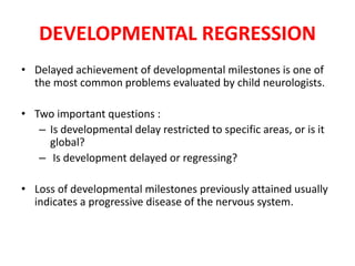
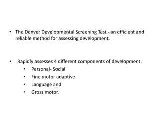
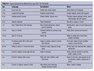
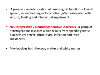
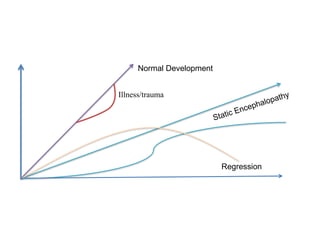
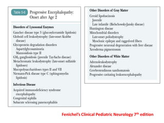
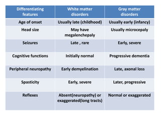
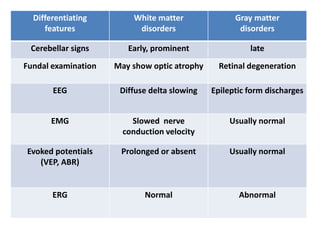
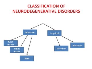
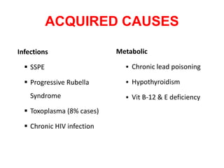
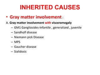
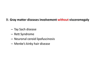
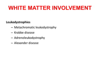
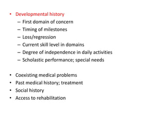
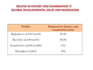
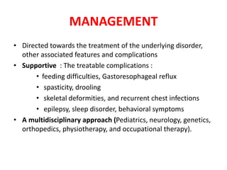
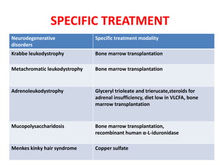
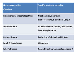
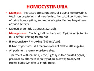
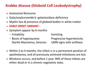
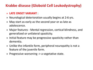
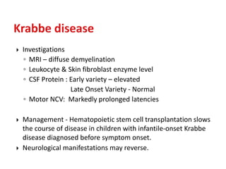
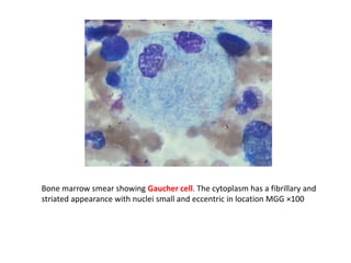
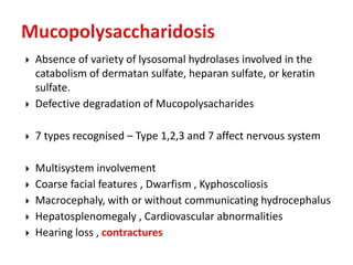
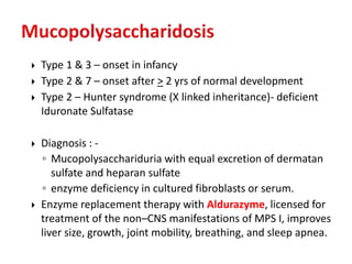
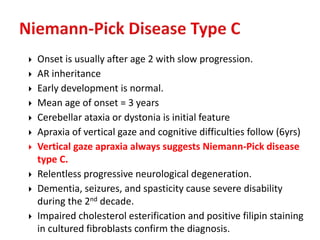
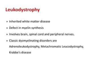
This document discusses the approach to evaluating children presenting with developmental regression. It defines developmental regression as the loss of developmental milestones previously attained, indicating a progressive nervous system disease. The evaluation involves a detailed history, developmental assessment, neurological exam, and targeted investigations to identify underlying genetic, metabolic, or acquired etiologies and guide management. A multidisciplinary approach is emphasized to address developmental delays, seizures, contractures, feeding issues, and provide genetic counseling.
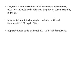
Presentation by Dr. Piyush Ojha on milestone regression in neurology, focusing on evaluation.
Discusses developmental regression in children, raising questions about delay specifics and implications.
Introduction to the Denver Developmental Screening Test, assessing personal-social, fine motor, language, and gross motor skills in children.
Explains severe neurological decline indicators, including loss of milestones and related disorders affecting nervous system.
Differentiates true regression from pseudo-regression, outlining factors leading to misinterpretation of regression.
Explains considerations in assessing children’s milestone regression, focusing on CNS involvement and age.
Differentiating clinical features of white and gray matter disorders, including age of onset and neurological symptoms.
Details acquired causes of regression including infections and developmental aspects in gray matter disorders.
Discusses inherited gray matter diseases and white matter leukodystrophies affecting children.
Outlines objectives in evaluating developmental regression in children to refer for necessary interventions.
Emphasizes importance of detailed birth, developmental, and family history for understanding regression causes.
Explains clinical examination findings including dysmorphism, neurological and systemic assessments relative to regression.
Details on eye examination findings linked to neurological disorders, including optic atrophy and imaging diagnostics.Various tests for determining underlying diagnoses and management plans for milestone regression conditions.
Encourages belief in comprehensive history-taking and examination for diagnosing developmental delays and regressions.
Outlines management focus on treating underlying conditions, co-morbidities, and supportive multidisciplinary care.
Presents targeted treatments for specific neurodegenerative disorders with examples of modalities.
Describes a rare disorder affecting development with specific management details and imaging findings.
Details on homocystinuria, its genetic basis, clinical features, and dietary management approaches.
Presents information on Krabbe disease variants, their clinical features, and management strategies.
Explains Tay-Sachs disease, including symptoms, progression, and diagnostic criteria for affected children.
Discusses mucopolysaccharidoses types, their multisystem impact, symptoms, and enzyme replacement options.
Explains inherited white matter disorders, symptoms, and disease progression patterns depending on the onset.
Introduces peroxisomal disorders, focusing on enzyme deficiencies and their resultant neurological symptoms.
Describes neuronal storage disorders such as Zellweger syndrome and associated limitations and symptoms.
Explains characteristics of Rett syndrome, symptoms, genetic aspects, and management of affected patients.
Discusses Wilson’s disease presentations including neurological effects and diagnostic indicators.
Details epidemiology, clinical features, risk factors for HIV-encephalopathy, and management considerations.Describes the chronic impact of measles on neurological health, emphasizing diagnostic and therapeutic approaches.
Concludes presentation thanking the audience and listing essential references for further understanding.
DEVELOPMENTAL REGRESSION
• Delayedachievement of developmental milestones is one of
the most common problems evaluated by child neurologists.
• Two important questions :
– Is developmental delay restricted to specific areas, or is it
global?
– Is development delayed or regressing?
• Loss of developmental milestones previously attained usually
indicates a progressive disease of the nervous system.
3.
• The DenverDevelopmental Screening Test - an efficient and
reliable method for assessing development.
• Rapidly assesses 4 different components of development:
• Personal- Social
• Fine motor adaptive
• Language and
• Gross motor.
5.
• A progressivedeterioration of neurological functions - loss of
speech, vision, hearing or locomotion ,often associated with
seizure, feeding and intellectual impairment.
• Neuroregressive / Neurodegenerative Disorders - a group of
heterogeneous diseases which results from specific genetic,
biochemical defect, chronic viral infection and toxic
substances.
• May involves both the gray matter and white matter
IMPORTANT CONSIDERATION
• RegressionAND NOT Delay ????
• Age above 2 Years OR Less than 2 Years ???
• Is only the Central nervous system involved, or are other
organs involved?
– Other organ involvement suggests lysosomal, peroxisomal,
and mitochondrial disorders.
10.
IMPORTANT CONSIDERATIONS
• Arethe clinical features referable only to the central nervous
system or to both the central and peripheral nervous
systems?
– Nerve or muscle involvement suggests mainly lysosomal
and mitochondrial disorders.
• Does the disease affect primarily the gray matter or the white
matter?
– Early features of
• Gray matter disease - personality change, seizures, and
dementia/cognitive decline.
• White matter disease - Focal neurological deficits, spasticity,
and blindness.
– Whether the process begins in the gray matter or white matter,
eventually clinical features of dysfunction develop in both.
Differentiating
features
White matter
disorders
Gray matter
disorders
Ageof onset Usually late (childhood) Usually early (infancy)
Head size May have
megalenchepaly
Usually microcepaly
Seizures Late , rare Early, severe
Cognitive functions Initially normal Progressive dementia
Peripheral neuropathy Early demyelination Late, axonal loss
Spasticity Early, severe Later, progressive
Reflexes Absent(neuropathy) or
exaggerated(long tracts)
Normal or exaggerated
15.
Differentiating
features
White matter
disorders
Gray matter
disorders
Cerebellarsigns Early, prominent late
Fundal examination May show optic atrophy Retinal degeneration
EEG Diffuse delta slowing Epileptic form discharges
EMG Slowed nerve
conduction velocity
Usually normal
Evoked potentials
(VEP, ABR)
Prolonged or absent Usually normal
ERG Normal Abnormal
• Developmental history:
–Detailed development history- decide whether there is
delayed development milestones or regression of
milestones
• Family history:
– Family history of neurological disorder
– Early or unexplained death
– Nature of the neurological manifestations should be
clarified
• H/o of consanguinity - ethnic heritage
– Origin from same village
– Same last names
24.
• Gestational history
–Prior pregnancies/ abortions/ Early postnatal deaths
– Adverse perinatal events – hypoxia/hypoglycemia
– Delivery details
– Apgar, birth weight
– Duration of hospital stay/ difficulties in Nutrition
– Possible neonatal encephalopathy : seizures, feeding
difficulty, obtundation
25.
• Developmental history
–First domain of concern
– Timing of milestones
– Loss/regression
– Current skill level in domains
– Degree of independence in daily activities
– Scholastic performance; special needs
• Coexisting medical problems
• Past medical history; treatment
• Social history
• Access to rehabilitation
26.
CLINICAL EXAMINATION
• Generalphysical examination
– Dysmorphism: Mitochondrial disorders, Neonatal adrenal
leukodystrophy ,coarse facial features (MPS)
– Occipito-Frontal Circumference –
• Microcepaly (gray matter disease)
• Megalenchepaly – certain white mater
disorder(Cannavan & Alexander)
– Jaundice
– Enlarged tongue
27.
• Skin &hair ( Hartnup Diseases-pellagra like skin rash, Menkes
disease-kinky hair)
• Examination of the spine- for associated complications
(scoliosis)
• Contractures of joints
• Systemic examination:
– Hepatosplenomegaly
– Chest deformity
– Cardiomyopathy
28.
NEUROLOGICAL EXAMINATION
• Highermental function, signs of raised ICP
• Speech, memory
• Cranial nerves
• Motor system:
– Tone
– Deep tendon reflexes
• Sensory loss /neuropathy
• Abnormal /involuntary movements
• Gait
29.
EYE EXAMINATION
• Opticatrophy (white matter- due to demyelination)
• Retinal degeneration (gray matter)- as the retinal receptors
are neuronal cells): Cherry red spot, retinitis pigmentosa
• Cataracts
• Telengiectasias
• K.F ring
• Urine
– Reducingsubstances, Organic acids,24 hr (MPS)
• Imaging
– Skull & Vertebrae, Long bones
– CT/MRI
• Biopsy
– Skin, Bone marrow, nerve, brain
• Diagnosis
– Important for genetic counseling
• Outcome - Prognosis poor
33.
BELIEVE IN HISTORYAND EXAMINATION !!!
(GLOBAL DEVELOPMENTAL DELAY AND REGRESSION)
34.
MANAGEMENT
• Directed towardsthe treatment of the underlying disorder,
other associated features and complications
• Supportive : The treatable complications :
• feeding difficulties, Gastoresophageal reflux
• spasticity, drooling
• skeletal deformities, and recurrent chest infections
• epilepsy, sleep disorder, behavioral symptoms
• A multidisciplinary approach (Pediatrics, neurology, genetics,
orthopedics, physiotherapy, and occupational therapy).
35.
SPECIFIC TREATMENT
Neurodegenerative
disorders
Specific treatmentmodality
Krabbe leukodystrophy Bone marrow transplantation
Metachromatic leukodystrophy Bone marrow transplantation
Adrenoleukodystrophy Glyceryl trioleate and trierucate,steroids for
adrenal insufficiency, diet low in VLCFA, bone
marrow transplantation
Mucopolysaccharidosis Bone marrow transplantation,
recombinant human α-L-iduronidase
Menkes kinky hair syndrome Copper sulfate
36.
Neurodegenerative
disorders
Specific treatment modality
Mitochondrialencephalopathies Nicotinamide, riboflavin,
dichloroacetate, L-carnitine, CoQ10
Wilson disease D- penicillamine, trietine, zinc acetate,
liver transplantation
Refsum disease Reduction of phytanic acid intake
Lesch-Nyhan disease Allopurinol
Fabry’s Disease Recombinant human α galactosidase A
37.
Guanidinoacetate Methyltransferase Deficiency
•Rare but treatable disorder
• Amidinotransferase converts glycine to guanidinoacetate and
guanidinoacetate methyltransferase converts
guanidinoacetate to creatine.
• Autosomal recessive inheritance (19p13.3)
• Affected children appear normal at birth and may develop
normally during infancy.
• By the end of the first year, development fails to progress and
hypotonia is noted.
• Regression of development follows and is associated with
dyskinesias, dystonia, and myoclonic jerks.
38.
• MRI -markeddemyelination
• MR Spectroscopy - creatine depletion and guanidinoacetate
phosphate accumulation.
• Management - Early oral administration of creatine
monohydrate significantly prevents and reverses all
symptoms, and late treatment provides some reduction of
abnormal movements.
39.
• Main defect- almost complete deficiency of the enzyme
cystathionine- b -synthase (Picker and Levy, 2006).
• Two variants –
– B 6 - responsive &
– B 6 –nonresponsive
• AR inheritance
HOMOCYSTINURIA
40.
HOMOCYSTINURIA
• Affected individualsappear normal at birth.
• Neurological features - mild to moderate MR, ectopia lentis,
and cerebral thromboembolism.
• Developmental delay ( 50% cases) and intelligence declines
progressively with age in untreated children.
• Most eventually function in the mildly retarded range.
• Intelligence generally higher in B 6 –responsive cases.
• Thromboembolism - first clue to the diagnosis in 15% cases.
• The presence of either thromboembolism or lens dislocation
strongly suggests homocystinuria.
41.
HOMOCYSTINURIA
• Diagnosis -increased concentrations of plasma homocystine,
total homocysteine, and methionine; increased concentration
of urine homocystine; and reduced cystathionine b-synthase
enzyme activity.
• Molecular genetic diagnosis available.
• Management. Challenge all patients with Pyridoxine (vitamin
B 6 ) before starting treatment.
• IF responsive – Pyridoxine (200 mg/day)
• IF Not responsive - still receive doses of 100 to 200 mg/day.
• All patients - protein-restricted diet.
• Treatment with betaine, 5 to 10 g/day in two divided doses,
provides an alternate remethylation pathway to convert
excess homocysteine to methionine.
42.
Autosomal Recessive
Galactosylceramide b -galactosidase deficiency
Myelin loss & presence of globoid bodies in white matter
EARLY ONSET VARIANT :
Symptom appear by 6 months
◦ Irritability Vomiting
◦ Bouts of hyperpyrexia Progressive hypertonicity
Startle Myoclonus, Seizures UMN signs with areflexia
Within 2 to 4 months, the infant is in a permanent position of
opisthotonos, and all previously achieved milestones are lost.
Blindness occurs, and before 1 year, 90% of these infants are
either dead or in a chronic vegetative state.
43.
LATE ONSETVARIANT :
Neurological deterioration usually begins at 2-6 yrs.
May start as early as the second year or as late as
adolescence.
Major features - Mental regression, cortical blindness, and
generalized or unilateral spasticity.
Initial feature may be progressive spasticity rather than
dementia.
Unlike the infantile form, peripheral neuropathy is not a
feature of the juvenile form.
Progressive worsening -> a vegetative state.
44.
Investigations
◦ MRI– diffuse demyelination
◦ Leukocyte & Skin fibroblast enzyme level
◦ CSF Protein : Early variety – elevated
Late Onset Variety - Normal
◦ Motor NCV: Markedly prolonged latencies
Management - Hematopoietic stem cell transplantation slows
the course of disease in children with infantile-onset Krabbe
disease diagnosed before symptom onset.
Neurological manifestations may reverse.
45.
TAY SACHS DISEASE
•N-acetyl- b -hexosaminidase deficiency
• Intralysosomal storage of the glycosphingolipid GM 2
ganglioside.
• Complete deficiency - Tay-Sachs disease
• CNS – the only affected organ.
• Partial deficiencies cause the juvenile and adult forms of GM 2
gangliosidosis.
46.
TAY SACHS DISEASE
•Typical initial symptom - between 3-6 months - an abnormal
startle reaction (Moro reflex) to noise or light.
• Motor regression begins between 4 and 6 months of age.
• The infant comes to medical attention because of either
delayed achievement of motor milestones or loss of
milestones previously attained.
• Cherry-red spot on macula present in almost every patient
(NOT Pathognomic)
• By 1 year - infant is severely retarded, unresponsive, and
spastic.
• 2nd year - the head enlarges and seizures develop.
• Most children die by 5 years of age.
47.
JUVENILE VARIANT :-
•Often begins with ataxia and incoordination between 2 and 10
years of age.
• Speech, life skills, and cognition decline.
• Spasticity and seizures present by end of 1st decade
• Loss of vision occurs much later than infantile form.
• Cherry-red spot not a consistent finding.
• Optic atrophy and retinitis pigmentosa occur late.
• A vegetative state with decerebrate rigidity by 10-15 years.
• Followed by death, usually due to infection.
• Sometimes may follow a aggressive course, culminating in
death in 2 to 4 years.
48.
TAY SACHS DISEASE
•Diagnosis - Suspect the diagnosis in any Jewish child with
psychomotor retardation and a cherry-red spot on the
macula.
• Demonstration of absent to nearly absent b -hexosaminidase
A enzymatic activity in the serum or white blood cells or
Fibroblast.
• Mutation analysis identifies the carrier state.
• Management - Treatment is supportive.
49.
Commonest
ARinheritance (Chr 1q21)
Deficiency of Glucocerebrosidase
5 types
◦ Type 1 – Perinatal lethal form – spares Brain
◦ Type 2 – onset < 2 years, rapidly progressive course
by 2-4 years
◦ Type 3 – onset > 2 years, slowly progressive course with
longer survival
◦ Type 4
◦ Type 5 – cardiovascular form
50.
Gaucher disease type2 – symptom onset usually < 6 months
• Initial features – motor regression and cranial nerve
dysfunction.
• Children are first hypotonic and then spastic.
• Head retraction - an early and characteristic sign - probably
due to meningeal irritation.
• Difficulties in sucking and swallowing, trismus, and
oculomotor palsies are typical.
• Mental deterioration is rapid , seizures uncommon.
• Splenomegaly >> hepatomegaly, jaundice not expected.
• Hypersplenism - anemia, thrombocytopenia, and leukopenia.
• Death usually occurs during the first year and always by the
second.
51.
Gaucher disease type3 –
• Age of onset – early childhood to adulthood
• Hepatosplenomegaly usually precedes neurological
deterioration.
• MC neurological manifestations - seizures and mental
regression (mild memory loss to severe dementia)
• Myoclonus and myoclonic seizures present in many patients.
• Combination of spasticity, ataxia, and cranial nerve
dysfunction may be present.
• Vertical oculomotor apraxia, as described in Niemann-Pick
disease, may occur in late-onset Gaucher disease as well.
52.
• Diagnosis –
–Assay of acid b –glucosylceramidase enzyme activity in
peripheral blood leukocytes or hepatocytes
– Gaucher cells in bone marrow - pathognomic
• Management - Symptomatic treatment includes partial or
total splenectomy, blood transfusion, analgesics and
supplemental treatment such as oral bisphosphonates for
severe osteopenia.
• Type 3 disease - may benefit from bone marrow
transplantation to correct the metabolic defect.
• Enzyme replacement therapy, using Imiglucerase (CEREZYME),
is effective in reversing the hematological and liver/spleen
involvement BUT NOT NEUROLOGICAL SYMPTOMS.
53.
Bone marrow smearshowing Gaucher cell. The cytoplasm has a fibrillary and
striated appearance with nuclei small and eccentric in location MGG ×100
54.
Absence ofvariety of lysosomal hydrolases involved in the
catabolism of dermatan sulfate, heparan sulfate, or keratin
sulfate.
Defective degradation of Mucopolysacharides
7 types recognised – Type 1,2,3 and 7 affect nervous system
Multisystem involvement
Coarse facial features , Dwarfism , Kyphoscoliosis
Macrocephaly, with or without communicating hydrocephalus
Hepatosplenomegaly , Cardiovascular abnormalities
Hearing loss , contractures
55.
Type 1& 3 – onset in infancy
Type 2 & 7 – onset after > 2 yrs of normal development
Type 2 – Hunter syndrome (X linked inheritance)- deficient
Iduronate Sulfatase
Diagnosis : -
◦ Mucopolysacchariduria with equal excretion of dermatan
sulfate and heparan sulfate
◦ enzyme deficiency in cultured fibroblasts or serum.
Enzyme replacement therapy with Aldurazyme, licensed for
treatment of the non–CNS manifestations of MPS I, improves
liver size, growth, joint mobility, breathing, and sleep apnea.
56.
Onset isusually after age 2 with slow progression.
AR inheritance
Early development is normal.
Mean age of onset = 3 years
Cerebellar ataxia or dystonia is initial feature
Apraxia of vertical gaze and cognitive difficulties follow (6yrs)
Vertical gaze apraxia always suggests Niemann-Pick disease
type C.
Relentless progressive neurological degeneration.
Dementia, seizures, and spasticity cause severe disability
during the 2nd decade.
Impaired cholesterol esterification and positive filipin staining
in cultured fibroblasts confirm the diagnosis.
57.
Inherited whitematter disease
Defect in myelin synthesis
Involves brain, spinal cord and peripheral nerves.
Classic dysmyelinating disorders are
Adrenoleukodystrophy, Metachromatic Leucodystrophy,
Krabbe's disease
58.
Autosomal Recessive
Arylsulfatase A deficiency deficiency
Infantile (50-60% cases) and Juvenile variety(20-30%)
Age at onset within a family usually similar.
All individuals eventually lose motor and intellectual
functions.
The disease course - 3 to 10 or more years in the late-
infantile-onset form and as long as 20 years or more in the
juvenile- and adult-onset forms.
Death – due to pneumonia or other infection.
Treatment is mainly supportive .
59.
Onset inlate infancy
• Hypotonia initially
• Peripheral neuropathy
• Dysarthria
• Optic atrophy
• Mental regression
• Decorticate posture
finally
Expected life span ~ 3.5
years after onset of
symptoms but can be as
long > 10 years
Onset at 5-10 yr
• Deterioration in school
performance
• Alteration in personality
• Incontinence
• Seizures
• Incoordination, Dysarthria
• Gait disturbances
• Spasticity
• No peripheral neuropathy
• Slow progression
• Spastic quadriplegia
eventually
•Death usually during 2nd decade.
60.
Life-threatening disorder
deficiency of a peroxisomal acyl coenzyme A synthetase.
occurring in males, affects white matter & adrenal gland.
Very long chain fatty acids accumulate in the cells & tissues
causing myelin sheath damage as well as dysfunction of the
adrenal gland.
X-linked form is the commonest
Diagnosis is made by estimation of very long-chain fatty acids
in plasma
61.
AR
Storageof auto fluorescent hydrophobic material in
lysosomes of neurons & other tissue
3 subtypes – Infantile / Late infantile / Juvenile
Symptoms/ Signs
◦ Myoclonic jerks , seizures (5-9 years)
◦ Visual symptoms
◦ Cerebellar ataxia
◦ Cognitive decline (5-9 years)
◦ Pigmentary abnormalities in retina (4-5 years)
Zellweger syndrome: dysmorphic features &severe
psychomotor retardation, sensory neuronal deafness,
peripheral neuropathy & hepatocellular degeneration.
Diagnosed by demonstrating the elevated very long chain
fatty acids, pipecolic acid and bile and derivatives.
64.
Females affectedonly
Mutations in the gene encoding methyl-CpG-binding protein-2,
located on chromosome Xq28
Normal during 1st year
Initial features - deceleration of head growth leading to
microcephaly, lack of interest in the environment & hypotonia.
Followed by rapid developmental regression characterized by
loss of language skills, gait ataxia, seizures, and autistic
behavior.
Characteristic feature - loss of purposeful hand movements
before the age of 3.
Stereotyped activity develops that looks like hand wringing or
washing.
65.
Repetitive blowsto the face – another stereotyped hand
movement.
Following the initial rapid progression - continued slower
progression of neurological deterioration.
Spastic paraparesis and quadriparesis - frequent endpoints.
Dementia is usually severe.
Stimulation produces an exaggerated stereotyped reaction
consisting of jerking movements of the trunk and limbs with
episodes of disorganized breathing and apnea followed by
hyperpnea.
66.
Seizures -most children before age 3 years.
After the period of rapid deterioration, the disease becomes
relatively stable, but dystonia may develop later.
Patients usually survive into early adulthood
An increased incidence of sudden, unexplained death.
Diagnosis - Clinical features & molecular genetic testing.
Management - Supportive.
67.
Wilson’s disease
AR
Degenerative disease of basal ganglia
Inborn error of copper metabolism
Academic deterioration
Behavioral changes , Dysarthria ,Dysphasia
Drooling , Dystonia
K F ring
68.
Acquired Immunodeficiency SyndromeEncephalopathy
• Human retroviral disease (Lentivirus subfamily)
• Transplacental or perinatal transmission of HIV from mothers
• Breast-feeding
• The mother may be asymptomatic when the child becomes
infected.
• Evidence of infection is apparent during the first year in 30%
of children born to AIDS-infected mothers.
• Outcome is worse when the onset of symptoms is early, and
the rate of progression in the child relates directly to the
severity of disease in the mother.
• 20% children present with severe symptoms or die in infancy.
69.
Acquired Immunodeficiency SyndromeEncephalopathy
• The spectrum of neurological and non-neurological
manifestations in HIV-infected children is somewhat different
from that of adults.
• Hepatosplenomegaly and bone marrow failure, lymphocytic
interstitial pneumonia, chronic diarrhea and failure to thrive,
acquired microcephaly, cerebral vasculopathy, and basal
ganglia calcification occur more frequently in children.
• Opportunistic infections (e.g., cerebral toxoplasmosis,
progressive multifocal leukoencephalopathy) are rare in
infants.
70.
Acquired Immunodeficiency SyndromeEncephalopathy
• AIDS encephalopathy - subacute or indolent
• Not necessarily associated with failure to thrive or
opportunistic infections.
• Onset of encephalopathy may occur from 2 months to 5 years
after exposure to the virus.
• 90 % affected infants show symptoms by 18 months of age.
• Progressive loss of developmental milestones, microcephaly,
dementia, and spasticity characterize the encephalopathy.
• Other features (< 50% cases) - ataxia, pseudobulbar palsy,
involuntary movement disorders, myoclonus, and seizures.
• Death usually occurs a few months after the onset of AIDS
encephalopathy.
71.
Acquired Immunodeficiency SyndromeEncephalopathy
• DNA PCR - preferred method for the diagnosis in infants and
children > 18 months of age.
• Combined treatment with zidovudine (azidothymidine),
didanosine, and nevirapine is well tolerated and may have
sustained efficacy against HIV-1.
72.
Form ofchronic measles encephalitis
In a nonimmunized population, the average age at onset is 8
years.
As a rule, children have experienced natural infection with the
rubeola virus at an early age, half of them before age 2 years.
First symptoms - Personality change and declining school
performance.
Personality change - aggressive behavior or withdrawal.
73.
• Retinal examinationduring early stage shows pigmentary
changes in the macula.
• Generalized seizures, usually myoclonic, develop next.
• EEG - characteristic pattern of periodic bursts of spike-wave
complexes (approximately every 5–7 sec) occurring
synchronously with the myoclonic jerk.
• After the onset of seizures, the child shows rapid neurological
deterioration characterized by spasticity, dementia, and
involuntary movements.
• Within 1 to 6 years from the onset of symptoms, the child is in
a chronic vegetative state.
74.
• Diagnosis –demonstration of an increased antibody titre,
usually associated with increased g –globulin concentrations,
in the CSF.
• Intraventricular interferon-alfa combined with oral
isoprinosine, 100 mg/kg/day.
• Repeat courses up to six times at 2- to 6-month intervals.