THE NERVOUS SYSTEM
• The nervous system coordinates all body functions, enabling
a person to adapt to changes in internal and external
environment.
• Stimulus: any change that results in a change in the
organism.
– temperature, light, pressure, sound, smell, etc.
• Response: any action resulting from a stimulus.
– contraction of muscle cells
– secretion by a gland
– stimulation of another nerve fiber.
• The nervous system is composed mainly of the nerve cells
(neurons) and supporting cells (neuroglia)
THE NERVOUS SYSTEM
•Function: environment is constantly
changing – nervous system detects
those changes and helps the organism
respond/adapt
•Irritability: ability to respond to a
stimulus
THE NERVOUS SYSTEM
•Nervous System detects (sensory
input), processes (integration), and
responds (motor output)
•Peripheral Nervous System detects
and responds
•Central Nervous System processes
information
NERVOUS SYSTEM
•Central nervous system
–brain & spinal cord
•Peripheral nervous
system
–nerves from senses
–nerves to muscles
cerebrum
cerebellum
spinal cord cervical
nerves
thoracic
nerves
lumbar
nerves
femoral
nerve
sciatic
nerve
tibial
nerve
THE NEURON (NERVE CELL)
• Three types of neurons:
–Sensory – carry impulses from the sense
organs (receptors) to the CNS
–Motor – carry impulses from the CNS to
the muscles or glands (effectors)
–Interneurons – connect and carry impulses
between sensory and motor neurons
• Messages carried by the nervous system are
electrical signals = impulses
• Nerve cells that transmit impulses = neurons
THREE COMPONENTS OF NEURONS
1. Cell body – largest part; most metabolic
activities take place here; contains
nucleus
2. Dendrites – carry impulses from the
environment or other neurons toward
the cell body
THREE COMPONENTS OF NEURONS
3. Axon – long fiber that carries
impulses away from the cell body
• Terminal branches – branching of
axon
• Synaptic knobs – ends of axon;
contain vesicles with
neurotransmitters
FUN FACTS ABOUT NEURONS
• Most specialized cell in
animals
• Longest cell
–blue whale neuron
• 10-30 meters
–giraffe axon
• 5 meters
–human neuron
• 1-2 meters
Nervous system allows for
1 millisecond response time
Nervous system allows for
1 millisecond response time
THE NEURON
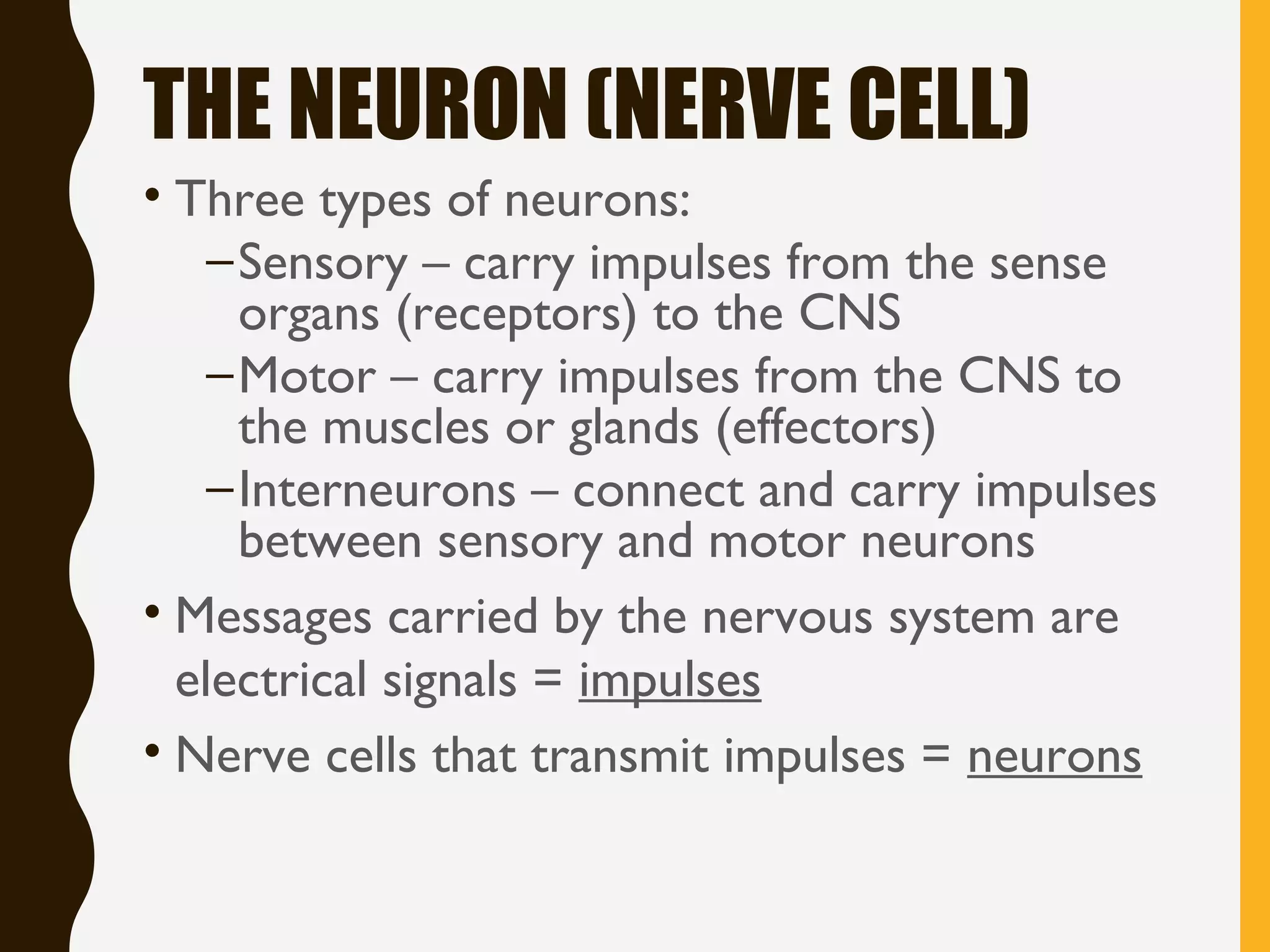
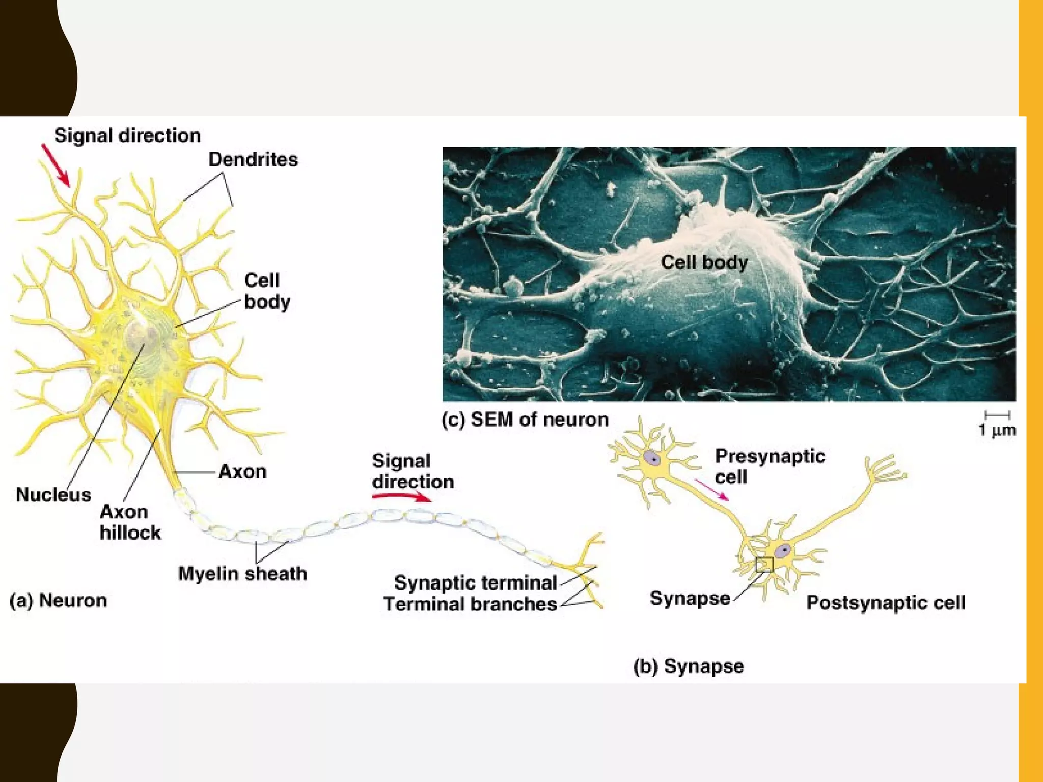
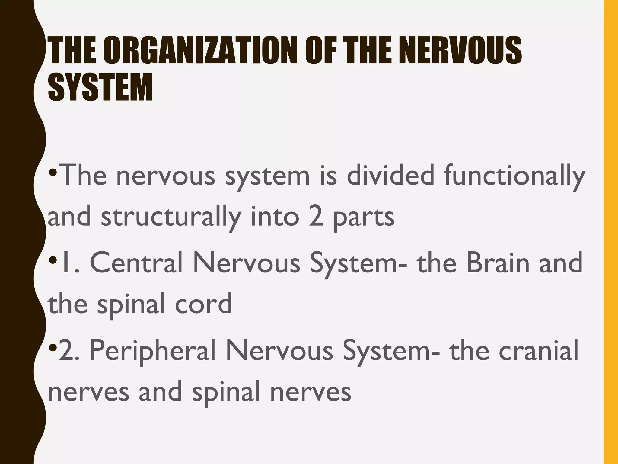
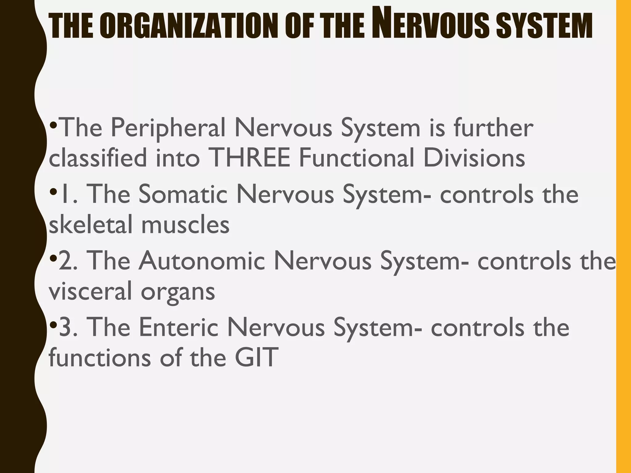
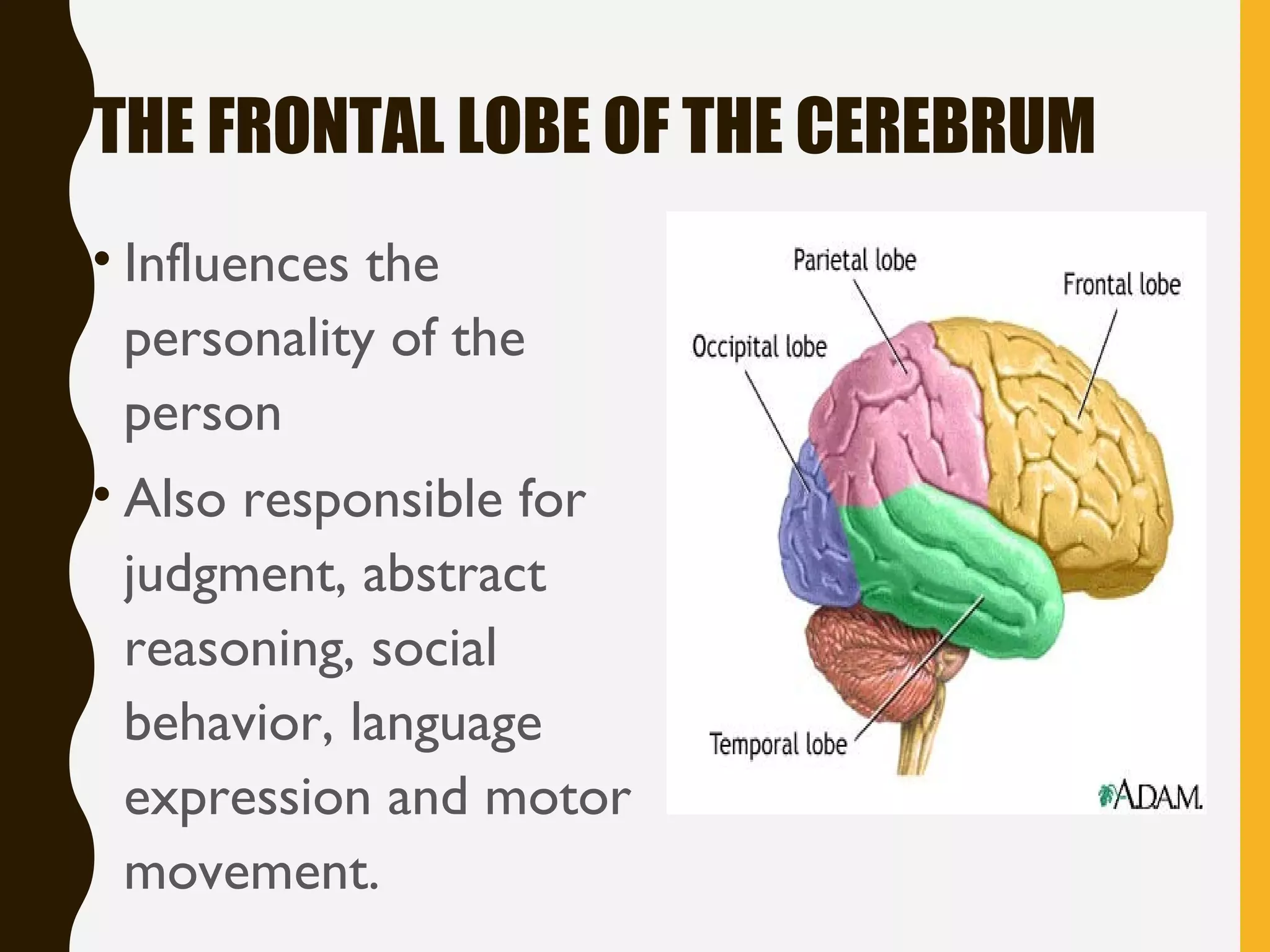
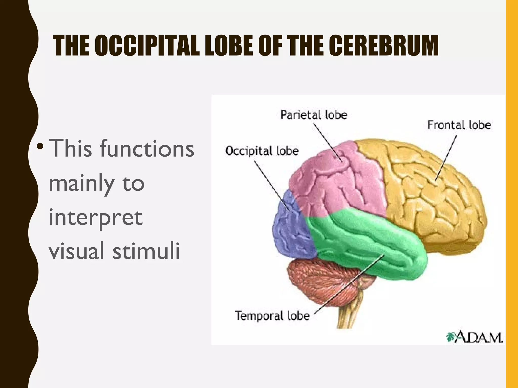
• The nervous
system is
composed of
neurons, which
produce and
conduct
electrochemical
impulses and
supporting cells,
which assist the
functions of
neurons.
THE NEUROGLIA
• The supporting cells
• They supply nutrients to the neurons and
help maintain the electrical potential
• They also form part of the blood-brain
barrier
• They are made up of macroglia, microglia
and ependymal cells
THE NEUROGLIA
•Oligodendrocytes produce myelin
sheath in the CN
•Scwhann cells or lemmocytes
produce myelin sheath in the
peripheral NS
THE ORGANIZATION OF THE NERVOUS
SYSTEM
•The nervous system is divided functionally
and structurally into 2 parts
•1. Central Nervous System- the Brain and
the spinal cord
•2. Peripheral Nervous System- the cranial
nerves and spinal nerves
THE ORGANIZATION OF THE NERVOUS SYSTEM
•The Peripheral Nervous System is further
classified into THREE Functional Divisions
•1. The Somatic Nervous System- controls the
skeletal muscles
•2. The Autonomic Nervous System- controls the
visceral organs
•3. The Enteric Nervous System- controls the
functions of the GIT
THE CENTRAL NERVOUS
SYSTEM
Composed of the brain
• The brain consists of the gross structures:
cerebrum, cerebellum, brainstem and the
diencephalon.
• Diencephalon - Thalamus. Hypothalamus and
pineal body
• Brainstem- Pons, medulla and Midbrain
THE CEREBRUM
• This is the largest part of the brain
• Consists of right and left hemisphere connected
by the corpus callosum
• Each cerebral hemisphere is composed of
different lobes- frontal, temporal, parietal and
occipital
• Embedded in the cerebrum is the BASAL ganglia
THE FRONTAL LOBE OF THE CEREBRUM
• Influences the
personality of the
person
• Also responsible for
judgment, abstract
reasoning, social
behavior, language
expression and motor
movement.
THE TEMPORAL LOBE OF THE CEREBRUM
• This part of the cerebrum
controls the hearing,
language comprehension,
storage and recall of
memories
• The LIMBIC system is
deeply located in the
temporal lobe. This
controls the basic drives
such as hunger, anger,
emotion and sexual drive.
THE PARIETAL LOBE OF THE CEREBRUM
• This is the principal
center for the reception
and interpretation of
Sensation
• This part interprets and
integrates the sensory
inputs like touch,
temperature and pain
• It interprets size, shape,
distance and texture
THE OCCIPITAL LOBE OF THE CEREBRUM
•This functions
mainly to
interpret
visual stimuli
THE CEREBELLUM
•The second largest brain region
•Has also two hemispheres
•Functions to maintain muscle tone,
coordinate muscle movement, posture
and control balance/equilibrium
•If this is damaged, muscle tone decreases
and fine motor movements become very
clumsy
THE BRAINSTEM
• Lies inferior to the cerebrum
• Continuous with the cerebrum and the
spinal cord
• It is composed of the midbrain, the pons
and the medulla oblongata
• Functions: houses the center for
respiration and cardiovascular system
THE MIDBRAIN
•This connects with the cerebrum
•Contains numerous ascending and
descending tracts and fibers
THE PONS
•Connects the cerebellum with the
cerebrum
•Houses the respiratory center and
cardiovascular center
•Exit points for cranial nerves
THE MEDULLA OBLONGATA
• The most inferior portion of the brainstem
• Serves as the center for autonomic reflexes
to maintain homeostasis, regulating
respiratory vasomotor and cardiac functions
• Serves as exit of cranial nerves 9,10,11 and
12
THE DIENCEPHALON
• The thalamus and the
hypothalamus
• The thalamus is the relay
station of all sensory stimuli
towards the brain
• The hypothalamus controls
body temperature, appetite,
water balance, pituitary
secretions and sleep-wake
cycle
THE SPINAL CORD
• The spinal cord carries out two main functions: It
connects a large part of the peripheral nervous system to
the brain. Information (nerve impulses) reaching the spinal
cord through sensory neurons are transmitted up into the
brain.
• A long cylindrical structure extending from the foramen
magnum to the L1 in adult, L3/L4 in pedia
• In the cross section of the spinal cord, we find the GRAY
matter- contains neurons; and WHITE matter-consists of
nerve fibers
• There are 31 pairs of spinal nerves that exit the spinal cord
THE MENINGES
• These are 3 connective tissue layers surrounding
the brain and spinal cord.
• 1. DURA MATER- the superficial, thickest layer.
The area above the dura mater is called epidural
space
• 2. ARACHNOID- second layer, thin and wispy.
• 3. PIA MATER- the deepest layer, adhered to
the brain and spinal cord substance
THE
MENINGES• The space in between
the arachnoid and pia
mater is called the
arachnoid space
• This arachnoid space
contains the cerebro-
spinal fluid (CSF)
• In this space, blood
vessels are also found
THE VENTRICLES
• The ventricular system is a set of
communicating cavities within the brain.
• These structures are responsible for the production,
transport and removal of cerebrospinal fluid, which
bathes the central nervous system.
• These are CSF filled cavities in the brain
• The lateral ventricle- found in the cerebrum
• The third ventricle- in the center of the thalamus and
hypothalamus
• The fourth ventricle- located at the base of the
cerebellum
THE CSF
• Cerebrospinal fluid (CSF) is a clear, colorless body fluid
found in the brain and spine. It is produced in the choroid
plexuses of the ventricles of the brain. It acts as a cushion or
buffer for the brain's cortex, providing basic mechanical and
immunological protection to the brain inside the skull.
• This is the fluid found inside the ventricles that bathe the
brain and spinal cord
• Function: provides protective cushion around the CNS
• Produced by the choroid plexus in the ventricles
• Absorbed by the arachnoid granulations
THE CRANIAL NERVES
• Cranial nerves are those nerves which arise from the brain and brain
stem rather than the spinal cord. Nerves arising from the spinal cord
are the spinal nerves. There are 12 pairs of cranial nerves and these
pairs of nerves passage through foramina in the skull, either
individually or in groups.
• 12 pairs of nerves that exit the brain. Can be classified as :
• Sensory
• Motor mixed (sensory and motor)
• The cranial nerves provide motor and sensory innervation mainly to
the structures within the head and neck. The sensory innervation
includes both "general" sensation such as temperature and touch, and
"special" innervation such as taste, vision, smell, balance and hearing
THE AUTONOMIC
NERVOUS SYSTEM
• The part of the peripheral nervous system that innervates (supply)
cardiac muscles, smooth muscles and glands
• It also relays visceral sensory information to the central nervous
system and processes it so that alterations can be made in the activity
of specific autonomic motor outflows, such as those that control
the heart, blood vessels, and other visceral organs.
• It also stimulates the release of certain hormones involved in energy
metabolism (e.g., insulin, glucagon, and epinephrine [also called
adrenaline]) or cardiovascular functions (e.g., renin and vasopressin).
These integrated responses maintain the normal internal
environment of the body in an equilibrium state called homeostasis.
• Functionally divided into
• Sympathetic Nervous System
• Parasympathetic Nervous System
THE SYMPATHETIC
SYSTEM• The sympathetic nervous system normally functions to produce
localized adjustments (such as sweating as a response to an increase
in temperature) and reflex adjustments of the cardiovascular system.
• Neurotransmitter agents are Epinephrine and Norepinephrine
(coming from the adrenal gland)
• ADRENERGIC system - a part of autonomic nervous system that
uses epinephrine or norepinephrine as its neurotransmitter
• Adrenergic means "working on adrenaline (epinephrine)
or noradrenaline (norepinephrine)"
– Epinephrine - hormone that is secreted mainly by the medulla
of the adrenal glands and that functions primarily to increase
cardiac output and to raise glucose levels in the blood.
Epinephrine typically is released during acute stress, and its
stimulatory effects fortify and prepare an individual for either
“fight or flight” 
– Norepinephrine - substance that is released predominantly
from the ends of sympathetic nerve fibres and that acts to
increase the force of skeletal muscle contraction and the rate
and force of contraction of the heart. The actions of
norepinephrine are vital to the fight-or-flight response, whereby
the body prepares to react to or retreat from an acute threat.
SYMPATHETIC
RESPONSES• Under conditions of stress, however, the entire sympathetic
nervous system is activated, producing an immediate,
widespread response called the fight-or-flight response
• This response is characterized by the release of large
quantities of epinephrine from the adrenal gland, an increase in
heart rate, an increase in cardiac output, skeletal muscle
vasodilation, cutaneous and gastrointestinal vasoconstriction,
pupillary dilation, bronchial dilation, and pilo erection. The
overall effect is to prepare the individual for imminent danger.
Sympathetic effects:
– dilates pupil
– accelerates heartbeat & respiration
– inhibits stomach & intestine activity
– relaxes urinary bladder
PARASYMPATHETIC
SYSTEM
• The parasympathetic nervous system sometimes called
the rest and digest system,
• Neurologically, cholinergic is the abbreviated term referring
to acetylcholine. The parasympathetic nervous system, which
uses acetylcholine almost exclusively send its messages, and
said to be almost entirely cholinergic. Neuromuscular
junctions, preganglionic neurons of the sympathetic nervous
system, the basal forebrain, and brain stem complexes are also
cholinergic
• Neurotransmitter is Acetylcholine
PARASYMPATHETIC
RESPONSES
The parasympathetic system conserves energy
as it slows the heart rate, increases intestinal and
gland activity, and relaxes sphincter muscles in
the gastrointestinal tract.
Parasympathetic effects:
• constricts pupil
• slows heartbeat & respiration
• stimulates stomach & intestine activity
• contracts urinary bladder
NERVE PHYSIOLOGY
• The nerve cells are excitable cells
• Any stimulus will change the membrane
potential and cause an action potential to
generate impulse transmission or action
potential
• The myelin sheath of the nerve cell is
responsible for the SALTATORY
conduction increases the nerve
transmission
FOUR STAGES OF THE MEMBRANE
POTENTIAL
1. Resting stage - Polarized stage
- this is the normal resting membrane
- charges are separated across the plasma membrane, so
the membrane has potential. Any time membrane potential
is other than 0 millivolts (mV).
- the magnitude of the potential is directly proportional to
the number of positive and negative charges separated by the
membrane and that the sign of the potential (+ or
–) always designates whether excess positive or excess
negative charges are present, respectively, on the inside of
the membrane.
- at resting potential, the membrane is polarized at –70 mV in a
typical neuron
2. Depolarization stage
- the membrane becomes less polarized
- the inside becomes less negative than at resting
potential, with the potential moving closer to 0 mV
(for example, a change from –70 to -60 mV); fewer
charges are separated than at resting potential.
- this term also refers to the inside even becoming
positive as it does during an action potential (a major
type of electrical signal) when the membrane
potential reverses itself (for example, becoming +30
mV)
3. Repolarization stage.
- The membrane returns to resting potential after
having been depolarized.
4. Hyperpolarization Stage.
- The membrane becomes more polarized; the inside
becomes more negative than at resting potential,
with the potential moving even farther from 0 mV
(for instance, a change from –70 to –80 mV); more
charges are separated than at resting potential.
Electrical signals are produced by changes in ion
movement across the plasma membrane
- Changes in membrane potential are brought about by
changes in ion movement across the membrane. For example, if
the net inward flow of positively charged ions increases compared to
the resting state (such as more Na+ moves in), the membrane
depolarizes (becomes less negative inside). By contrast, if the net
outward flow of positively charged ions increases compared to the
resting state (such as more K+ moves out), the membrane
hyperpolarizes (becomes more negative inside).
- Changes in ion movement are brought about by
changes in membrane permeability in response to triggering
events.
- A triggering event triggers a change in membrane potential
by altering membrane permeability and consequently altering ion flow
across the membrane. These ion movements redistribute charge across
the membrane, causing membrane potential to fluctuate.
• Because the water-soluble ions responsible for carrying
charge cannot penetrate the plasma membrane’s lipid
bilayer,
• Therefore these charges can cross the membrane only
through channels specific for them or by carrier-mediated
transport.
• Membrane channels may be either leak channels or gated
channels.
• Leak channels, which are open all the time, permit
unregulated leakage of their specific ion across the
membrane through the channels. Gated channels, in
contrast, have gates that can be open or closed,
permitting ion passage through the channels when open
and preventing ion passage through the channels when
closed
FOUR KINDS OF GATED
CHANNELS
• (1) voltage-gated channels open or close in
response to changes in membrane potential,
• (2) chemically gated channels change shape in
response to binding of a specifi c extracellular
chemical messenger to a surface membrane
receptor,
• (3) mechanically gated channels respond to
stretching or other mechanical deformation, and
• (4) thermally gated channels respond to
local changes in temperature (heat or cold).
TWO BASIC FORMS OF
ELECTRICAL SIGNALS
• Graded potentials are local changes in membrane
potential that occur in varying grades or degrees of
magnitude or strength. For example, membrane potential
could change from –70 to –60 mV (a 10-mV graded
potential) or from –70 to –50 mV (a 20-mV graded
potential)
• Action potentials are brief, rapid, large (100 mV)
changes in membrane potential during which the potential
actually reverses so that the inside of the excitable cell
transiently becomes more positive than the outside.
MYELIN
•Is composed of 80% lipid and 20% protein
•Used for insulation and to help speed up
the nerve impulse
•Wraps around the axon of some neurons
MYELIN
•Gaps in myelin sheath cells called
Nodes of Ranvier – allow impulses to
move more quickly down neurons
MYELIN
•In Saltatory Conduction, only the
Nodes of Ranvier depolarize and
therefore conduct an impulse faster
Myelin is a fatty substance that wraps around
nerve fibers and serves to increase the speed of
electrical communication between neurons.
THE SYNAPSE
• SYNAPSE: the space between the axon of one
neuron and the dendrite of another
• Axon terminals have vesicles containing
chemicals: NEUROTRANSMITTERS
• These chemicals are secreted from the axon of
one neuron  stimulates receptor sites on the
effector or the dendrite of the next neuron
SYNAPSE
• Neurons have specialized projections called
dendrites and axons. Dendrites bring information to
the cell body and axons take information away from the
cell body.
• Information from one neuron flows to another neuron
across a synapse. The synapse contains a small gap
separating neurons. The synapse consists of:
• 1.a presynaptic ending that contains neurotransmitters,
mitochondria and other cell organelles
• 2.a postsynaptic ending that contains receptor sites for
neurotransmitters
• 3.a synaptic cleft or space between the presynaptic and
postsynaptic endings
SYNAPSE
Junction between nerve cells
–1st cell releases chemical to trigger
next cell
–where drugs affect nervous system
synapsesynapse
NEUROTRANSMITTER ACTION AT SYNAPSE
1. Action potential arrives at axon
terminal of presynaptic neuron
2. Synaptic vesicles rupture, releasing
neurotransmitter into synapse
3. Neurotransmitter diffuses across
synapse & binds to receptor protein
on postsynaptic cell
4. Postsynaptic cell is excited or
inhibited
5. Neurotransmitter in synapse is
deactivated
• Neurotransmitters are the chemicals
which allow the transmission of signals
from one neuron to the next across
synapses.
• They are also found at the axon endings of
motor neurons, where they stimulate the
muscle fibers.
• They and their close relatives are
produced by some glands such as the
pituitary and the adrenal glands.
• They are chemicals that communicate
information throughout our brain and
body. 
• The brain uses neurotransmitters to tell
your heart to beat, your lungs to breathe,
and your stomach to digest. 
• They can also affect mood, sleep,
concentration, weight, and can cause
adverse symptoms when they are out of
balance.
• Neurotransmitter levels can be
depleted many ways.
• Stress, poor diet, neurotoxins, genetic
predisposition, drug (prescription and
recreational), alcohol and caffeine
usage can cause these levels to be out
of optimal range.
TYPES OF NEUROTRANSMITTERS
• Two kinds of neurotransmitters –
INHIBITORY and EXCITATORY. 
• Excitatory neurotransmitters are not
necessarily exciting
–they are what stimulate the brain. 
• Inhibitory - calm the brain and help create
balance are called . 
– balance mood and are easily depleted when
the excitatory neurotransmitters are
overactive. 
Small molecule neurotransmitters
Type Neurotransmitter Postsynaptic effect
Acetylcholine Excitatory
Amino acids Gamma aminobutyric
acidGABA
Inhibitory
Glycine Inhibitory
Glutamate Excitatory
Aspartate Excitatory
Biogenic amines Dopamine Inhibitory
Noradrenaline Excitatory
Serotonin Inhibitory
Histamine Excitatory
ACETYLCHOLINE
• Acetylcholine was the first
neurotransmitter to be discovered.
• It is responsible for much of the
stimulation of muscles, including the
muscles of the gastro-intestinal system. 
• It is also found in sensory neurons and in
the autonomic nervous system, and has a
part in scheduling REM (dream) sleep.
• There is a link between acetylcholine and Alzheimer's
disease:  There is something on the order of a 90% loss
of acetylcholine in the brains of people suffering from
Alzheimer's, which is a major cause of senility.
• Outside the brain, acetylcholine is the main
neurotransmitter in the parasympathetic nervous system
– the system that controls functions such as heart rate,
digestion, secretion of saliva and bladder function.
• The plant poisons curare cause paralysis by blocking the
acetylcholine receptor sites of muscle cells. 
• The well-known poison botulin works by preventing the
vesicles in the axon ending from releasing acetylcholine,
causing paralysis. 
SEROTONIN
• SEROTONIN is an inhibitory neurotransmitter –
which means that it does not stimulate the brain. 
• Adequate amounts of serotonin are necessary
for a stable mood and to balance any excessive
excitatory (stimulating) neurotransmitter firing in
the brain. 
• If you use stimulant medications or caffeine in
your daily regimen – it can cause a depletion of
serotonin over time. 
• Low serotonin levels leads to an increased appetite for
carbohydrates (starchy foods) and trouble sleeping, which
are also associated with depression and other emotional
disorders.  It has also been tied to migraines, irritable
bowel syndrome, and fibromyalgia.
• Low serotonin levels are also associated with decreased
immune system function.
• In addition to mood control, serotonin has been linked
with a wide variety of functions, including the regulation
of sleep, pain perception, body temperature, blood
pressure and hormonal activity
• Largest amount of serotonin is found in the intestinal
mucosa.
• Although the CNS contains less than 2% of the total serotonin
in the body, serotonin plays a very important role in a range of
brain functions. It is synthesized from the amino acid
tryptophan.
• Gamma amino butyric acid(GABA) is the major inhibitory
neurotransmitter that is often referred to as “nature’s
VALIUM-like substance”.  When GABA is out of range (high or
low excretion values), it is likely that an excitatory
neurotransmitter is firing too often in the brain.  GABA will be
sent out to attempt to balance this stimulating over-firing.
• People with too little GABA tend to suffer from anxiety
disorders, and drugs like Valium work by enhancing the effects
of GABA.  Lots of other drugs influence GABA receptors,
including alcohol and barbiturates.  If GABA is lacking in
certain parts of the brain, epilepsy results.
HISTAMINE
• Amino acid Histidine is the precursor of an
important neurotransmitter histamine.
• Histamine is present in venom and other stinging
secretions.
• Histamine is a biogenic amine involved in local
immune responses.
• Regulate physiological function in the gut
• Act as a neurotransmitter.
• Triggers the inflammatory response.
REFLEX ARC
A reflex arc is a neural pathway that controls an action
reflex. In higher animals, most sensory neurons do not pass
directly into the brain, but synapse in the spinal cord.
This characteristic allows reflex actions to occur relatively
quickly by activating spinal motor neurons without the delay
of routing signals through the brain, although the brain will
receive sensory input while the reflex action occurs. There
are two types of reflex arcs: autonomic reflex arc (affecting
inner organs) and somatic reflex arc (affecting muscles)
TYPES OF SENSORY
RECEPTORS
• Thermoreceptors – detect heat and cold
• Pain receptors (nocioceptors) – detect chemicals released
from injured cells
• Mechanoreceptors – detect mechanical energy (touch,
pressure, vibration)
TYPES OF SENSORY
RECEPTORS
•Chemoreceptors – detect chemicals
•Photoreceptors – detect light energy
•Electroreceptors – detect electrical
fields
HOW ARE SOUNDS SENSED?
• The ear captures, transmits, and
converts sound into electrical signals
• Ear has three basic parts:
1. Outer ear
2. Middle ear
3. Inner ear
HOW ARE SOUNDS SENSED?
•Outer ear: external ear (pinna) and
auditory canal
–Funnels sound
–Sound waves vibrate the tympanic
membrane
HOW ARE SOUNDS SENSED?
• Middle ear
–Tympanic membrane (ear drum)
–Three tiny bones: malleus (hammer), incus
(anvil), stapes (stirrup); transfer vibrations to
the oval window on the cochlea
–Eustachian tube – equalize pressure; connects
middle ear to pharynx
HOW ARE SOUNDS SENSED?
•Inner ear: cochlea
–converts vibrations into electrical
signals
–As the oval window vibrates, it sets
the cochlear fluid in motion
–Moving fluid brushes over hairs
–Bending of hairs is sensed by
mechanoreceptors and sends the signal
to the brain (auditory nerve)
FISH “HEARING” – LATERAL LINES
• Contains mechanoreceptors that function
similarly to mammalian inner ear
• Gives info about direction and velocity of
water flowing over fish’s body
HOW IS LIGHT SENSED?
• Light waves travel at a speed of 186,000 miles per second.
Light is reflected into the eyes by objects within the field of
vision.
• In order to achieve clear vision, light reflected from objects
within the visual field is focused in to the retina of both eyes.
• The processes involved in producing a clear image are
refraction of the light rays and accommodation of the eyes.
• The eye is made up of three layers
– Fibrous layer- sclerae and cornea
– Uvea- choroid and iris and
ciliary bodies
– Nervous coat- retina
• Sclera – tough, white layer
• Conjunctiva – external cover of sclera; keeps eye moist;
HOW IS LIGHT SENSED?
•Cornea – transparent covering in front of
eye
•Choroid – thin, pigmented layer lining
interior surface of the sclera; prevents light
rays from scattering and distorting the
image
•Iris - regulates size of pupil/amount of light
into eye
HOW IS LIGHT SENSED?
•Lens focuses light on retina
•Retina – Contains photoreceptors (Except
at the optic disk where the optic nerve
attaches)
–Rods: Black and White
–Cones: Color
•Optic nerve takes electric signals from eye
to brain
HOW ARE SCENTS SENSED?
•Insects smell through their legs
and antennae
Male silkworm moth
Bombyx mori
Sensory hairs on
antennae detect
pheromones
released by female
HOW ARE SCENTS SENSED?
•Olfactory nerves are stimulated when
chemicals touch them
•Different chemicals create different
responses in the olfactory nerves;
hence we detect different smells
HOW ARE TASTES SENSED?
• Taste buds on tongue act just like the
olfactory nerves
–Different chemicals stimulate the
nerves in the taste buds differently;
hence we detect different tastes
• Four “primary” tastes are bitter, sour,
salty, and sweet
THE GUSTATORY
APPARATUS
•The receptor for taste are cells in
the tongue group together called
the taste buds
•They are numerous in the vallate
and fungiform papillae
THE GUSTATORY APPARATUS
Basic taste modalities
• Sweet- tip of the tongue
• Salty- over the dorsum of the tongue
• Sour- sides of the tongue
• Bitter- back of the tongue
THANK YOU

nervous system

  • 1.
  • 2.
    • The nervoussystem coordinates all body functions, enabling a person to adapt to changes in internal and external environment. • Stimulus: any change that results in a change in the organism. – temperature, light, pressure, sound, smell, etc. • Response: any action resulting from a stimulus. – contraction of muscle cells – secretion by a gland – stimulation of another nerve fiber. • The nervous system is composed mainly of the nerve cells (neurons) and supporting cells (neuroglia)
  • 3.
    THE NERVOUS SYSTEM •Function:environment is constantly changing – nervous system detects those changes and helps the organism respond/adapt •Irritability: ability to respond to a stimulus
  • 4.
    THE NERVOUS SYSTEM •NervousSystem detects (sensory input), processes (integration), and responds (motor output) •Peripheral Nervous System detects and responds •Central Nervous System processes information
  • 5.
    NERVOUS SYSTEM •Central nervoussystem –brain & spinal cord •Peripheral nervous system –nerves from senses –nerves to muscles cerebrum cerebellum spinal cord cervical nerves thoracic nerves lumbar nerves femoral nerve sciatic nerve tibial nerve
  • 7.
    THE NEURON (NERVECELL) • Three types of neurons: –Sensory – carry impulses from the sense organs (receptors) to the CNS –Motor – carry impulses from the CNS to the muscles or glands (effectors) –Interneurons – connect and carry impulses between sensory and motor neurons • Messages carried by the nervous system are electrical signals = impulses • Nerve cells that transmit impulses = neurons
  • 8.
    THREE COMPONENTS OFNEURONS 1. Cell body – largest part; most metabolic activities take place here; contains nucleus 2. Dendrites – carry impulses from the environment or other neurons toward the cell body
  • 9.
    THREE COMPONENTS OFNEURONS 3. Axon – long fiber that carries impulses away from the cell body • Terminal branches – branching of axon • Synaptic knobs – ends of axon; contain vesicles with neurotransmitters
  • 11.
    FUN FACTS ABOUTNEURONS • Most specialized cell in animals • Longest cell –blue whale neuron • 10-30 meters –giraffe axon • 5 meters –human neuron • 1-2 meters Nervous system allows for 1 millisecond response time Nervous system allows for 1 millisecond response time
  • 12.
    THE NEURON • Thenervous system is composed of neurons, which produce and conduct electrochemical impulses and supporting cells, which assist the functions of neurons.
  • 13.
    THE NEUROGLIA • Thesupporting cells • They supply nutrients to the neurons and help maintain the electrical potential • They also form part of the blood-brain barrier • They are made up of macroglia, microglia and ependymal cells
  • 14.
    THE NEUROGLIA •Oligodendrocytes producemyelin sheath in the CN •Scwhann cells or lemmocytes produce myelin sheath in the peripheral NS
  • 17.
    THE ORGANIZATION OFTHE NERVOUS SYSTEM •The nervous system is divided functionally and structurally into 2 parts •1. Central Nervous System- the Brain and the spinal cord •2. Peripheral Nervous System- the cranial nerves and spinal nerves
  • 19.
    THE ORGANIZATION OFTHE NERVOUS SYSTEM •The Peripheral Nervous System is further classified into THREE Functional Divisions •1. The Somatic Nervous System- controls the skeletal muscles •2. The Autonomic Nervous System- controls the visceral organs •3. The Enteric Nervous System- controls the functions of the GIT
  • 22.
    THE CENTRAL NERVOUS SYSTEM Composedof the brain • The brain consists of the gross structures: cerebrum, cerebellum, brainstem and the diencephalon. • Diencephalon - Thalamus. Hypothalamus and pineal body • Brainstem- Pons, medulla and Midbrain
  • 24.
    THE CEREBRUM • Thisis the largest part of the brain • Consists of right and left hemisphere connected by the corpus callosum • Each cerebral hemisphere is composed of different lobes- frontal, temporal, parietal and occipital • Embedded in the cerebrum is the BASAL ganglia
  • 25.
    THE FRONTAL LOBEOF THE CEREBRUM • Influences the personality of the person • Also responsible for judgment, abstract reasoning, social behavior, language expression and motor movement.
  • 26.
    THE TEMPORAL LOBEOF THE CEREBRUM • This part of the cerebrum controls the hearing, language comprehension, storage and recall of memories • The LIMBIC system is deeply located in the temporal lobe. This controls the basic drives such as hunger, anger, emotion and sexual drive.
  • 27.
    THE PARIETAL LOBEOF THE CEREBRUM • This is the principal center for the reception and interpretation of Sensation • This part interprets and integrates the sensory inputs like touch, temperature and pain • It interprets size, shape, distance and texture
  • 28.
    THE OCCIPITAL LOBEOF THE CEREBRUM •This functions mainly to interpret visual stimuli
  • 29.
    THE CEREBELLUM •The secondlargest brain region •Has also two hemispheres •Functions to maintain muscle tone, coordinate muscle movement, posture and control balance/equilibrium •If this is damaged, muscle tone decreases and fine motor movements become very clumsy
  • 30.
    THE BRAINSTEM • Liesinferior to the cerebrum • Continuous with the cerebrum and the spinal cord • It is composed of the midbrain, the pons and the medulla oblongata • Functions: houses the center for respiration and cardiovascular system
  • 31.
    THE MIDBRAIN •This connectswith the cerebrum •Contains numerous ascending and descending tracts and fibers
  • 32.
    THE PONS •Connects thecerebellum with the cerebrum •Houses the respiratory center and cardiovascular center •Exit points for cranial nerves
  • 33.
    THE MEDULLA OBLONGATA •The most inferior portion of the brainstem • Serves as the center for autonomic reflexes to maintain homeostasis, regulating respiratory vasomotor and cardiac functions • Serves as exit of cranial nerves 9,10,11 and 12
  • 34.
    THE DIENCEPHALON • Thethalamus and the hypothalamus • The thalamus is the relay station of all sensory stimuli towards the brain • The hypothalamus controls body temperature, appetite, water balance, pituitary secretions and sleep-wake cycle
  • 35.
    THE SPINAL CORD •The spinal cord carries out two main functions: It connects a large part of the peripheral nervous system to the brain. Information (nerve impulses) reaching the spinal cord through sensory neurons are transmitted up into the brain. • A long cylindrical structure extending from the foramen magnum to the L1 in adult, L3/L4 in pedia • In the cross section of the spinal cord, we find the GRAY matter- contains neurons; and WHITE matter-consists of nerve fibers • There are 31 pairs of spinal nerves that exit the spinal cord
  • 36.
    THE MENINGES • Theseare 3 connective tissue layers surrounding the brain and spinal cord. • 1. DURA MATER- the superficial, thickest layer. The area above the dura mater is called epidural space • 2. ARACHNOID- second layer, thin and wispy. • 3. PIA MATER- the deepest layer, adhered to the brain and spinal cord substance
  • 37.
    THE MENINGES• The spacein between the arachnoid and pia mater is called the arachnoid space • This arachnoid space contains the cerebro- spinal fluid (CSF) • In this space, blood vessels are also found
  • 38.
    THE VENTRICLES • The ventricularsystem is a set of communicating cavities within the brain. • These structures are responsible for the production, transport and removal of cerebrospinal fluid, which bathes the central nervous system. • These are CSF filled cavities in the brain • The lateral ventricle- found in the cerebrum • The third ventricle- in the center of the thalamus and hypothalamus • The fourth ventricle- located at the base of the cerebellum
  • 39.
    THE CSF • Cerebrospinalfluid (CSF) is a clear, colorless body fluid found in the brain and spine. It is produced in the choroid plexuses of the ventricles of the brain. It acts as a cushion or buffer for the brain's cortex, providing basic mechanical and immunological protection to the brain inside the skull. • This is the fluid found inside the ventricles that bathe the brain and spinal cord • Function: provides protective cushion around the CNS • Produced by the choroid plexus in the ventricles • Absorbed by the arachnoid granulations
  • 40.
    THE CRANIAL NERVES •Cranial nerves are those nerves which arise from the brain and brain stem rather than the spinal cord. Nerves arising from the spinal cord are the spinal nerves. There are 12 pairs of cranial nerves and these pairs of nerves passage through foramina in the skull, either individually or in groups. • 12 pairs of nerves that exit the brain. Can be classified as : • Sensory • Motor mixed (sensory and motor) • The cranial nerves provide motor and sensory innervation mainly to the structures within the head and neck. The sensory innervation includes both "general" sensation such as temperature and touch, and "special" innervation such as taste, vision, smell, balance and hearing
  • 42.
    THE AUTONOMIC NERVOUS SYSTEM •The part of the peripheral nervous system that innervates (supply) cardiac muscles, smooth muscles and glands • It also relays visceral sensory information to the central nervous system and processes it so that alterations can be made in the activity of specific autonomic motor outflows, such as those that control the heart, blood vessels, and other visceral organs. • It also stimulates the release of certain hormones involved in energy metabolism (e.g., insulin, glucagon, and epinephrine [also called adrenaline]) or cardiovascular functions (e.g., renin and vasopressin). These integrated responses maintain the normal internal environment of the body in an equilibrium state called homeostasis. • Functionally divided into • Sympathetic Nervous System • Parasympathetic Nervous System
  • 43.
    THE SYMPATHETIC SYSTEM• Thesympathetic nervous system normally functions to produce localized adjustments (such as sweating as a response to an increase in temperature) and reflex adjustments of the cardiovascular system. • Neurotransmitter agents are Epinephrine and Norepinephrine (coming from the adrenal gland) • ADRENERGIC system - a part of autonomic nervous system that uses epinephrine or norepinephrine as its neurotransmitter • Adrenergic means "working on adrenaline (epinephrine) or noradrenaline (norepinephrine)" – Epinephrine - hormone that is secreted mainly by the medulla of the adrenal glands and that functions primarily to increase cardiac output and to raise glucose levels in the blood. Epinephrine typically is released during acute stress, and its stimulatory effects fortify and prepare an individual for either “fight or flight”  – Norepinephrine - substance that is released predominantly from the ends of sympathetic nerve fibres and that acts to increase the force of skeletal muscle contraction and the rate and force of contraction of the heart. The actions of norepinephrine are vital to the fight-or-flight response, whereby the body prepares to react to or retreat from an acute threat.
  • 44.
    SYMPATHETIC RESPONSES• Under conditionsof stress, however, the entire sympathetic nervous system is activated, producing an immediate, widespread response called the fight-or-flight response • This response is characterized by the release of large quantities of epinephrine from the adrenal gland, an increase in heart rate, an increase in cardiac output, skeletal muscle vasodilation, cutaneous and gastrointestinal vasoconstriction, pupillary dilation, bronchial dilation, and pilo erection. The overall effect is to prepare the individual for imminent danger. Sympathetic effects: – dilates pupil – accelerates heartbeat & respiration – inhibits stomach & intestine activity – relaxes urinary bladder
  • 45.
    PARASYMPATHETIC SYSTEM • The parasympathetic nervoussystem sometimes called the rest and digest system, • Neurologically, cholinergic is the abbreviated term referring to acetylcholine. The parasympathetic nervous system, which uses acetylcholine almost exclusively send its messages, and said to be almost entirely cholinergic. Neuromuscular junctions, preganglionic neurons of the sympathetic nervous system, the basal forebrain, and brain stem complexes are also cholinergic • Neurotransmitter is Acetylcholine
  • 46.
    PARASYMPATHETIC RESPONSES The parasympathetic system conserves energy asit slows the heart rate, increases intestinal and gland activity, and relaxes sphincter muscles in the gastrointestinal tract. Parasympathetic effects: • constricts pupil • slows heartbeat & respiration • stimulates stomach & intestine activity • contracts urinary bladder
  • 49.
    NERVE PHYSIOLOGY • Thenerve cells are excitable cells • Any stimulus will change the membrane potential and cause an action potential to generate impulse transmission or action potential • The myelin sheath of the nerve cell is responsible for the SALTATORY conduction increases the nerve transmission
  • 50.
    FOUR STAGES OFTHE MEMBRANE POTENTIAL 1. Resting stage - Polarized stage - this is the normal resting membrane - charges are separated across the plasma membrane, so the membrane has potential. Any time membrane potential is other than 0 millivolts (mV). - the magnitude of the potential is directly proportional to the number of positive and negative charges separated by the membrane and that the sign of the potential (+ or –) always designates whether excess positive or excess negative charges are present, respectively, on the inside of the membrane. - at resting potential, the membrane is polarized at –70 mV in a typical neuron
  • 51.
    2. Depolarization stage -the membrane becomes less polarized - the inside becomes less negative than at resting potential, with the potential moving closer to 0 mV (for example, a change from –70 to -60 mV); fewer charges are separated than at resting potential. - this term also refers to the inside even becoming positive as it does during an action potential (a major type of electrical signal) when the membrane potential reverses itself (for example, becoming +30 mV) 3. Repolarization stage. - The membrane returns to resting potential after having been depolarized.
  • 52.
    4. Hyperpolarization Stage. -The membrane becomes more polarized; the inside becomes more negative than at resting potential, with the potential moving even farther from 0 mV (for instance, a change from –70 to –80 mV); more charges are separated than at resting potential.
  • 53.
    Electrical signals areproduced by changes in ion movement across the plasma membrane - Changes in membrane potential are brought about by changes in ion movement across the membrane. For example, if the net inward flow of positively charged ions increases compared to the resting state (such as more Na+ moves in), the membrane depolarizes (becomes less negative inside). By contrast, if the net outward flow of positively charged ions increases compared to the resting state (such as more K+ moves out), the membrane hyperpolarizes (becomes more negative inside). - Changes in ion movement are brought about by changes in membrane permeability in response to triggering events. - A triggering event triggers a change in membrane potential by altering membrane permeability and consequently altering ion flow across the membrane. These ion movements redistribute charge across the membrane, causing membrane potential to fluctuate.
  • 54.
    • Because thewater-soluble ions responsible for carrying charge cannot penetrate the plasma membrane’s lipid bilayer, • Therefore these charges can cross the membrane only through channels specific for them or by carrier-mediated transport. • Membrane channels may be either leak channels or gated channels. • Leak channels, which are open all the time, permit unregulated leakage of their specific ion across the membrane through the channels. Gated channels, in contrast, have gates that can be open or closed, permitting ion passage through the channels when open and preventing ion passage through the channels when closed
  • 55.
    FOUR KINDS OFGATED CHANNELS • (1) voltage-gated channels open or close in response to changes in membrane potential, • (2) chemically gated channels change shape in response to binding of a specifi c extracellular chemical messenger to a surface membrane receptor, • (3) mechanically gated channels respond to stretching or other mechanical deformation, and • (4) thermally gated channels respond to local changes in temperature (heat or cold).
  • 56.
    TWO BASIC FORMSOF ELECTRICAL SIGNALS • Graded potentials are local changes in membrane potential that occur in varying grades or degrees of magnitude or strength. For example, membrane potential could change from –70 to –60 mV (a 10-mV graded potential) or from –70 to –50 mV (a 20-mV graded potential) • Action potentials are brief, rapid, large (100 mV) changes in membrane potential during which the potential actually reverses so that the inside of the excitable cell transiently becomes more positive than the outside.
  • 57.
    MYELIN •Is composed of80% lipid and 20% protein •Used for insulation and to help speed up the nerve impulse •Wraps around the axon of some neurons
  • 58.
    MYELIN •Gaps in myelinsheath cells called Nodes of Ranvier – allow impulses to move more quickly down neurons
  • 59.
    MYELIN •In Saltatory Conduction,only the Nodes of Ranvier depolarize and therefore conduct an impulse faster
  • 60.
    Myelin is afatty substance that wraps around nerve fibers and serves to increase the speed of electrical communication between neurons.
  • 61.
    THE SYNAPSE • SYNAPSE:the space between the axon of one neuron and the dendrite of another • Axon terminals have vesicles containing chemicals: NEUROTRANSMITTERS • These chemicals are secreted from the axon of one neuron  stimulates receptor sites on the effector or the dendrite of the next neuron
  • 62.
    SYNAPSE • Neurons havespecialized projections called dendrites and axons. Dendrites bring information to the cell body and axons take information away from the cell body. • Information from one neuron flows to another neuron across a synapse. The synapse contains a small gap separating neurons. The synapse consists of: • 1.a presynaptic ending that contains neurotransmitters, mitochondria and other cell organelles • 2.a postsynaptic ending that contains receptor sites for neurotransmitters • 3.a synaptic cleft or space between the presynaptic and postsynaptic endings
  • 63.
    SYNAPSE Junction between nervecells –1st cell releases chemical to trigger next cell –where drugs affect nervous system synapsesynapse
  • 64.
    NEUROTRANSMITTER ACTION ATSYNAPSE 1. Action potential arrives at axon terminal of presynaptic neuron 2. Synaptic vesicles rupture, releasing neurotransmitter into synapse 3. Neurotransmitter diffuses across synapse & binds to receptor protein on postsynaptic cell 4. Postsynaptic cell is excited or inhibited 5. Neurotransmitter in synapse is deactivated
  • 65.
    • Neurotransmitters arethe chemicals which allow the transmission of signals from one neuron to the next across synapses. • They are also found at the axon endings of motor neurons, where they stimulate the muscle fibers. • They and their close relatives are produced by some glands such as the pituitary and the adrenal glands.
  • 66.
    • They arechemicals that communicate information throughout our brain and body.  • The brain uses neurotransmitters to tell your heart to beat, your lungs to breathe, and your stomach to digest.  • They can also affect mood, sleep, concentration, weight, and can cause adverse symptoms when they are out of balance.
  • 67.
    • Neurotransmitter levelscan be depleted many ways. • Stress, poor diet, neurotoxins, genetic predisposition, drug (prescription and recreational), alcohol and caffeine usage can cause these levels to be out of optimal range.
  • 68.
    TYPES OF NEUROTRANSMITTERS •Two kinds of neurotransmitters – INHIBITORY and EXCITATORY.  • Excitatory neurotransmitters are not necessarily exciting –they are what stimulate the brain.  • Inhibitory - calm the brain and help create balance are called .  – balance mood and are easily depleted when the excitatory neurotransmitters are overactive. 
  • 69.
    Small molecule neurotransmitters TypeNeurotransmitter Postsynaptic effect Acetylcholine Excitatory Amino acids Gamma aminobutyric acidGABA Inhibitory Glycine Inhibitory Glutamate Excitatory Aspartate Excitatory Biogenic amines Dopamine Inhibitory Noradrenaline Excitatory Serotonin Inhibitory Histamine Excitatory
  • 70.
    ACETYLCHOLINE • Acetylcholine wasthe first neurotransmitter to be discovered. • It is responsible for much of the stimulation of muscles, including the muscles of the gastro-intestinal system.  • It is also found in sensory neurons and in the autonomic nervous system, and has a part in scheduling REM (dream) sleep.
  • 71.
    • There isa link between acetylcholine and Alzheimer's disease:  There is something on the order of a 90% loss of acetylcholine in the brains of people suffering from Alzheimer's, which is a major cause of senility. • Outside the brain, acetylcholine is the main neurotransmitter in the parasympathetic nervous system – the system that controls functions such as heart rate, digestion, secretion of saliva and bladder function. • The plant poisons curare cause paralysis by blocking the acetylcholine receptor sites of muscle cells.  • The well-known poison botulin works by preventing the vesicles in the axon ending from releasing acetylcholine, causing paralysis. 
  • 72.
    SEROTONIN • SEROTONIN isan inhibitory neurotransmitter – which means that it does not stimulate the brain.  • Adequate amounts of serotonin are necessary for a stable mood and to balance any excessive excitatory (stimulating) neurotransmitter firing in the brain.  • If you use stimulant medications or caffeine in your daily regimen – it can cause a depletion of serotonin over time. 
  • 73.
    • Low serotoninlevels leads to an increased appetite for carbohydrates (starchy foods) and trouble sleeping, which are also associated with depression and other emotional disorders.  It has also been tied to migraines, irritable bowel syndrome, and fibromyalgia. • Low serotonin levels are also associated with decreased immune system function. • In addition to mood control, serotonin has been linked with a wide variety of functions, including the regulation of sleep, pain perception, body temperature, blood pressure and hormonal activity • Largest amount of serotonin is found in the intestinal mucosa.
  • 74.
    • Although theCNS contains less than 2% of the total serotonin in the body, serotonin plays a very important role in a range of brain functions. It is synthesized from the amino acid tryptophan. • Gamma amino butyric acid(GABA) is the major inhibitory neurotransmitter that is often referred to as “nature’s VALIUM-like substance”.  When GABA is out of range (high or low excretion values), it is likely that an excitatory neurotransmitter is firing too often in the brain.  GABA will be sent out to attempt to balance this stimulating over-firing. • People with too little GABA tend to suffer from anxiety disorders, and drugs like Valium work by enhancing the effects of GABA.  Lots of other drugs influence GABA receptors, including alcohol and barbiturates.  If GABA is lacking in certain parts of the brain, epilepsy results.
  • 75.
    HISTAMINE • Amino acidHistidine is the precursor of an important neurotransmitter histamine. • Histamine is present in venom and other stinging secretions. • Histamine is a biogenic amine involved in local immune responses. • Regulate physiological function in the gut • Act as a neurotransmitter. • Triggers the inflammatory response.
  • 76.
    REFLEX ARC A reflexarc is a neural pathway that controls an action reflex. In higher animals, most sensory neurons do not pass directly into the brain, but synapse in the spinal cord. This characteristic allows reflex actions to occur relatively quickly by activating spinal motor neurons without the delay of routing signals through the brain, although the brain will receive sensory input while the reflex action occurs. There are two types of reflex arcs: autonomic reflex arc (affecting inner organs) and somatic reflex arc (affecting muscles)
  • 78.
    TYPES OF SENSORY RECEPTORS •Thermoreceptors – detect heat and cold • Pain receptors (nocioceptors) – detect chemicals released from injured cells • Mechanoreceptors – detect mechanical energy (touch, pressure, vibration)
  • 79.
    TYPES OF SENSORY RECEPTORS •Chemoreceptors– detect chemicals •Photoreceptors – detect light energy •Electroreceptors – detect electrical fields
  • 80.
    HOW ARE SOUNDSSENSED? • The ear captures, transmits, and converts sound into electrical signals • Ear has three basic parts: 1. Outer ear 2. Middle ear 3. Inner ear
  • 81.
    HOW ARE SOUNDSSENSED? •Outer ear: external ear (pinna) and auditory canal –Funnels sound –Sound waves vibrate the tympanic membrane
  • 82.
    HOW ARE SOUNDSSENSED? • Middle ear –Tympanic membrane (ear drum) –Three tiny bones: malleus (hammer), incus (anvil), stapes (stirrup); transfer vibrations to the oval window on the cochlea –Eustachian tube – equalize pressure; connects middle ear to pharynx
  • 83.
    HOW ARE SOUNDSSENSED? •Inner ear: cochlea –converts vibrations into electrical signals –As the oval window vibrates, it sets the cochlear fluid in motion –Moving fluid brushes over hairs –Bending of hairs is sensed by mechanoreceptors and sends the signal to the brain (auditory nerve)
  • 85.
    FISH “HEARING” –LATERAL LINES • Contains mechanoreceptors that function similarly to mammalian inner ear • Gives info about direction and velocity of water flowing over fish’s body
  • 86.
    HOW IS LIGHTSENSED? • Light waves travel at a speed of 186,000 miles per second. Light is reflected into the eyes by objects within the field of vision. • In order to achieve clear vision, light reflected from objects within the visual field is focused in to the retina of both eyes. • The processes involved in producing a clear image are refraction of the light rays and accommodation of the eyes. • The eye is made up of three layers – Fibrous layer- sclerae and cornea – Uvea- choroid and iris and ciliary bodies – Nervous coat- retina • Sclera – tough, white layer • Conjunctiva – external cover of sclera; keeps eye moist;
  • 87.
    HOW IS LIGHTSENSED? •Cornea – transparent covering in front of eye •Choroid – thin, pigmented layer lining interior surface of the sclera; prevents light rays from scattering and distorting the image •Iris - regulates size of pupil/amount of light into eye
  • 88.
    HOW IS LIGHTSENSED? •Lens focuses light on retina •Retina – Contains photoreceptors (Except at the optic disk where the optic nerve attaches) –Rods: Black and White –Cones: Color •Optic nerve takes electric signals from eye to brain
  • 89.
    HOW ARE SCENTSSENSED? •Insects smell through their legs and antennae Male silkworm moth Bombyx mori Sensory hairs on antennae detect pheromones released by female
  • 90.
    HOW ARE SCENTSSENSED? •Olfactory nerves are stimulated when chemicals touch them •Different chemicals create different responses in the olfactory nerves; hence we detect different smells
  • 92.
    HOW ARE TASTESSENSED? • Taste buds on tongue act just like the olfactory nerves –Different chemicals stimulate the nerves in the taste buds differently; hence we detect different tastes • Four “primary” tastes are bitter, sour, salty, and sweet
  • 93.
    THE GUSTATORY APPARATUS •The receptorfor taste are cells in the tongue group together called the taste buds •They are numerous in the vallate and fungiform papillae
  • 94.
    THE GUSTATORY APPARATUS Basictaste modalities • Sweet- tip of the tongue • Salty- over the dorsum of the tongue • Sour- sides of the tongue • Bitter- back of the tongue
  • 96.

Editor's Notes

  • #5 CNS – brain and spinal cord PNS – Cranial and spinal nerves Somatic – supplies the skeletal muscles and skin
  • #9 Neurons This is the basic conducting cell of the nervous system Highly specialized but cannot reproduce itself Main parts are the cell body (soma), the fibers: axon and dendrites.
  • #14 Neuroglia or glial cells are non-neuronal cells that maintain homeostasis, form myelin, and provide support and protection for neurons in the central and peripheral nervous systems.
  • #15 1.In the central nervous system, glial cells include oligodendrocytes, astrocytes,ependymal cells and microglia, 2. While in the peripheral nervous system glial cells include Schwann cells and (myosatellite) satellite cells.
  • #17 Neural tissue in the CNS showing the relationship neuroglia and neurons
  • #22 Division of the Nervous System
  • #37 The meninges is a series of membranes that cover the central nervous system. The meninges consist of three layers: the dura mater, the arachnoid mater, and the pia mater. These layers cover the brain and spinal cord with the primary function of protecting and nourishing the central nervous system.
  • #42 I Olfactory (Smell) II Optic (Sight) III Oculomotor (Moves eyelid and eyeball and adjusts the pupil and lens of the eye) IV Trochlear (Moves eyeballs) V Trigeminal (Facial muscles incl. chewing; Facial sensations) VI Abducens (Moves eyeballs) VII Facial (Taste, tears, saliva, facial expressions) VIII Vestibulocochlear (Auditory) IX Glossopharyngeal (Swallowing, saliva, taste) X Vagus (Control of PNS e.g. smooth muscles of GI tract) XI Accessory (Moving head & shoulders, swallowing) XII Hypoglossal (Tongue muscles - speech & swallowing)
  • #50 constant membrane potential present when a cell is electrically at rest, that is, not producing electrical signals, is referred to as the resting membrane potential. Two types of cells, neurons (nerve cells) and muscle cells, have developed a specialized use for membrane potential. They can undergo transient, rapid fluctuations in their membrane potentials, which serve as electrical signals. because they produce electrical signals when excited. Neurons use these electrical signals to receive, process, initiate, and transmit messages
  • #51 When a neuron is not sending a signal, it is "at rest." When a neuron is at rest, the inside of the neuron is negative relative to the outside. Although the concentrations of the different ions attempt to balance out on both sides of the membrane, they cannot because the cell membrane allows only some ions to pass through channels (ion channels). At rest, potassium ions (K+) can cross through the membrane easily. Also at rest, sodium ions (Na+) have a more difficult time crossing. The negatively charged protein molecules (A-) inside the neuron cannot cross the membrane. In addition to these selective ion channels, there is a pump that uses energy to move three sodium ions out of the neuron for every two potassium ions it puts in. Finally, when all these forces balance out, and the difference in the voltage between the inside and outside of the neuron is measured, you have the resting potential. The resting membrane potential of a neuron is about -70 mV (mV=millivolt) - this means that the inside of the neuron is 70 mV less than the outside. At rest, there are relatively more sodium ions outside the neuron and more potassium ions inside that neuron.
  • #52 The resting potential tells about what happens when a neuron is at rest. An action potentially occurs when a neuron sends information down an axon, away from the cell body. Neuroscientists use other words, such as a "spike" or an "impulse" for the action potential. The action potential is an explosion of electrical activity that is created by a depolarizing current. This means that some event (a stimulus) causes the resting potential to move toward 0 mV. When the depolarization reaches about -55 mV a neuron will fire an action potential. This is the threshold. If the neuron does not reach this critical threshold level, then no action potential will fire. Also, when the threshold level is reached, an action potential of a fixed sized will always fire...for any given neuron, the size of the action potential is always the same. Action potentials are caused when different ions cross the neuron membrane. A stimulus first causes sodium channels to open. Because there are many more sodium ions on the outside, and the inside of the neuron is negative relative to the outside, sodium ions rush into the neuron. Remember, sodium has a positive charge, so the neuron becomes more positive and becomes depolarized. It takes longer for potassium channels to open. When they do open, potassium rushes out of the cell, reversing the depolarization. Also at about this time, sodium channels start to close. This causes the action potential to go back toward -70 mV (a repolarization). The action potential actually goes past -70 mV (a hyperpolarization) because the potassium channels stay open a bit too long. Gradually, the ion concentrations go back to resting levels and the cell returns to -70 mV.
  • #53 During a depolarization, when the inside becomes less negative than at resting, this decrease in the magnitude of the potential is represented as an upward deflection. During a hyperpolarization, when the inside becomes more negative than at resting, this increase in the magnitude of the potential is represented by a downward deflection.
  • #55 Leak channels by definition are always letting ions leak through. They can be made to leak faster or slower depending on ion concentrations, but they're independent of resting membrane potential. Voltage-gated channels can only be opened when the membrane potential reaches a set point (usually by depolarization). Thus, they are inactivated when the membrane is at resting potential.
  • #56 There are four kinds of gated channels, depending on the factor that causes the channel to change shape
  • #57 (1) graded potentials, which serve as short-distance signals (2) action potentials, which signal over long distances.
  • #60 An action potential at one node of Ranvier causes inwards currents that move down the axon, depolarizing the membrane and stimulating a new action potential at the next node of Ranvier.
  • #61 In this image, myelin can be seen on either end of the nerve fibers. The gaps in the middle of the fibers are called nodes, which help transmit electrical signals in neurons.
  • #62 This is the region where communication occurs between 2 neurons or between a neuron and a target cell A neurotransmitter is released from the nerve cell towards the other cell with receptor
  • #64 At the synaptic terminal (the presynaptic ending), an electrical impulse will trigger the migration of vesicles (the red dots in the figure to the left) containing neurotransmitters toward the presynaptic membrane. The vesicle membrane will fuse with the presynaptic membrane releasing the neurotransmitters into the synaptic cleft. Until recently, it was thought that a neuron produced and released only one type of neurotransmitter.
  • #69 Excitatory NeurotransmittersDopamine – daydreaming, forgetfulDopamine is also responsible for our drive or desire to get things done – or motivation.  Stimulants such as medications for ADD/ADHD and caffeine cause dopamine to be pushed into the synapse so that focus is improved.  Unfortunately, stimulating dopamine consistently can cause a depletion of dopamine over time.NOREPINEPHRINE is an excitatory neurotransmitter that is responsible for stimulatory processes in the body.  Norepinephrine helps to make epinephrine as well.  This neurotransmitter can cause ANXIETY at elevated excretion levels as well as some “MOOD DAMPENING” effects.  Low levels of norepinephrine are associated with LOW ENERGY, DECREASED FOCUS ability and sleep cycle problems. EPINEPHRINE is an excitatory neurotransmitter that is reflective of stress.  This neurotransmitter will often be elevated when ADHD like symptoms are present.  Long term STRESS or INSOMNIA can cause epinephrine levels to be depleted (low).  Epinephrine also regulates HEART RATE and BLOOD PRESSURE.
  • #77 Some of the actions of our bodies take place without any awareness on our part. For example: Breathing, the beating of the heart, digestion – work all the time without any conscious thought. These voluntary actions are controlled by reflexes that leave the brain free for more complex coordination. Situations when you are at risk, you react so quickly that there is no time for conscious thought. If you touch something hot, you pull away before you consciously feel the pain. These unconscious, fast responses help us to avoid danger or damage and they are also controlled by reflexes.
  • #81 Sound waves cause movements of the tympanic membrane and these movements are both conveyed and amplified by the middle ear ossicles (malleus, incus, and stapes).
  • #85 Outer ear - funnels sound, sound waves vibrate the tympanic membrane Travels to Tympanic membrane or the ear drum Three tiny bones – transfer vibrations to the oval window on the cochlea Eustachian tube – equalize the pressure, connects to middle ear to pharynx Inner ear – converts vibrations into electrical signals and as the oval vibrates its sets the cochlear fluid in motion Then moving fluid brushes over hairs bending of hairs is sensed by mechanoreceptors and sends the signal to the brain
  • #89 Vision is made possible by the stimulation of the photoreceptor cells in the retina Receptor cells are the RODS and CONES
  • #91 The sense of smell is perceived when odorous materials in the air are carried into the nose and stimulate the olfactory cells. Perception (sensitivity) of odor decreases and eventually ceases due to smell adaptation The sense of smell may affect the appetite
  • #92 Stimulation from the olfactory nerves will reach the limbic system of the brain Consists of the nose and the olfactory nerve