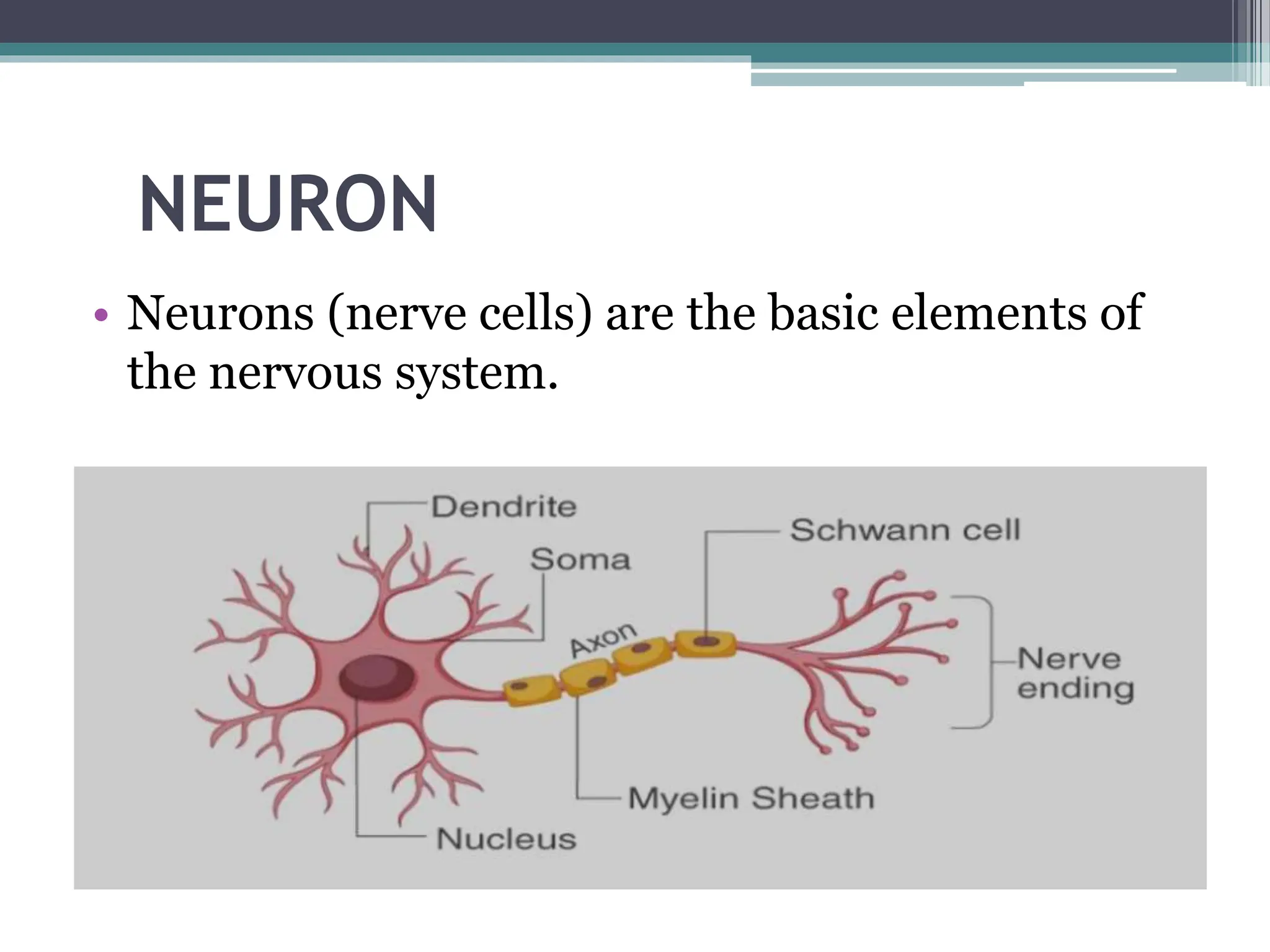
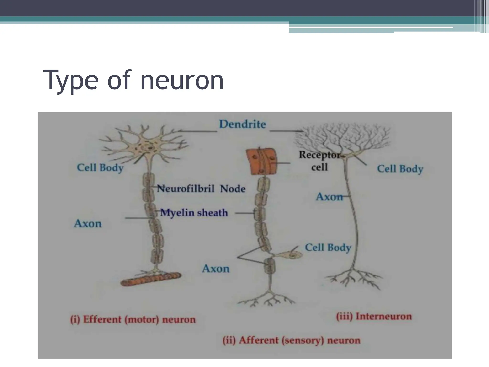
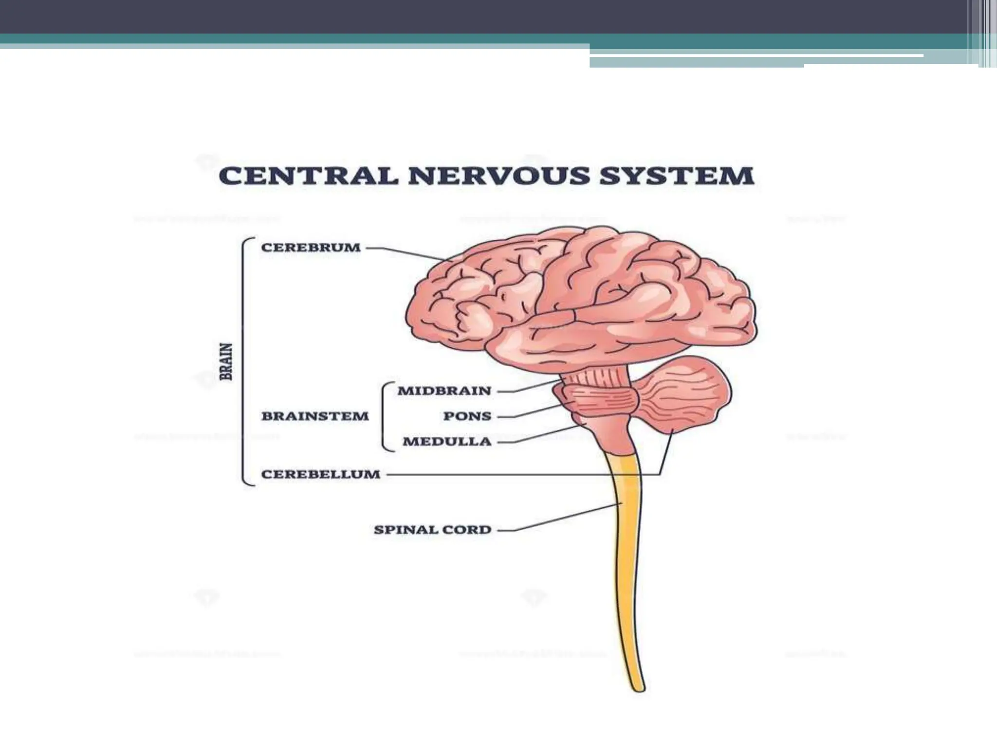
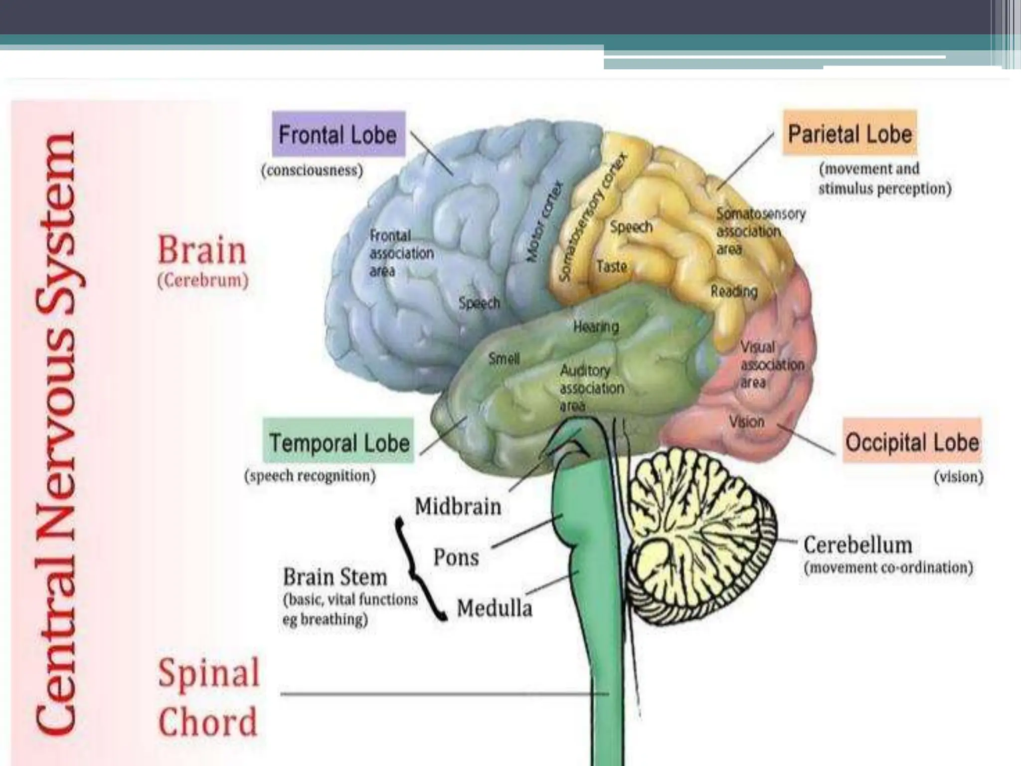
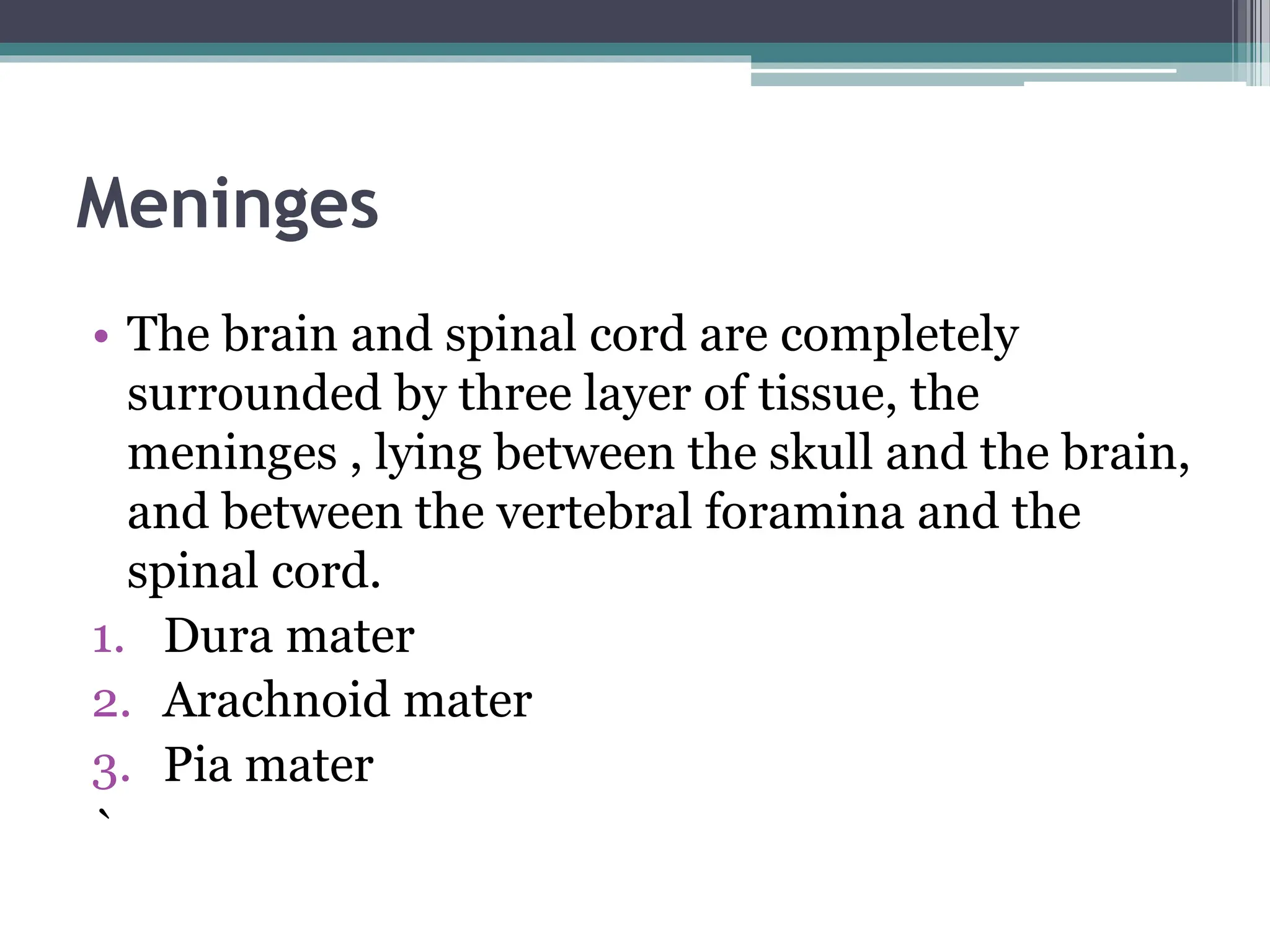
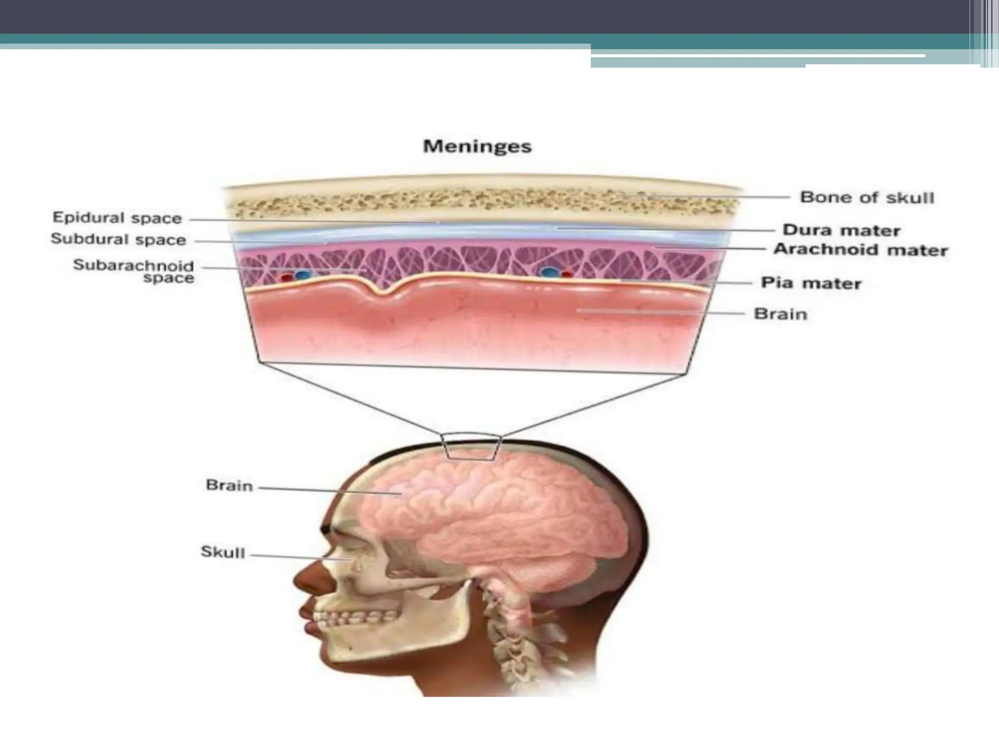
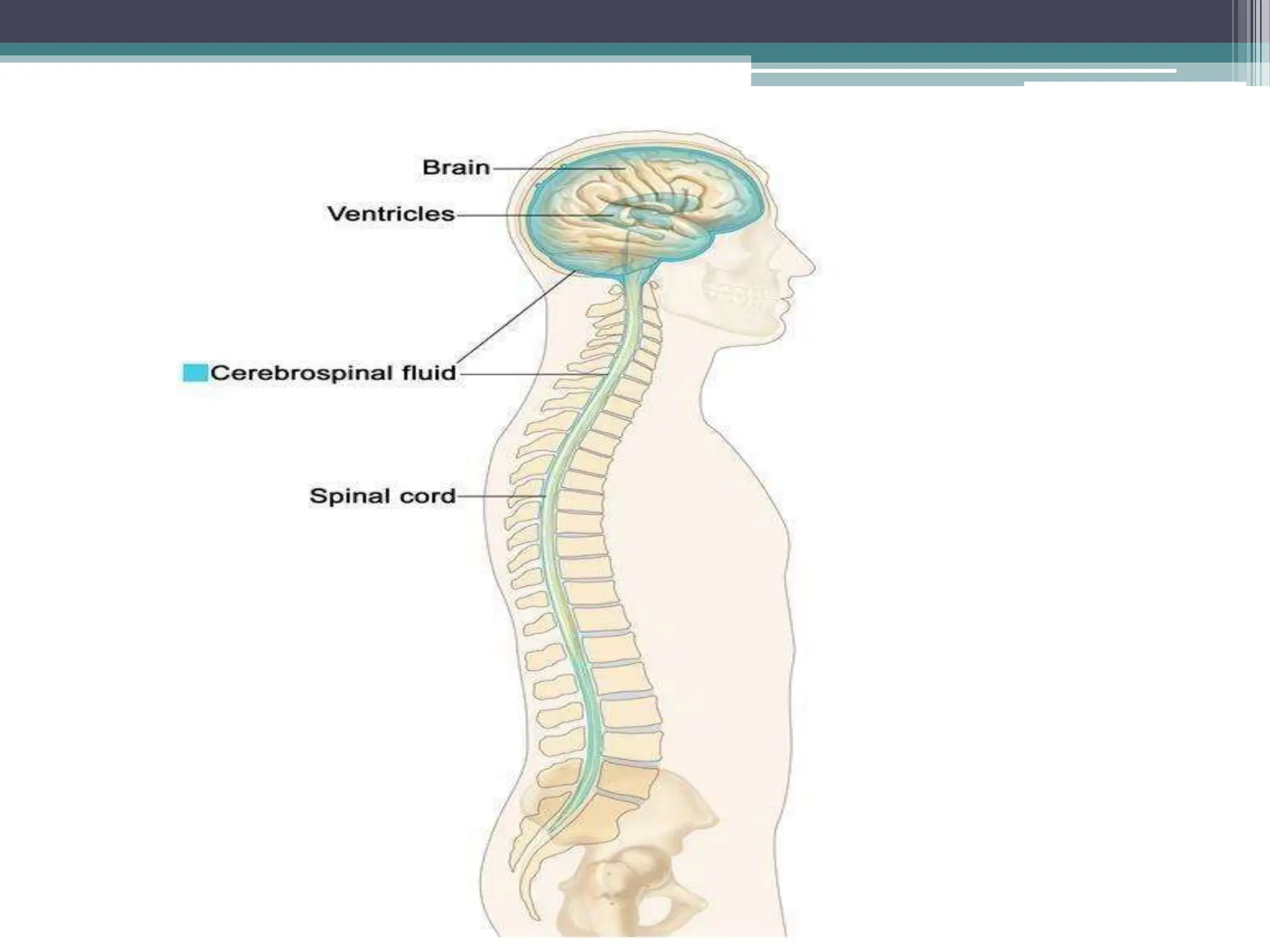
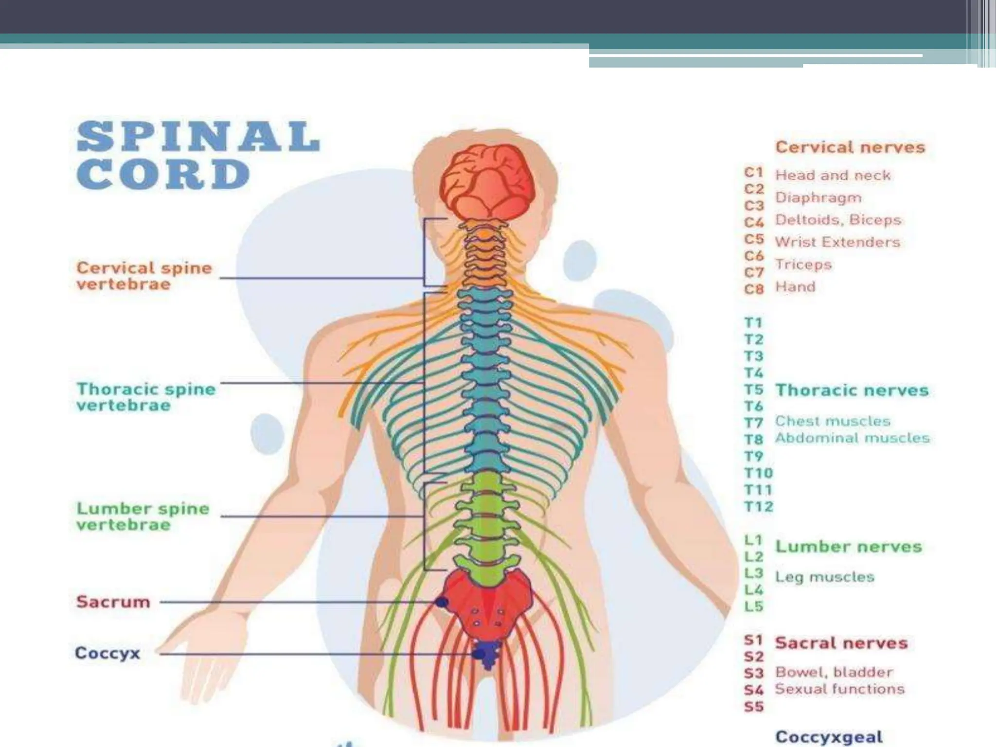
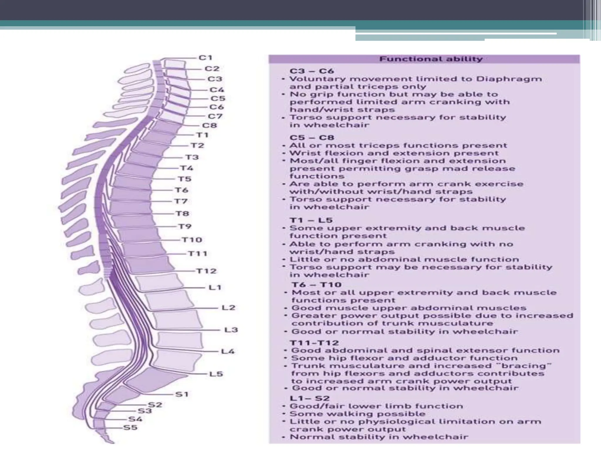
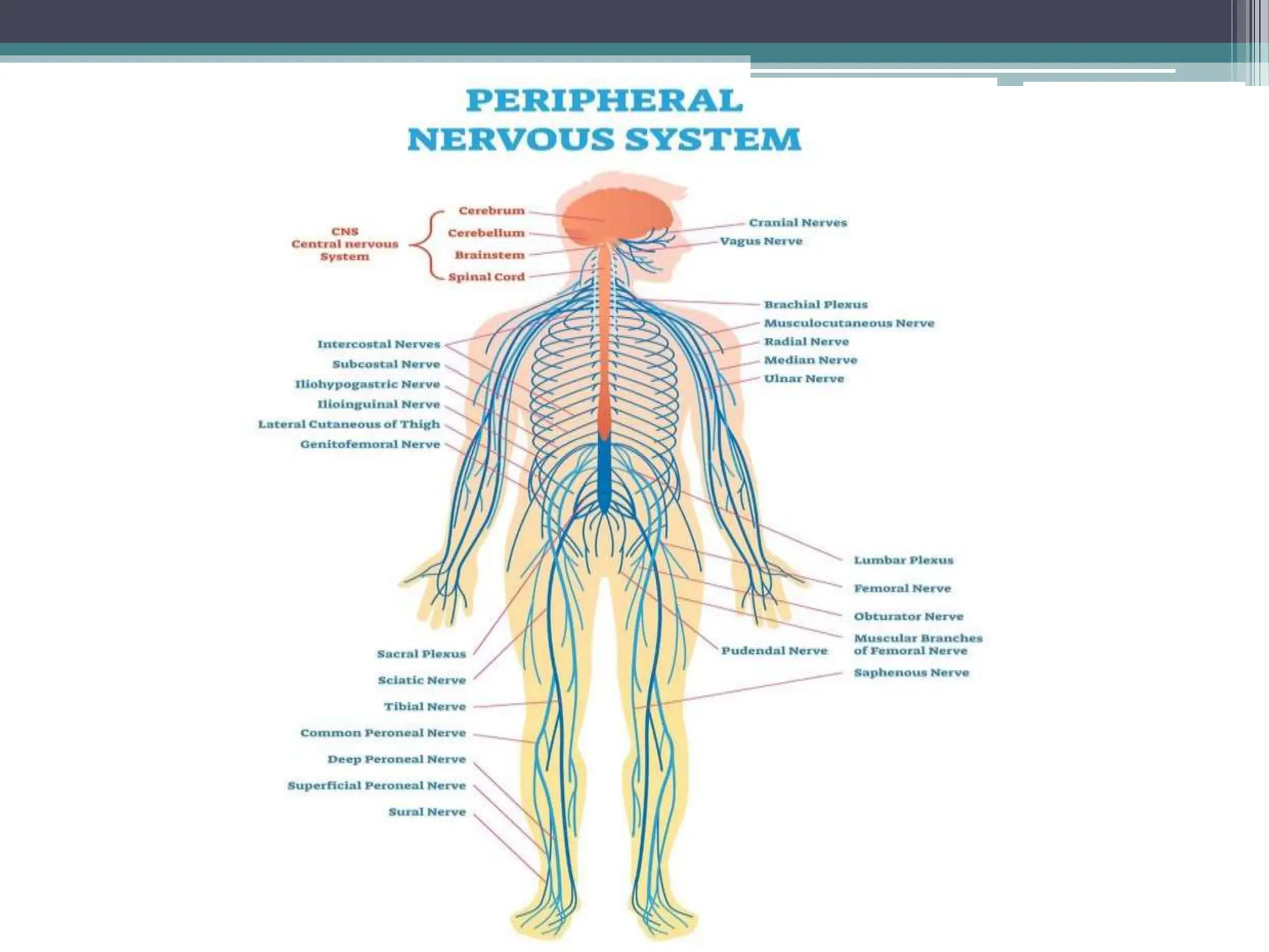
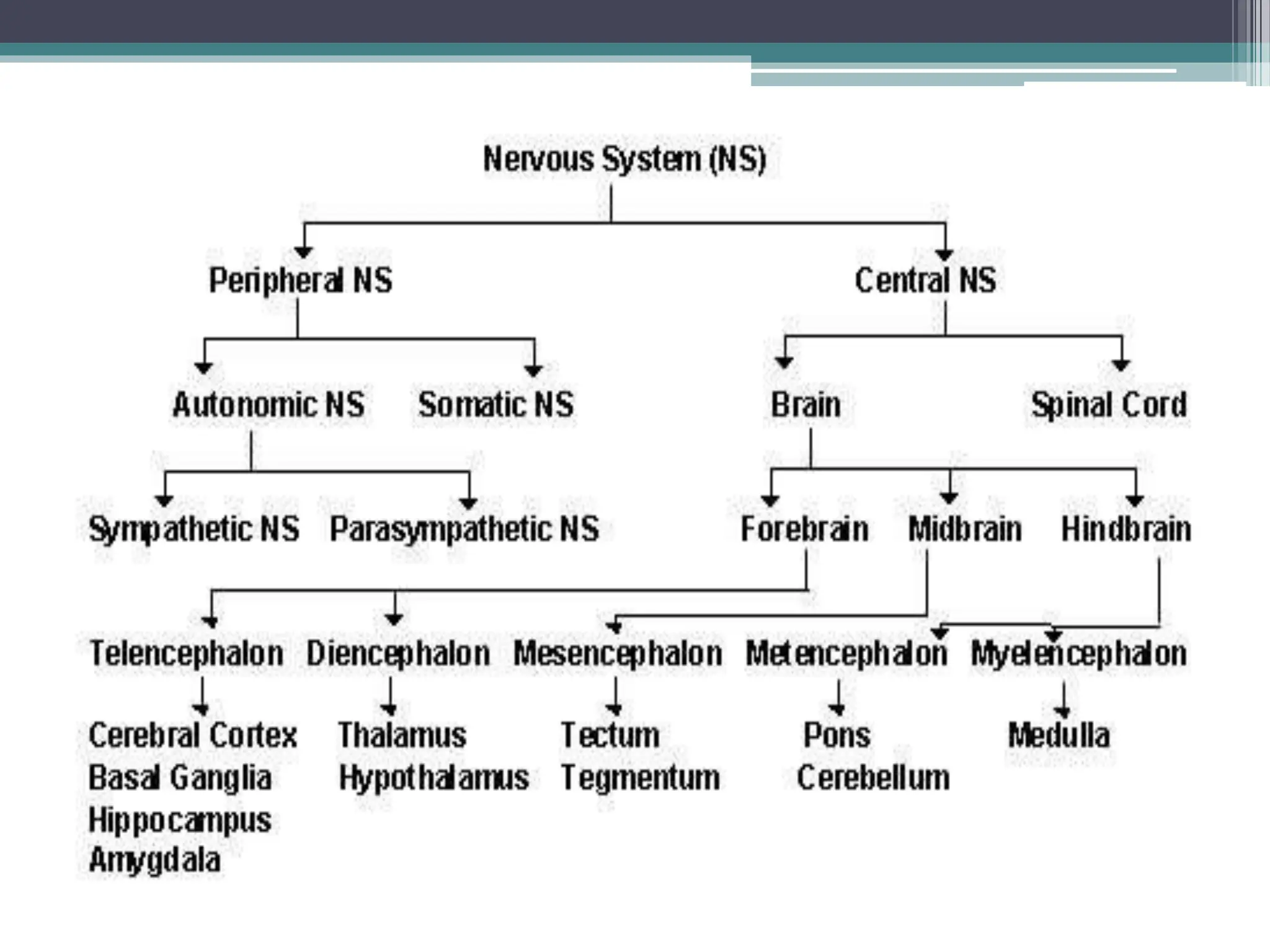
NERVOUS SYSTEM
BY : TANVI VAGHELA
INTRODUCTION
• The nervous system is very important in helping to
maintain the homeostasis (balance) of the human
body.
• A series of sensory receptors work with the nervous
system to provide information about change in both
the internal and external environment.
• The human nervous system is a complex of
interconnected systems in which large system are
comprised of smaller subsystems each of which have
specific structures with specific function.
• Nervous system also divided in two part.
1. Central nervous system (CNS)
2. Peripheral nervous system (PNS)
• The PNS consisting of all the nerves outside
the brain.
• The CNS consisting of the brain and the spinal
cord.
NEURON
• Neurons (nerve cells) are the basic elements of
the nervous system.
• NEURONS
- Cell body : The main processing center of the
cell.
- Dendrites : Thin branching extension of cell
body that conduct nerve impulses toward the
cell body.
- Axon : A single branch (in most neurons) which
conducts nerve impulses away from the cell
body.
- Myelin sheath and neurilemma are coverings.
- Impulse transmission : Terminal end fibers are
located at the ends of the axon and they transmit
impulses leaving the neuron across a synapse to
the next neuron.
Three types of neuron
• Efferent (motor) : conveys information from the
CNS to muscles and glands.
• Afferent (sensory) : carry information from
sensory rewceptors to the CNS.
• Interneuron : carry and process sensory
information.
Type of neuron
CENTRAL NERVES SYSTEM
• The CNS consists of the brain and the spinal
cord . These essential structures are both well
protected from damage and injury ; the brain is
enclosed within the skull and the spinal cord by
the vertebrae that from the spinal column.
Membranous coverings called the meninges
provide further protection. The structure and
function of the meninges , brain and spinal cord
are explored in this section.
Meninges
• The brain and spinal cord are completely
surrounded by three layer of tissue, the
meninges , lying between the skull and the brain,
and between the vertebral foramina and the
spinal cord.
1. Dura mater
2. Arachnoid mater
3. Pia mater
`
• In the brain, two spaces are associated with the
meninges :
1. The subdural space : this is a potential space
that lies between the dural and arachnoid
mater, and contains a very small amount of
serous fluid.
2. The subarachnoid space : this separates the
arachnoid and pia mater, and contains
cerebrospinal fluid (CSF).
VENTRICLES OF THE BRAIN AND
THE CEREBROSPINAL FLUID
• The brain contains four irregular shaped
cavities, or ventricles, containing CSF.
1. Right and left lateral ventricles
2. Third ventricle
3. Fourth ventricle
CEREBROSPINAL FLUID (CSF)
• CSF circulates constantly from the ventricles through the
subarachnoid space around the brain and spinal cord.
• CSF is a clear, slightly alkaline fluid with a specific gravity,
consisting of :
• Water
• Mineral salts
• Glucose
• Plasma protein : small amount of albumin and globulin
• A few leukocytes
• Small amount of creatinine and urea
• CSF is secreted into each ventricle of the brain
by choroid plexuses.
• These are vascular areas that are rich in blood
vessels and surrounded by ependymal cells in
the lining of ventricle walls.
• CSF passes back into the blood through tiny
finger like projections of arachnoid mater, called
arachnoid villi.
• From the roof of the fourth ventricle, CSF flows
through foramina into the subarachnoid space
and completely surrounded the brain and spinal
cord.
• There is no discrete CSF pump, but its movement
is aided by pulsating blood vessels, respiration
and changes of posture.
• CSF is secreted continuously at a rate of about 0.5
ml/min,720ml/day.
• The volume remains fairly constant at about
150ml, as absorption keeps pace with secretion.
• CSF may be sampled by inserting a needle into
the subarachnoid space above or below the 4th
lumbar vertebra, which is about 2 cm below the
end of the spinal cord.
• This procedure is known as a lumbar puncture,
CSF pressure can be measured by attaching a
vertical tube to the needle.
Function
• CSF supports and protects the brain and spinal cord
by maintaining a uniform pressure around these
vital structure and by acting as a cushion or shock
absorber between the brain and skull.
• CSF keeps the brain and spinal cord moist and these
may be exchange of nutrients and waste products
between CSF and the interstitial fluid of the brain.
• CSF is involved is regulation of breathing, as it
bathes the surface of the medulla where the central
respiratory chemoreceptors are located.
BRAIN
• The brain is a large organ weighing around 1.4kg
which lies within the cranial cavity. Its parts are ;
• Cerebrum
• Thalamus
• Hypothalamus
• Midbrain
• Pons
• Medulla oblongata
• Cerebellum
BLOOD SUPPLY AND VENOUS DRAINAGE
• The circulus arteriouus and its contributing arteries
play a vital role in maintaining a constant supply of
oxygen and glucose to the brain when the head is moved
and also when a contributing artery is narrowed.
• The brain receives about 15% of cardiac output,
approximately 750 ml of blood/min.
• Autoregulation of cerebral arteriolar diameter maintain
a constant blood flow to the brain, compensating for
fluctuations in systemic blood pressure
• This mechanism protects the brain, provide that
systemic blood pressure remains within the 65-140
mmHg rang.
• CEREBRUM
• This is the large part of the brain and it occupies
the anterior and middle cranial fossae .
• It is divided by a deep cleft, the longitudinal
cerebral fissure, into right and left cerebral
hemispheres, each containing one of the
ventricles.
• The hemispheres are connected by a mass of
white matter (nerve fibres) called the corpus
callosum.
• The falx cerebri is formed by the dura mater.
• It seprates the two cerebral hemispheres and
penetrates to the depth of the corpus callosum.
• The superficial part of cerebrum is composed of
nerve cell bodies (grey matter), forming the cerebral
cortex and the deeper layer consist of nerve fibres
(axons, white matter) .
• Each hemisphere of the cerebrum is divided into
lobes, which take the names of the bones of the
cranium above them;
- Frontal
- Parietal
- Temporal
- Occipital
FUNCTION OF CEREBRALCORTEX
• There are the main three type of activity associated
with the cerebral cortex.
1. Higher order functions, e.g. language, memory,
sense of responsibility, thinking, reasoning, moral
decision making and learning.
2. Sensory perception, including the perception of
the pain, temperature, touch, sight, hearing, taste
and smell.
3. Initiation and control of skeletal muscle
contraction and therefore voluntary movement.
Functional area of the cerebral cortex
• The different area active in both hemispheres.
There are the different types of functional area;
• Motor, which direct skeletal (voluntary) muscle
movements.
• Sensory, which receive and decode sensory
impulses, enabling sensory perception.
• Association, which are concerned with
integration and processing of complex mental
function such as intelligence, memory,
reasoning, judgement and emotions.
Motor area of cerebral cortex
1. Primary motor area
2. Motor speech area
3. Association area
Sensory area of the cerebral cortex
• Somatosensory arean : sensation of pain, temp.,
pressure, touch.
• Auditory (hearing) area
• Olfactory (smell) area
• Taste area
• Visual area
Association area
• Premotor area
• Prefrontal area
• Sensory speech area
• Parieto-occipital area
• Diencephalon
• The diencephalon connected with cerebrum and
mid brain.
• The deep portion of the brain containing :
1. Thalamus
2. Hypothalamus
3. Epithalamus
4. Ventral thalamus
• BRAINSTEM
• Made up of the midbrain , pons and the medulla
oblongata.
1. Midbrain : involved with visual reflexes
2. Pons : Located between the midbrain and
medulla oblongata
- controls certain respiratory function.
3. Medulla oblongata : contains centers that
regulates heart and lung functioning,
swallowing, coughing, vomiting and sneezing.
• Serves as center for sensations relay like:
• Heart rate
• Blood pressure
• Temperature control
• Behavioral responses
• Digestive functions
• Water and electrolyte balance
• CEREBELLUM
• Area that coordinates musculoskeletal
movement to maintain posture, balance and
muscle tone.
• Inferior to the occipital lobes of the cerebrum.
• Posterior to the pons and medulla oblongata.
SPINAL CORD
• Extends from the medulla oblongata of the body
of the brain to the area round the first lumbar
vertebra in the lower back
• Nerves from the peripheral nervous system
extend out from the spinal cord.
• Protected by :
1. Vertebral column
2. Cerebrospinal fluid
3. meninges
PERIPHERAL NERVOUS SYSTEM
• The peripheral nervous system (PNS) is a
collection of peripheral nerves, ganglia and
specialized sensory structures that, as a system,
carries sensory and motor information between
the central nervous system and all other organs
and tissues of the body.
• The peripheral nervous system is functionally
divided into two major division :
- The sensory or afferent division
- The motor or afferent division
1. The somatic nervous system
2. The autonomic nerves system (visceral) system
• PNS consists of 12 pairs of cranial nerves and
31 pairs of spinal nerves
Somatic nervous system
• Responsible for receiving and processing
sensory input from the skin, muscle, tendons,
joints, eyes, tongue, nose, and ears as well as
excite the voluntary contraction of skeletal
muscles.
Autonomic nervous system
• Carries impulses from the central nervous
system to glands, various muscles, cardiac
muscle and various membranes.
• Stimulates organs, glands and senses.
THANK YOU

NERVOUS SYSTEM BY MS. TANVI P. VAGHELA..

  • 1.
    NERVOUS SYSTEM BY :TANVI VAGHELA
  • 2.
    INTRODUCTION • The nervoussystem is very important in helping to maintain the homeostasis (balance) of the human body. • A series of sensory receptors work with the nervous system to provide information about change in both the internal and external environment. • The human nervous system is a complex of interconnected systems in which large system are comprised of smaller subsystems each of which have specific structures with specific function.
  • 3.
    • Nervous systemalso divided in two part. 1. Central nervous system (CNS) 2. Peripheral nervous system (PNS) • The PNS consisting of all the nerves outside the brain. • The CNS consisting of the brain and the spinal cord.
  • 4.
    NEURON • Neurons (nervecells) are the basic elements of the nervous system.
  • 5.
    • NEURONS - Cellbody : The main processing center of the cell. - Dendrites : Thin branching extension of cell body that conduct nerve impulses toward the cell body. - Axon : A single branch (in most neurons) which conducts nerve impulses away from the cell body. - Myelin sheath and neurilemma are coverings. - Impulse transmission : Terminal end fibers are located at the ends of the axon and they transmit impulses leaving the neuron across a synapse to the next neuron.
  • 6.
    Three types ofneuron • Efferent (motor) : conveys information from the CNS to muscles and glands. • Afferent (sensory) : carry information from sensory rewceptors to the CNS. • Interneuron : carry and process sensory information.
  • 7.
  • 8.
    CENTRAL NERVES SYSTEM •The CNS consists of the brain and the spinal cord . These essential structures are both well protected from damage and injury ; the brain is enclosed within the skull and the spinal cord by the vertebrae that from the spinal column. Membranous coverings called the meninges provide further protection. The structure and function of the meninges , brain and spinal cord are explored in this section.
  • 11.
    Meninges • The brainand spinal cord are completely surrounded by three layer of tissue, the meninges , lying between the skull and the brain, and between the vertebral foramina and the spinal cord. 1. Dura mater 2. Arachnoid mater 3. Pia mater `
  • 13.
    • In thebrain, two spaces are associated with the meninges : 1. The subdural space : this is a potential space that lies between the dural and arachnoid mater, and contains a very small amount of serous fluid. 2. The subarachnoid space : this separates the arachnoid and pia mater, and contains cerebrospinal fluid (CSF).
  • 14.
    VENTRICLES OF THEBRAIN AND THE CEREBROSPINAL FLUID • The brain contains four irregular shaped cavities, or ventricles, containing CSF. 1. Right and left lateral ventricles 2. Third ventricle 3. Fourth ventricle
  • 16.
    CEREBROSPINAL FLUID (CSF) •CSF circulates constantly from the ventricles through the subarachnoid space around the brain and spinal cord. • CSF is a clear, slightly alkaline fluid with a specific gravity, consisting of : • Water • Mineral salts • Glucose • Plasma protein : small amount of albumin and globulin • A few leukocytes • Small amount of creatinine and urea
  • 17.
    • CSF issecreted into each ventricle of the brain by choroid plexuses. • These are vascular areas that are rich in blood vessels and surrounded by ependymal cells in the lining of ventricle walls. • CSF passes back into the blood through tiny finger like projections of arachnoid mater, called arachnoid villi. • From the roof of the fourth ventricle, CSF flows through foramina into the subarachnoid space and completely surrounded the brain and spinal cord.
  • 18.
    • There isno discrete CSF pump, but its movement is aided by pulsating blood vessels, respiration and changes of posture. • CSF is secreted continuously at a rate of about 0.5 ml/min,720ml/day. • The volume remains fairly constant at about 150ml, as absorption keeps pace with secretion. • CSF may be sampled by inserting a needle into the subarachnoid space above or below the 4th lumbar vertebra, which is about 2 cm below the end of the spinal cord. • This procedure is known as a lumbar puncture, CSF pressure can be measured by attaching a vertical tube to the needle.
  • 19.
    Function • CSF supportsand protects the brain and spinal cord by maintaining a uniform pressure around these vital structure and by acting as a cushion or shock absorber between the brain and skull. • CSF keeps the brain and spinal cord moist and these may be exchange of nutrients and waste products between CSF and the interstitial fluid of the brain. • CSF is involved is regulation of breathing, as it bathes the surface of the medulla where the central respiratory chemoreceptors are located.
  • 20.
    BRAIN • The brainis a large organ weighing around 1.4kg which lies within the cranial cavity. Its parts are ; • Cerebrum • Thalamus • Hypothalamus • Midbrain • Pons • Medulla oblongata • Cerebellum
  • 22.
    BLOOD SUPPLY ANDVENOUS DRAINAGE • The circulus arteriouus and its contributing arteries play a vital role in maintaining a constant supply of oxygen and glucose to the brain when the head is moved and also when a contributing artery is narrowed. • The brain receives about 15% of cardiac output, approximately 750 ml of blood/min. • Autoregulation of cerebral arteriolar diameter maintain a constant blood flow to the brain, compensating for fluctuations in systemic blood pressure • This mechanism protects the brain, provide that systemic blood pressure remains within the 65-140 mmHg rang.
  • 23.
    • CEREBRUM • Thisis the large part of the brain and it occupies the anterior and middle cranial fossae . • It is divided by a deep cleft, the longitudinal cerebral fissure, into right and left cerebral hemispheres, each containing one of the ventricles. • The hemispheres are connected by a mass of white matter (nerve fibres) called the corpus callosum.
  • 25.
    • The falxcerebri is formed by the dura mater. • It seprates the two cerebral hemispheres and penetrates to the depth of the corpus callosum. • The superficial part of cerebrum is composed of nerve cell bodies (grey matter), forming the cerebral cortex and the deeper layer consist of nerve fibres (axons, white matter) . • Each hemisphere of the cerebrum is divided into lobes, which take the names of the bones of the cranium above them; - Frontal - Parietal - Temporal - Occipital
  • 26.
    FUNCTION OF CEREBRALCORTEX •There are the main three type of activity associated with the cerebral cortex. 1. Higher order functions, e.g. language, memory, sense of responsibility, thinking, reasoning, moral decision making and learning. 2. Sensory perception, including the perception of the pain, temperature, touch, sight, hearing, taste and smell. 3. Initiation and control of skeletal muscle contraction and therefore voluntary movement.
  • 28.
    Functional area ofthe cerebral cortex • The different area active in both hemispheres. There are the different types of functional area; • Motor, which direct skeletal (voluntary) muscle movements. • Sensory, which receive and decode sensory impulses, enabling sensory perception. • Association, which are concerned with integration and processing of complex mental function such as intelligence, memory, reasoning, judgement and emotions.
  • 29.
    Motor area ofcerebral cortex 1. Primary motor area 2. Motor speech area 3. Association area
  • 30.
    Sensory area ofthe cerebral cortex • Somatosensory arean : sensation of pain, temp., pressure, touch. • Auditory (hearing) area • Olfactory (smell) area • Taste area • Visual area
  • 31.
    Association area • Premotorarea • Prefrontal area • Sensory speech area • Parieto-occipital area
  • 32.
    • Diencephalon • Thediencephalon connected with cerebrum and mid brain. • The deep portion of the brain containing : 1. Thalamus 2. Hypothalamus 3. Epithalamus 4. Ventral thalamus
  • 34.
    • BRAINSTEM • Madeup of the midbrain , pons and the medulla oblongata. 1. Midbrain : involved with visual reflexes 2. Pons : Located between the midbrain and medulla oblongata - controls certain respiratory function. 3. Medulla oblongata : contains centers that regulates heart and lung functioning, swallowing, coughing, vomiting and sneezing.
  • 35.
    • Serves ascenter for sensations relay like: • Heart rate • Blood pressure • Temperature control • Behavioral responses • Digestive functions • Water and electrolyte balance
  • 36.
    • CEREBELLUM • Areathat coordinates musculoskeletal movement to maintain posture, balance and muscle tone. • Inferior to the occipital lobes of the cerebrum. • Posterior to the pons and medulla oblongata.
  • 39.
    SPINAL CORD • Extendsfrom the medulla oblongata of the body of the brain to the area round the first lumbar vertebra in the lower back • Nerves from the peripheral nervous system extend out from the spinal cord. • Protected by : 1. Vertebral column 2. Cerebrospinal fluid 3. meninges
  • 43.
    PERIPHERAL NERVOUS SYSTEM •The peripheral nervous system (PNS) is a collection of peripheral nerves, ganglia and specialized sensory structures that, as a system, carries sensory and motor information between the central nervous system and all other organs and tissues of the body. • The peripheral nervous system is functionally divided into two major division : - The sensory or afferent division - The motor or afferent division
  • 47.
    1. The somaticnervous system 2. The autonomic nerves system (visceral) system • PNS consists of 12 pairs of cranial nerves and 31 pairs of spinal nerves
  • 49.
    Somatic nervous system •Responsible for receiving and processing sensory input from the skin, muscle, tendons, joints, eyes, tongue, nose, and ears as well as excite the voluntary contraction of skeletal muscles.
  • 50.
    Autonomic nervous system •Carries impulses from the central nervous system to glands, various muscles, cardiac muscle and various membranes. • Stimulates organs, glands and senses.
  • 55.