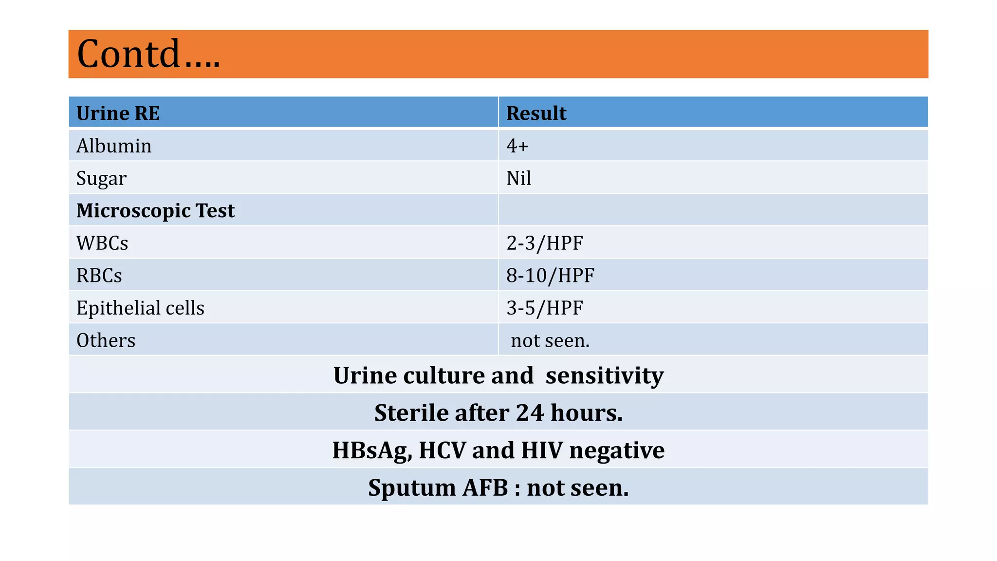
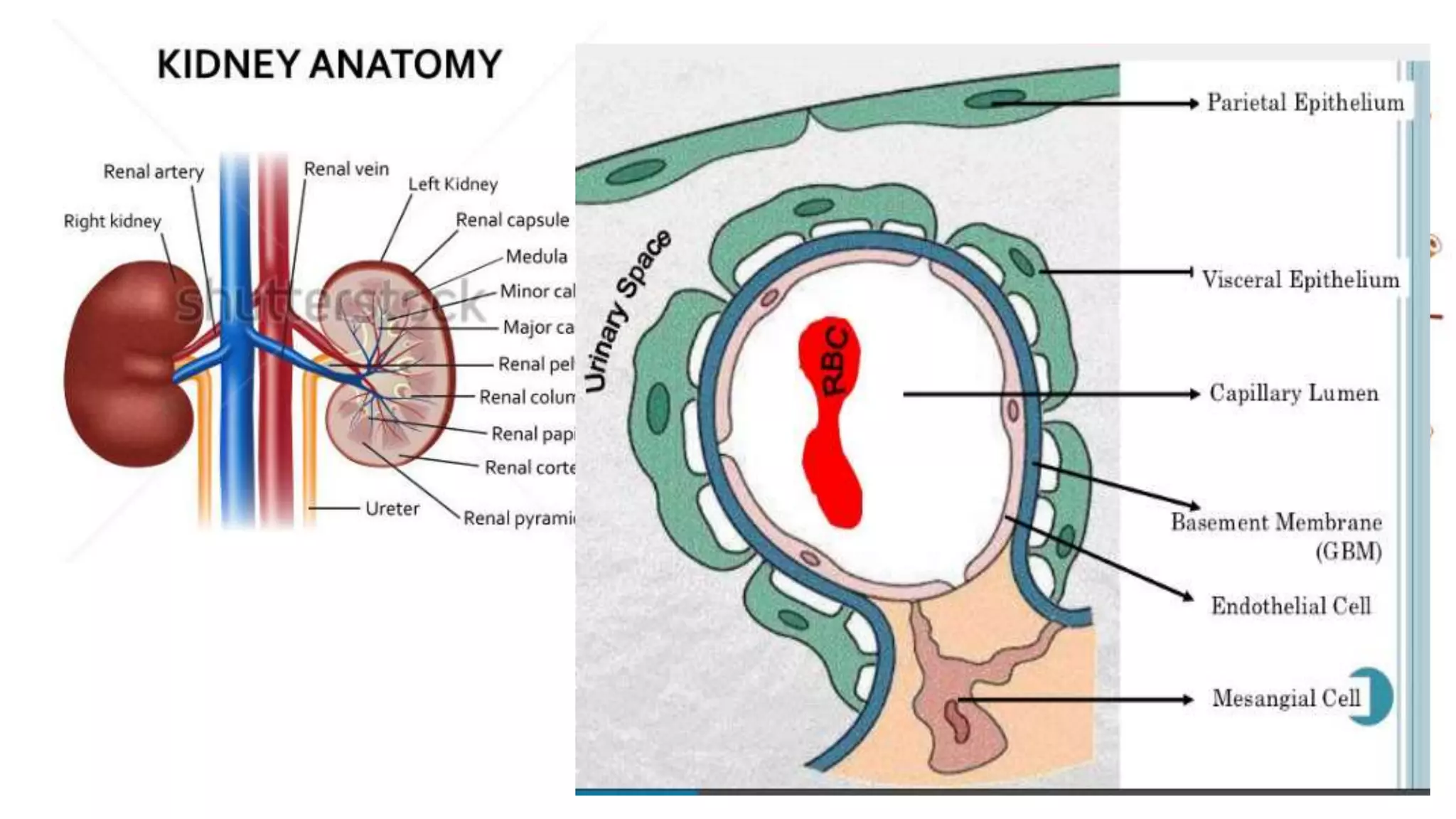
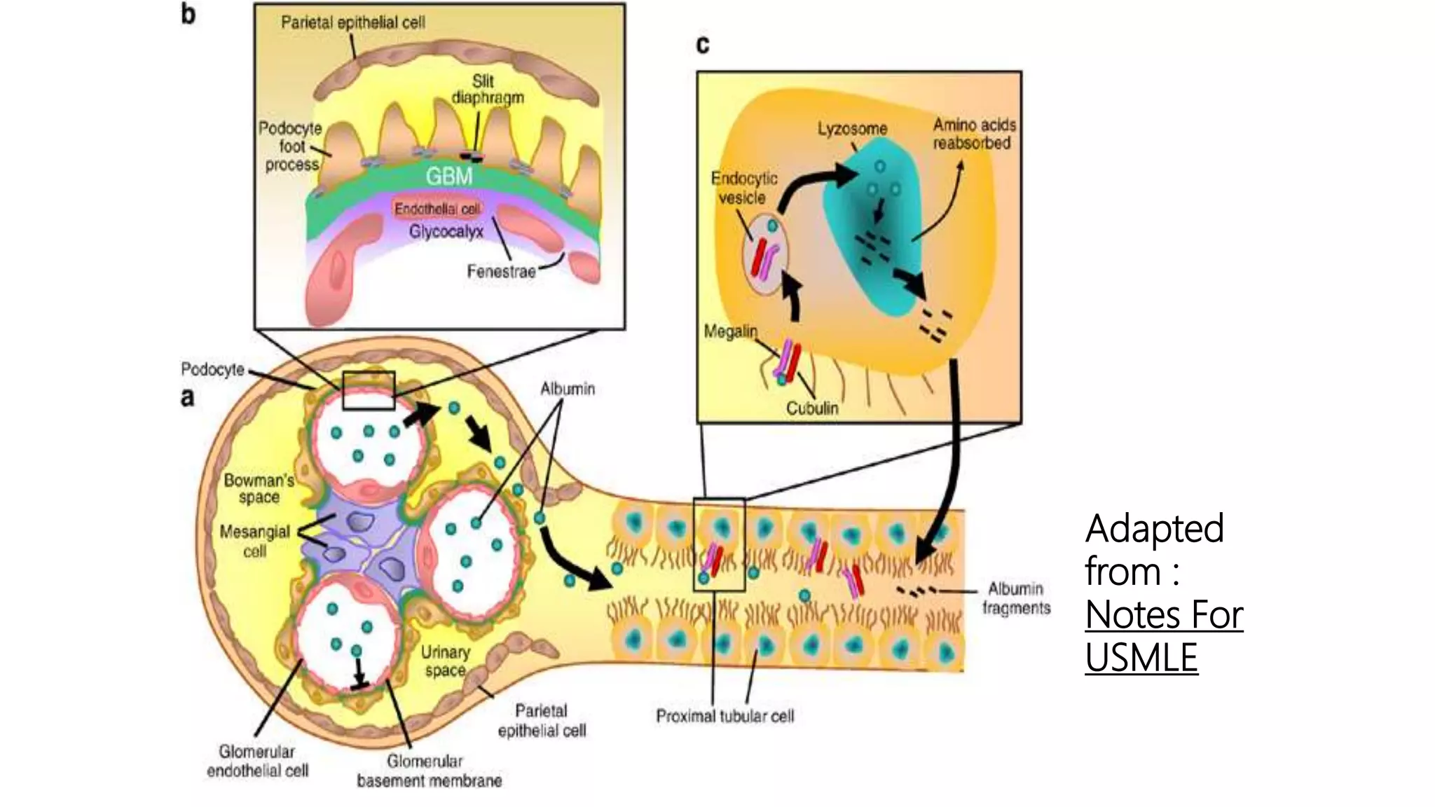
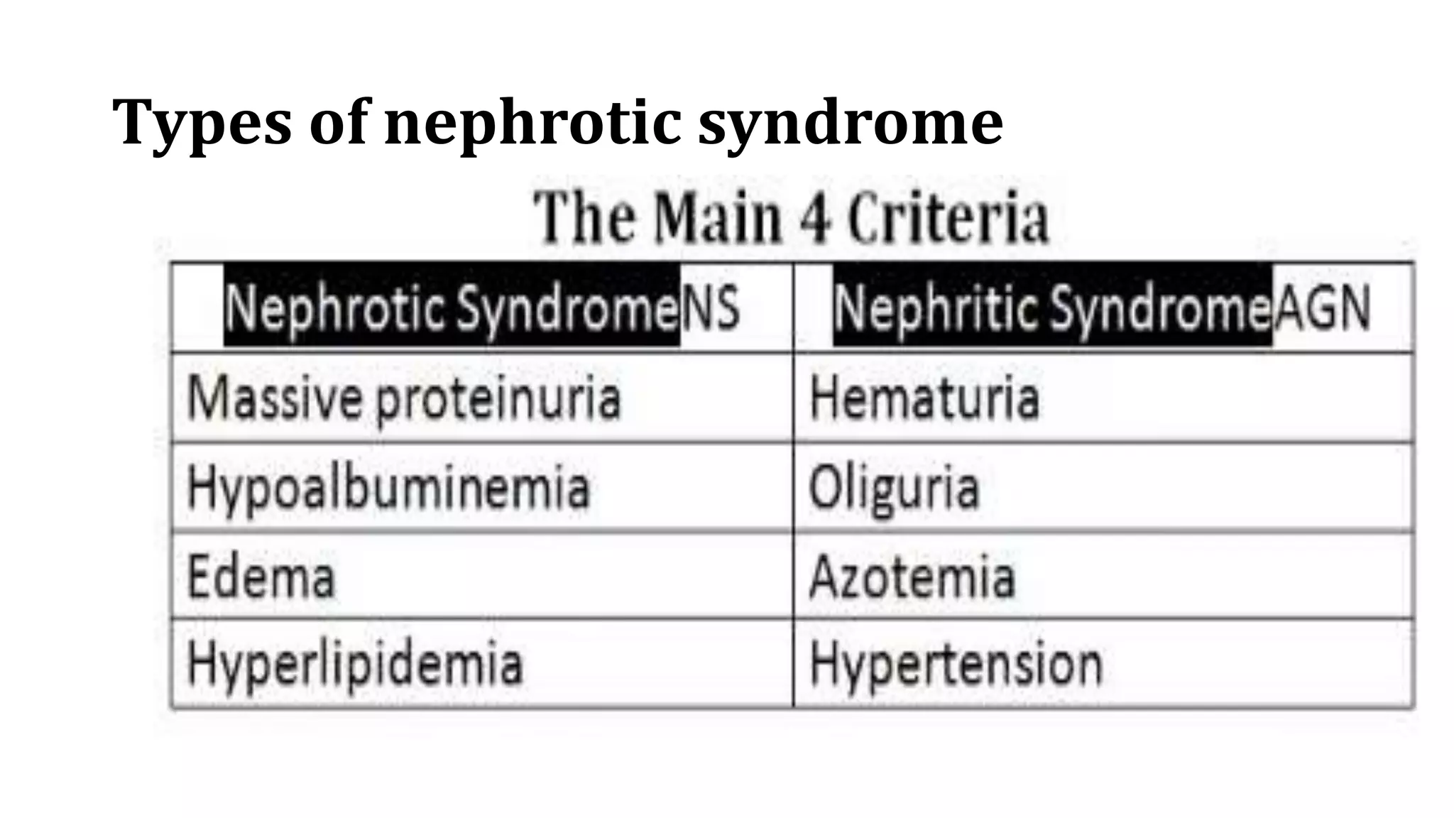
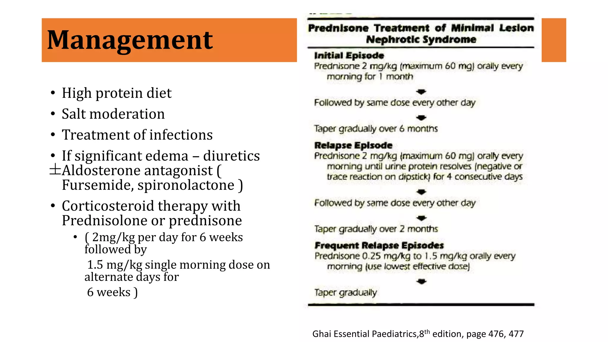
A CASE STUDY
Moderator
Dr. Jouslin k Baranwal
Asst. Prof
Department of
Biochemistry, BPKIHS
Presenter
Binaya Tamang
MSc. 3rd year
Department of
Biochemistry, BPKIHS
History
Name: Rhythm Rai
Age: 2.5 years
Sex: Male
Religion: Hindu
From: Chulachuli
Patient Id:2724478
• On 74/ 2/18, 4: 05 PM
• Reported to the pediatric emergency with the chief complains of
Generalized swelling of the body X 5 days .
Swelling started from the face followed by swelling of abdomen , upper and
lower limbs.
No h/o of rash , fever, sore throat, fast breathing
Patient was taken to the local hospital and some medicine was given, a USG
was done and referred to BPKIHS.
History of Present Illness
• The patient was apparently well 5 days back when his mother noticed
swelling of her face, which was acute in onset and gradually progressing
towards the abdomen and bilateral upper and lower limbs.
• The swelling is painless and pitting in nature.
• The overlying skin was normal and there is no history of itching and rashes.
• No h/o frothy urine ,yellowish discoloration of urine
• No h/o cough, chest pain
• No h/o abdominal pain, loss of appetite, vomiting.
• No h/o fever , joint pain, photosensitivity and oral ulcers
Contd,,,
• No h/o altered bowel habit and bladder
• No h/o passage of frank blood in the urine
• No h/o crying at micturition , yellowish discoloration of the body
• No h/o any skin infection
• No h/o petechia, purpura.
Past history:
• h/o throat infection 2-3 months back
• No h/o of similar illness
• No h/o TB, asthma, HTN, DM
• No h/o fever with rash, neck swelling.
Antenatal:
• No any ANC visits
• No TT vaccines
• No iron and calcium taken
• No h/o fever, rashes, lymphadenopathy
• No h/o excessive vomiting, increased frequency of
micturition
• No h/o burning micturition, increased urgency
• No h/o PV bleeding, spotting
• No h/o headache, blurring of vision, epigastric
pain.
Perinatal history:
• Normal term, spontaneous vaginal delivery at (
patient not sure of exact date)
• Baby cried at birth.
• Baby weight:-2.0 kg
• Breast feeding at 4 hours of life.
Post natal history:
• No h/o Excessive bleeding
• No other complications
Family history:
• No h/o similar illness in family
• No h/o consanguineous marriage
• No h/o TB, DM, Kidney deases
• No h/o HTN
Dietary History:
• Tea+ 1 kotori rice+ 1 kotori daal+ 1
kotori potato curry
• Not significant history given by
mother.
Immunization history:
• Immunization as per EPI schedule
with strictly all vaccines given to child
Developmental History
• H/o normal developments
• Explore drawers, runs ups and
downstairs
• Vertical and circular strokes
• Asks for food , toilets
• 2-3 word sentence, short sentences
On Examination
General condition:
• facial puffiness +ve
• Edema +ve, bilateral pitting
edema of limbs
• No any scar marks of infection
Vitals
• Pulse: 110/min; regular, normal
vol and character,
• RR: 22/min
• Temp: 98 F
• BP: 90/60 on rt arm in sitting
position
 GPE
• Pallor
• Icterus
• Lymphadenopathy
• Cyanosis
• Dehydration
Anthropometry
• Weight: 13.5 kg
GIT:
Abdomen distended, umbilicus central, all quadrant moving symmetrically
with respiration, no venous prominence, no scar marks, hernia site are intact.
On palpation: no local rise in temp
no tenderness
no any lump and organ
On percussion: shifting dullness present
 Chest: Bilateral Equal Chest Expansion;B/L Normal
vesicular breath sounds ; no added sounds, Trachea Central
CVS: S1S2,Mo, No added sounds.
Preliminary Diagnosis.
Probably a case of nephrotic syndrome. Mostly Minimal change Disease (
as per incidence and geography).
Investigation
• Biochemical Parameters
Parameters Result Reference range
UREA 14mg/dl 10-50
Creatinine 0.2mg/dl 0.5-1.4
Sodium 131 mmol/L 136-145
Potassium 4.1mmol/L 3.5-5.0
Total Protein in 24 hrs
urine
360 ml/ 0.27 g/day 1500-2500
<0.15
Serum
cholesterol(Total)
423 mg/dl <200
Serum albumin 2.3 g/dl 3.5-5.3
Urinary protein
creatinine ratio
0.51
3.06
<0.2mg/g
Contd….
Urine RE Result
Albumin 4+
Sugar Nil
Microscopic Test
WBCs 2-3/HPF
RBCs 8-10/HPF
Epithelial cells 3-5/HPF
Others not seen.
Urine culture and sensitivity
Sterile after 24 hours.
HBsAg, HCV and HIV negative
Sputum AFB : not seen.
Hematological parameters.
Parameters Result Reference
Blood group O positive
CBC
Hemoglobin 12.8 gm/dl 11-16gm/dl
PCV 35.9 36-48%
TLC 9400 4000-11000 cell/mm cu
Neutrophil 25 40-75 %
Lymphocyte 60 20-45%
Monocyte 06 2-10%
Eosinophil 09 1-6%
Platelet 324000 150000-400000cell/mm cu
ESR 54 1st hour
Final Diagnosis
• With the clinical sign and symptoms , lab diagnosis, age as well as other no
secondary diseases associated.
• Nephrotic syndrome with some hematuria. Most probably minimal change
disease nephrotic syndrome for the confirmation renal biopsy is preferred.
• Most (>85%) of children MCN.
Treatment Given :
Sryp Ritocef (5/50) 5ml x PO x BD
Tab Emsolone 25 mg x PO X OD Orally allowed
Sryp Digene 5ml x PO x BD
Glomerular Membrane
 Modified capillary wall comprising endothelium (50-100
nm pores)
 A cell-less basement membrane and
an outer specialized epithelial cell layer
(55 nm slit diaphragm)
 The whole of the glomerular membrane carries a fixed net
negative charge:
 Due to a Glycosialoprotein coat
 Charge increases in density from the Lamina Interna
towards the Lamina rara Externa with the greatest
density at the slit diaphragm of the epithelium
Adapted
from :
Notes For
USMLE
Nephrotic Syndrome
It is a common glomerular disease, characterized by
nephrotic range proteinuria and a triad of clinical findings
associated with large urinary losses of protein :
hypoalbuminaemia , edema and hyperlipidemia
It is clinical face of glomerular injury.
Why ‘nephrotic
range’
Defined as
protein excretion of > 40 mg/m2/hr
and >3.5 gm/day.
Incidence ( paediatric ) ?
• 2 – 7 cases per 100,000 children per year
• Higher in underdeveloped countries ( South east Asia )
• Occurs at all ages but is most prevalent in children
between the ages 1.5-6 years.
• It affects more boys than girls, 2:1 ratio
http://www.kidney.org/site/107/pdf/NephroticSyndrome.pdf
Etiology
• Genetic
• Secondary
•Idiopathic or Primary
Types of nephrotic syndrome
Causes prevalence(%)
children adults
Primary glomerular diseases
Membranous nephropathy 3 30
Minimal change disease 75 8
Focalsegmental glomerulosclerosis 10 35
Membranoproliferative glomerulonephritis and dense disease 10 10
Other proliferative glomerulonephritis(focal, pure mesangial, IgA nephropathy 2 17
Systemic Disease
Diabetes mellitus, amyloidosis, systemic lupus erythematous
Drugs ( NSAIDS, penicillamine, heroin), infection ( malaria, syphilis, hepatitis B and C, HIV), Malignant disease
( carcinoma, lymphoma), Miscellaneous ( bee-sting, hereditary nephritis)
Pathophysiology :- Minimal change disease
• This relatively benign disorder is characterized by diffuse effacement of
foot processes of visceral epithelial cells( podocytes), detectable only by
electron microscope, appear virtually normal by light microscope.
• It is the most common cause of nephrotic syndrome in children(2-6
years)
Pathogenesis: idiopathic but current leading hypothesis involves some
immune dysfunction that results in the elaboration of factors that
damage visceral epithelial cells and cause proteinuria.
• Animal model=angiopoietin –like-4 but not have been proven to cause in
human.
• Primary visceral epithelial cell injury (podocytopathy) and loss of
polyanions.
• Protein traverse capillary wall remains enigma.
http://www.highlands.edu/academics/divisions/scipe/biology/faculty/harnden/2122/images/renalcorpuscle.jpg
Complex disturbances in
immune system
Genetic Mutations /
Mutations in proteins
Extensive effacement of podocyte foot processes
Increased permeability of the glomerular capillary wall
Massive proteinuria
Hypoalbuminaemia
Edema
How changes occurs?
The glomerular capillary wall, with its endothelium , GBM and visceral
epithelial cells, acts as size and charge barrier through which plasma filter
passes.
Increased permeability from either structural and physiochemical alterations
in this barrier allows proteins to escape from plasma in to urine 
proteinuria.
Heavy proteinuria depletes serum albumin level at rate beyond the
compensatory synthetic capacity of liverhypoalbunemia
The generalized edema is a direct consequence of decreased intravascular colloid
osmotic pressure .
There is also sodium and water retention, which aggravates the edema.
• Several factors including compensatory secretion of aldosterone mediated by
hypovolemia-enhanced renin secretion; stimulation of sympathetic system; and
a reduction in the secretion of natriuretic factors such as atrial peptides
Edema ; soft and pitting, and is most marked in periorbital regions.
The genesis of the hyper lipidemia is complex.
 Increased synthesis of lipoproteins in the liver
 Abnormal transport of circulating lipids particles
 Decreased lipid catabolism.
 Finally lipiduria seen as oval fat bodies.
Clinical features
•Edema
• Mild to start with – peri orbital puffiness, lower extremities
• Progression to generalized edema, ascites, pleural effusion, genital edema
•Decreased urine output
• Anorexia, Irritability, Abdominal pain and diarrhea
• Absence of
• Hypertension
• Gross hematuria
Vulnerable of
• Infection , especially staphylococcal and pneumococcal infection
probably due to loss of immunoglobulins.
• Thrombotic and thromboembolic complication : antithrombin III
• Anemia due to loss of transferrin.
Lab Investigations
• Urine Examination
• Complete Blood Count & Blood
picture
• Renal parameters :
• Spot Urine Protein : Creatinine
ratio
• Urinary protein excretion
• protein selectivity ratio
• Liver Function Test
• Renal Biopsy ???
• Urinalysis - 3+ to 4+ proteinuria
• Renal Function
• Spot UPC ratio > 2.0
• UPE > 40 mg/m2/hr
• Serum Creatinine – normal or
elevated
• Serum albumin - < 2.5 gm/dl
• Serum Cholesterol/ TGA levels –
elevated
• Serum Complement levels –
Normal or low
Additional Tests
• C3 and antistreptolysin O
• Chest X ray and tuberculin test
• ANA
• Hepatitis B surface antigen
• Genetic analysis
Ghai Essential Paediatrics,8th edition, page 478
Indications for Biopsy
• Age below 12 months
• Gross or persistent microscopic hematuria
• Low blood C3
• Hypertension
• Impaired renal Function
• Failure of steroid therapy
Management
• High protein diet
• Salt moderation
• Treatment of infections
• If significant edema – diuretics
Aldosterone antagonist (
Fursemide, spironolactone )
• Corticosteroid therapy with
Prednisolone or prednisone
• ( 2mg/kg per day for 6 weeks
followed by
1.5 mg/kg single morning dose on
alternate days for
6 weeks )
Ghai Essential Paediatrics,8th edition, page 476, 477
Complications
• Edema
• Infections
• Thrombotic complications
• Hypovolemia and Acute renal Failure
• Steroid Toxicity
Ghai Essential Paediatrics,8th edition, page 480, 481
Parent education.
• Should be explained about diseases and the usual outcomes
• Their cooperation is ensured
• If possible they are taught how to examine urine for protein,
• Which should be done periodically to detect relapse early.
Nephrotic syndrome case presentation

Nephrotic syndrome case presentation

  • 1.
    A CASE STUDY Moderator Dr.Jouslin k Baranwal Asst. Prof Department of Biochemistry, BPKIHS Presenter Binaya Tamang MSc. 3rd year Department of Biochemistry, BPKIHS
  • 2.
    History Name: Rhythm Rai Age:2.5 years Sex: Male Religion: Hindu From: Chulachuli Patient Id:2724478 • On 74/ 2/18, 4: 05 PM • Reported to the pediatric emergency with the chief complains of Generalized swelling of the body X 5 days . Swelling started from the face followed by swelling of abdomen , upper and lower limbs. No h/o of rash , fever, sore throat, fast breathing Patient was taken to the local hospital and some medicine was given, a USG was done and referred to BPKIHS.
  • 3.
    History of PresentIllness • The patient was apparently well 5 days back when his mother noticed swelling of her face, which was acute in onset and gradually progressing towards the abdomen and bilateral upper and lower limbs. • The swelling is painless and pitting in nature. • The overlying skin was normal and there is no history of itching and rashes. • No h/o frothy urine ,yellowish discoloration of urine • No h/o cough, chest pain • No h/o abdominal pain, loss of appetite, vomiting. • No h/o fever , joint pain, photosensitivity and oral ulcers
  • 4.
    Contd,,, • No h/oaltered bowel habit and bladder • No h/o passage of frank blood in the urine • No h/o crying at micturition , yellowish discoloration of the body • No h/o any skin infection • No h/o petechia, purpura.
  • 5.
    Past history: • h/othroat infection 2-3 months back • No h/o of similar illness • No h/o TB, asthma, HTN, DM • No h/o fever with rash, neck swelling. Antenatal: • No any ANC visits • No TT vaccines • No iron and calcium taken • No h/o fever, rashes, lymphadenopathy • No h/o excessive vomiting, increased frequency of micturition • No h/o burning micturition, increased urgency • No h/o PV bleeding, spotting • No h/o headache, blurring of vision, epigastric pain. Perinatal history: • Normal term, spontaneous vaginal delivery at ( patient not sure of exact date) • Baby cried at birth. • Baby weight:-2.0 kg • Breast feeding at 4 hours of life. Post natal history: • No h/o Excessive bleeding • No other complications
  • 6.
    Family history: • Noh/o similar illness in family • No h/o consanguineous marriage • No h/o TB, DM, Kidney deases • No h/o HTN Dietary History: • Tea+ 1 kotori rice+ 1 kotori daal+ 1 kotori potato curry • Not significant history given by mother. Immunization history: • Immunization as per EPI schedule with strictly all vaccines given to child Developmental History • H/o normal developments • Explore drawers, runs ups and downstairs • Vertical and circular strokes • Asks for food , toilets • 2-3 word sentence, short sentences
  • 7.
    On Examination General condition: •facial puffiness +ve • Edema +ve, bilateral pitting edema of limbs • No any scar marks of infection Vitals • Pulse: 110/min; regular, normal vol and character, • RR: 22/min • Temp: 98 F • BP: 90/60 on rt arm in sitting position  GPE • Pallor • Icterus • Lymphadenopathy • Cyanosis • Dehydration Anthropometry • Weight: 13.5 kg
  • 8.
    GIT: Abdomen distended, umbilicuscentral, all quadrant moving symmetrically with respiration, no venous prominence, no scar marks, hernia site are intact. On palpation: no local rise in temp no tenderness no any lump and organ On percussion: shifting dullness present  Chest: Bilateral Equal Chest Expansion;B/L Normal vesicular breath sounds ; no added sounds, Trachea Central CVS: S1S2,Mo, No added sounds. Preliminary Diagnosis. Probably a case of nephrotic syndrome. Mostly Minimal change Disease ( as per incidence and geography).
  • 10.
    Investigation • Biochemical Parameters ParametersResult Reference range UREA 14mg/dl 10-50 Creatinine 0.2mg/dl 0.5-1.4 Sodium 131 mmol/L 136-145 Potassium 4.1mmol/L 3.5-5.0 Total Protein in 24 hrs urine 360 ml/ 0.27 g/day 1500-2500 <0.15 Serum cholesterol(Total) 423 mg/dl <200 Serum albumin 2.3 g/dl 3.5-5.3 Urinary protein creatinine ratio 0.51 3.06 <0.2mg/g
  • 11.
    Contd…. Urine RE Result Albumin4+ Sugar Nil Microscopic Test WBCs 2-3/HPF RBCs 8-10/HPF Epithelial cells 3-5/HPF Others not seen. Urine culture and sensitivity Sterile after 24 hours. HBsAg, HCV and HIV negative Sputum AFB : not seen.
  • 12.
    Hematological parameters. Parameters ResultReference Blood group O positive CBC Hemoglobin 12.8 gm/dl 11-16gm/dl PCV 35.9 36-48% TLC 9400 4000-11000 cell/mm cu Neutrophil 25 40-75 % Lymphocyte 60 20-45% Monocyte 06 2-10% Eosinophil 09 1-6% Platelet 324000 150000-400000cell/mm cu ESR 54 1st hour
  • 13.
    Final Diagnosis • Withthe clinical sign and symptoms , lab diagnosis, age as well as other no secondary diseases associated. • Nephrotic syndrome with some hematuria. Most probably minimal change disease nephrotic syndrome for the confirmation renal biopsy is preferred. • Most (>85%) of children MCN.
  • 14.
    Treatment Given : SrypRitocef (5/50) 5ml x PO x BD Tab Emsolone 25 mg x PO X OD Orally allowed Sryp Digene 5ml x PO x BD
  • 16.
    Glomerular Membrane  Modifiedcapillary wall comprising endothelium (50-100 nm pores)  A cell-less basement membrane and an outer specialized epithelial cell layer (55 nm slit diaphragm)  The whole of the glomerular membrane carries a fixed net negative charge:  Due to a Glycosialoprotein coat  Charge increases in density from the Lamina Interna towards the Lamina rara Externa with the greatest density at the slit diaphragm of the epithelium
  • 17.
  • 18.
    Nephrotic Syndrome It isa common glomerular disease, characterized by nephrotic range proteinuria and a triad of clinical findings associated with large urinary losses of protein : hypoalbuminaemia , edema and hyperlipidemia It is clinical face of glomerular injury. Why ‘nephrotic range’ Defined as protein excretion of > 40 mg/m2/hr and >3.5 gm/day.
  • 19.
    Incidence ( paediatric) ? • 2 – 7 cases per 100,000 children per year • Higher in underdeveloped countries ( South east Asia ) • Occurs at all ages but is most prevalent in children between the ages 1.5-6 years. • It affects more boys than girls, 2:1 ratio http://www.kidney.org/site/107/pdf/NephroticSyndrome.pdf
  • 20.
  • 21.
    Types of nephroticsyndrome Causes prevalence(%) children adults Primary glomerular diseases Membranous nephropathy 3 30 Minimal change disease 75 8 Focalsegmental glomerulosclerosis 10 35 Membranoproliferative glomerulonephritis and dense disease 10 10 Other proliferative glomerulonephritis(focal, pure mesangial, IgA nephropathy 2 17 Systemic Disease Diabetes mellitus, amyloidosis, systemic lupus erythematous Drugs ( NSAIDS, penicillamine, heroin), infection ( malaria, syphilis, hepatitis B and C, HIV), Malignant disease ( carcinoma, lymphoma), Miscellaneous ( bee-sting, hereditary nephritis)
  • 22.
    Pathophysiology :- Minimalchange disease • This relatively benign disorder is characterized by diffuse effacement of foot processes of visceral epithelial cells( podocytes), detectable only by electron microscope, appear virtually normal by light microscope. • It is the most common cause of nephrotic syndrome in children(2-6 years) Pathogenesis: idiopathic but current leading hypothesis involves some immune dysfunction that results in the elaboration of factors that damage visceral epithelial cells and cause proteinuria. • Animal model=angiopoietin –like-4 but not have been proven to cause in human. • Primary visceral epithelial cell injury (podocytopathy) and loss of polyanions. • Protein traverse capillary wall remains enigma. http://www.highlands.edu/academics/divisions/scipe/biology/faculty/harnden/2122/images/renalcorpuscle.jpg
  • 25.
    Complex disturbances in immunesystem Genetic Mutations / Mutations in proteins Extensive effacement of podocyte foot processes Increased permeability of the glomerular capillary wall Massive proteinuria Hypoalbuminaemia Edema
  • 26.
    How changes occurs? Theglomerular capillary wall, with its endothelium , GBM and visceral epithelial cells, acts as size and charge barrier through which plasma filter passes. Increased permeability from either structural and physiochemical alterations in this barrier allows proteins to escape from plasma in to urine  proteinuria. Heavy proteinuria depletes serum albumin level at rate beyond the compensatory synthetic capacity of liverhypoalbunemia
  • 27.
    The generalized edemais a direct consequence of decreased intravascular colloid osmotic pressure . There is also sodium and water retention, which aggravates the edema. • Several factors including compensatory secretion of aldosterone mediated by hypovolemia-enhanced renin secretion; stimulation of sympathetic system; and a reduction in the secretion of natriuretic factors such as atrial peptides Edema ; soft and pitting, and is most marked in periorbital regions. The genesis of the hyper lipidemia is complex.  Increased synthesis of lipoproteins in the liver  Abnormal transport of circulating lipids particles  Decreased lipid catabolism.  Finally lipiduria seen as oval fat bodies.
  • 28.
    Clinical features •Edema • Mildto start with – peri orbital puffiness, lower extremities • Progression to generalized edema, ascites, pleural effusion, genital edema •Decreased urine output • Anorexia, Irritability, Abdominal pain and diarrhea • Absence of • Hypertension • Gross hematuria
  • 29.
    Vulnerable of • Infection, especially staphylococcal and pneumococcal infection probably due to loss of immunoglobulins. • Thrombotic and thromboembolic complication : antithrombin III • Anemia due to loss of transferrin.
  • 30.
    Lab Investigations • UrineExamination • Complete Blood Count & Blood picture • Renal parameters : • Spot Urine Protein : Creatinine ratio • Urinary protein excretion • protein selectivity ratio • Liver Function Test • Renal Biopsy ??? • Urinalysis - 3+ to 4+ proteinuria • Renal Function • Spot UPC ratio > 2.0 • UPE > 40 mg/m2/hr • Serum Creatinine – normal or elevated • Serum albumin - < 2.5 gm/dl • Serum Cholesterol/ TGA levels – elevated • Serum Complement levels – Normal or low
  • 31.
    Additional Tests • C3and antistreptolysin O • Chest X ray and tuberculin test • ANA • Hepatitis B surface antigen • Genetic analysis Ghai Essential Paediatrics,8th edition, page 478 Indications for Biopsy • Age below 12 months • Gross or persistent microscopic hematuria • Low blood C3 • Hypertension • Impaired renal Function • Failure of steroid therapy
  • 32.
    Management • High proteindiet • Salt moderation • Treatment of infections • If significant edema – diuretics Aldosterone antagonist ( Fursemide, spironolactone ) • Corticosteroid therapy with Prednisolone or prednisone • ( 2mg/kg per day for 6 weeks followed by 1.5 mg/kg single morning dose on alternate days for 6 weeks ) Ghai Essential Paediatrics,8th edition, page 476, 477
  • 33.
    Complications • Edema • Infections •Thrombotic complications • Hypovolemia and Acute renal Failure • Steroid Toxicity Ghai Essential Paediatrics,8th edition, page 480, 481
  • 34.
    Parent education. • Shouldbe explained about diseases and the usual outcomes • Their cooperation is ensured • If possible they are taught how to examine urine for protein, • Which should be done periodically to detect relapse early.

Editor's Notes

  • #5 a small red or purple spot caused by bleeding into the skin. any of several hemorrhagic states characterized by patches of purplish discoloration resulting from extravasation of blood into the skin and mucous membranes
  • #6 a disease affecting the lymph nodes. Abnormal Vaginal Bleeding. Many women experience abnormal vaginal bleeding or spotting between periodssometime in their lives. =spotting Antenatal care is the care you receive from healthcare professionals during your pregnancy.  Perinatal: Pertaining to the period immediately before and after birth. The perinatalperiod is defined in diverse ways. Depending on the definition, it starts at the 20th to 28th week of gestation and ends 1 to 4 weeks after birth. A postpartum period or postnatal period is the periodbeginning immediately after the birth of a child and extending for about six weeks. Less frequently used are the terms puerperium or puerperal period.
  • #7 EPI expanded program on immunization
  • #8 Distribution of edema: unilateral leg edema is generally due to a local cause such as deep vein thrombosis, venous insufficiency, or lymphedema. Bilateral edemacan be due to a local cause or systemic disease, such as heart failure or kidney disease
  • #11 Clearly hypoalbunemia, proteinuria, hypercholesterolemia, with edema. Liver finction test ?????
  • #13 Children (Westergren method) Newborn: 0-2 mm/hr Newborn to puberty: 3-13 mm/hr The erythrocyte sedimentation rate (ESR) is elevated in almost all patients with the nephrotic syndrome or end-stage renal disease. It is likely that plasma factors, particularly increased levels of fibrinogen, and possibly decreased albumin concentration contribute to the elevation in ESR in patients 
  • #19 The resultant fall in plasma oncotic pressure leads to interstitial edema and hypovolemia. This leads to stimulation of reni-Ag –aldosterone axis and ADH secretion enhances the sodim and water retention. Hypoalbunemia also induces hepatic synthesis of beta lipoproteins resulting hypercholesterimia Nephrotic-range proteinuria is the loss of 3 grams or more per day of protein into the urine or on a single spot urine collection, the presence of 2 g of protein per gram of urine creatinine. Nephrotic syndrome is the combination of nephrotic-range proteinuria with a low serum albumin level and edema
  • #23 Mcd= children Membranous nephron= older adults………….images pathology>diffuse thickening of G. cappilaries wall due to accumulation of deposists conating Igalong the subthelial side of basement membrane. Focal segmental=at all ages
  • #25 Segmental=only a portion of capillary tuft is involved. NPHS1 >>nephrine>>chr 19>>key component of slit diaphragm>>mutation congenital nep syndrome>>Finnish Type>>MCD NPHS2>>podocin>>chr 1>>another compo of slit diaphgram>>steroid resistant of neph. Syndrome. 3rd set podocyte actin-binding protein alfa-actin 4 >>autosomal FSGS TRPC6 >>protein expressd in podocyte>>pathologic may pertub influx of calcium in these cell. Mem pro.glo nep is best example of immune mediated injury rather than specific disease.
  • #28 Edema if severe leads to pleural effusion and ascites. Pitting; lymphatic vessel not damage and pressure leads to press away from area ,, since no protein is there water cannot be hold and pitting is seen. Periorbital aarea loose attachment of the skin to under line tissue and can accumulate more water with out increasing local pressure.
  • #30 C3b loss,,cannot attack by forming MAC.. Antithrombin loss,,more fibrinogen form,,, hyperlipedimia > platelet and endothelial wall is damaged and aggregation start,, hemoconcentartio >> stasis occur adding up thrombosis. Trnasferrin>> iron transpoting ..iron deficiency anemia
  • #31 The ratio of low to high mol. Wt protein in the urine is selectivity of protein uria. A highly selective protein uria consists mostly of lower mol. Wt protein ( alb, 68kd and transferrin 76kd) whereas poorly selective proteinuria consists of higher mol wt globulins in addition to albmn. To know whether NS was due to MCD and to avoid the need of biopsies to make diagnosis. But nowadays renal physisans treat with steroid in the first instance and biopsies for non responders.
  • #32 erum complement levels in idiopathic NS (MCD, FSGS, membranous nephropathy) are normal. C3 levels are low in MPGN, postinfectious glomerulonephritis, and SLE. If the C3 level is low, additional tests should be performed to differentiate these conditions, including ANA, anti-double-stranded DNA antibody, antistreptolysin O antibody, anti-DNase antibody, and CH50.
  • #34 Common side effects of prednisolone include infections, heartburn, trouble sleeping (insomnia), hunger, nausea, headache, dizziness, menstrual period changes, increased sweating, acne, and nervousness.