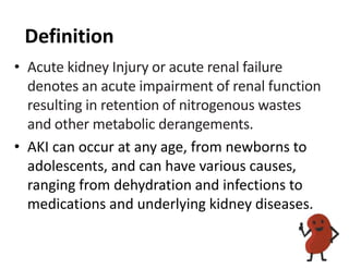
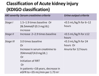
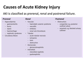
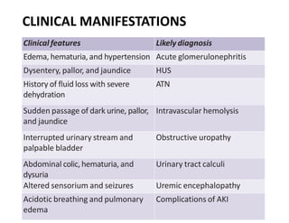
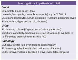
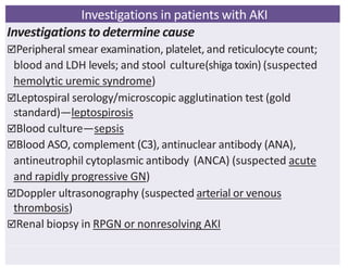
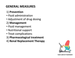
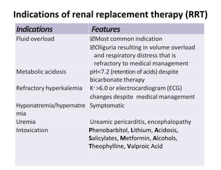
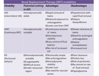
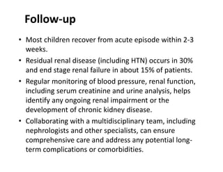
Acute Kidney Injury (AKI)
Definition
• Acute kidney Injury or acute renal failure
denotes an acute impairment of renal function
resulting in retention of nitrogenous wastes
and other metabolic derangements.
• AKI can occur at any age, from newborns to
adolescents, and can have various causes,
ranging from dehydration and infections to
medications and underlying kidney diseases.
AKI severity Serum creatinine criteria Urine output criteria
Stage I 1.5–1.9 times baseline Or
26.5mmol/l (0.3 mg/dL)
increase
<0.5 mL/kg/h for 6–12
hours
Stage II Increase 2–2.9 times baseline <0.5 mL/kg/h for ≥12
hours
Stage III 3.0 times baseline
Or
Increase in serum creatinine to
354mmol/l(4.0mg/dL )
Or
Initiation of RRT
Or
In patients <18 years, decrease in
eGFR to <35 mL/min per 1.73 m2
<0.3 mL/kg/h for 24
hours Or
Anuria for 12 hours
Classification of Acute kidney injury
(KDIGO classification)
AKI is classified as prerenal, renal and postrenal failure.
Causes of Acute Kidney Injury
Clinical features Likelydiagnosis
Edema, hematuria,and hypertension Acute glomerulonephritis
Dysentery, pallor, and jaundice HUS
History of fluid loss with severe
dehydration
ATN
Sudden passage of dark urine, pallor,
and jaundice
Intravascular hemolysis
Interrupted urinary stream and
palpable bladder
Obstructive uropathy
Abdominal colic,hematuria, and
dysuria
Urinary tract calculi
Altered sensorium and seizures Uremic encephalopathy
Acidotic breathing and pulmonary
edema
Complications of AKI
CLINICAL MANIFESTATIONS
Investigations in patients with AKI
Blood
Complete blood counts (any
anemia,leucopenia,thromobocytopenia) e.g. In SLE/HUS
Urea and Electrolytes/Serum Creatinine + Calcium, phosphate levels
Venous blood gas (pH and bicarbonate)
Urine
Urinalysis; culture (if symptoms of urinary infection)
Sodium, osmolality, fractional excretion of sodium (if available to
differentiate prerenal from intrinsic AKI)
Radiology
Chest X-ray (for fluid overload and cardiomegaly)
Ultrasonography (identify obstruction and dilatation)
ECG for hyperkalemia (peaked T waves,wide QRS complex)
Investigations in patients with AKI
Investigations to determine cause
Peripheral smear examination, platelet, and reticulocyte count;
blood and LDH levels; and stool culture(shiga toxin) (suspected
hemolytic uremic syndrome)
Leptospiral serology/microscopic agglutination test (gold
standard)—leptospirosis
Blood culture—sepsis
Blood ASO, complement (C3), antinuclear antibody (ANA),
antineutrophil cytoplasmic antibody (ANCA) (suspected acute
and rapidly progressive GN)
Doppler ultrasonography (suspected arterial or venous
thrombosis)
Renal biopsy in RPGN or nonresolving AKI
GENERAL MEASURES
1) Prevention
• Fluid administration
• Adjustment of drug dosing
2) Management
• Fluid management
• Nutritional support
• Treat complications
3) Pharmacological treatment
4) Renal Replacement Therapy
Prevention of AKI
• Fluid administration
Adequate hydration is essential, particularly during
periods of illness or high-risk situations (e.g., surgery),
Prerenal AKI due to hypovolemia – Administration of IV
fluid bolus with N/S(10-20ml/kg over 30 minutes) may
prevent more severe intrinsic AKI.
• Adjustment of drug dosing
Drugs that increase severity of renal damage, delay
recovery of renal function or reduce renal perfusion, e.g.
aminoglycosides, radiocontrast media, NSAIDs,
amphotericin B, ACE inhibitors and indomethacin should
be avoided.
Fluid management
• Depends on volume status, urine output and extra-renal losses.
• Never use a potassium-containing solution in an anuric patient.
• Only use parenteral fluids if oral intake is not possible.
• Fluid management according to fluid status:
• Insensible water loss is calculated as:
• Neonates and young babies: 30 - 40 mL/kg/day
• Older children: 25 mL/kg/day (400 mL/m2/day)
• Normally hydrated plus normal urine output: Give normal fluid intake.
• Normally hydrated + oliguria: Oral fluid intake to replace insensible water loss
plus urine output of previous 24 hours
• Hydrated anuric patient without extra-renal fluid losses: Oral fluid to replace
insensible water losses only.
• Dehydrated, oliguric and ongoing extra-renal fluid losses: Replace fluid losses
with an appropriate solution which mirrors losses e.g.:
for diarrhoea: IV or oral rehydration solution;
for vomiting/gastric fluid losses: sodium chloride 0.9%/dextrose 5%.
• Pulmonary oedema + oliguria/anuria: Do not give fluid.
Nutritional support
• Patients with AKI are usually catabolic and have increased metabolic
needs. Adequate nutritional support is desirable with maximization
of caloric intake.
• However, volume restriction necessary during the oliguric phase
often imposes limitations. A diet containing 0.8–1.2 g/kg of
protein in infants and 0.6–0.8 g/ kg in older children and a
minimum of 50–60 Cal/kg should be given.
• The latter requirement can be met by adding liberal amounts of
carbohydrates and fats to the diet. Once dialysis is initiated, dietary
fluid and electrolyte restrictions can be made more liberal.
Management ofcomplications
Complications Management Remarks
Fluid overload  Fluid restriction—insensible losses to be
replaced by 5% Dextrose
 Urine Output to be replaced by NS
 Replace other losses
 Consider dialysis
 0.5–1% weight loss per day
Pulmonary
edema
 Oxygen ; IV furosemide (2–4 mg/kg)  Chest X-ray
 IVC assessment and ejection fraction
 Monitor by CVP line
 Dialysis (for fluid removal)
Hypertension  Symptomatic
–Labetalol infusion @ 0.25–1 mg/kg/h
–IV furosemide 2–4 mg/kg
 Asymptomatic—oral nifedipine/
amlodipine 0.3–0.5 mg/kg
 SodiumNitroprusside—0.5–8 µg/kg/min
(not to be given for >48 hours due to
risk of toxicity)
 Maintenance with amlodipine/
hydralazine
Metabolic
acidosis
IV or oral Sodium Bicarbonate To monitor for fluid overload and
hypernatremia
Hyperkalemia  Stopallpotassium inIVFandmedications
 IV 10% Ca gluconate 1 mL/kg over 5–10
minutes
 Salbutamol (2.5–5 mg) nebulization.
 IV Dextrose (10%) 0.5–1 g/kg and insulin
0.1–0.2 U/kg over 30 minutes.
 Sodium Bicarbonate (7.5%) —1–2 mL/kg over
15–20 minutes
 Calcium or sodium resonium (karyexalate)
1g/kg
 Monitor blood glucose for
hypoglycemia
 Continuous ECG monitoring
 Repeat potassium level
Anemia Packed Red cells transfusion at 5–10 mL/kg Monitor bp, fluid overload
Hyper-
phosphatemia
Phosphate binders such as calcium carbonate Avoid milk pdts
Pharmacological
• The KDIGO guideline recommend N-acetylcysteine as prophylaxis
to prevent contrast-induced AKI in high-risk patients. However,
two large, well-designed studies reported no benefit of NAC in
reducing the incidence of contrast-induced AKI.
• Diuretics are not recommended for the prevention of AKI
because their use does not alter outcomes in those with
established AKI. Diuretics should be used only to control fluid
overload.
• Low-dose dopamine causes renal vasodilatation and may induce
a modest natriuresis and diuresis. However, it has no beneficial
effect on the outcome of AKI, and may be associated with
transient tachyarrhythmia or tissue ischemia.
• The role of other medications, including fenoldopam, atrial
natriuretic peptide, calcium channel blockers and other
medications is investigational. Mannitol is not recommended for
children.
Indications of renal replacement therapy (RRT)
Indications Features
Fluid overload Most common indication
Oliguria resulting in volume overload
and respiratory distress that is
refractory to medical management
Metabolic acidosis pH<7.2 (retention of acids) despite
bicarbonate therapy
Refractory hyperkalemia K+
>6.0 or electrocardiogram (ECG)
changes despite medical management
Hyponatremia/hypernatre
mia
Symptomatic
Uremia Ureamic pericarditis, encephalopathy
Intoxication Phenobarbitol, Lithium, Acidosis,
Salicylates, Metformin, Alcohols,
Theophylline, Valproic Acid
Renal Replacement Therapy (RRT) modalities
Modality Potential setting
inAKI
Advantages Disadvantages
IHD
(intermittent HD)
Hemodynamically
stable
Rapid removal of
toxins
Reduced exposure to
anticoagulants
Lower cost than CRRT
Hypotension with
rapid fluid removal
Dialysis
disequilibrium
CRRT
(Continuous RRT)
Hemodynamically
unstable
Continuous removal
of toxins
Hemodynamic
stability
Easy control of fluid
balance
No risk of increased
ICP
Slower clearance of
toxins
Need for prolonged
anticoagulation
Patient
immobilization
Increased cost
PD
(Peritoneal
dialysis)
Hemodynamically
unstable
Coagulopathy
Difficult access
Under resourced
region
Technically simple
No anticoagulation
Hemodynamic
stability
Lower cost
No need for vascular
access
Poor clearance
Protein loss
Risk of peritonitis
No control on rate
of fluid removal
Hyperglycemia
Follow-up
• Most children recover from acute episode within 2-3
weeks.
• Residual renal disease (including HTN) occurs in 30%
and end stage renal failure in about 15% of patients.
• Regular monitoring of blood pressure, renal function,
including serum creatinine and urine analysis, helps
identify any ongoing renal impairment or the
development of chronic kidney disease.
• Collaborating with a multidisciplinary team, including
nephrologists and other specialists, can ensure
comprehensive care and address any potential long-
term complications or comorbidities.
THANK YOU

AKI in children

  • 1.
  • 2.
    Definition • Acute kidneyInjury or acute renal failure denotes an acute impairment of renal function resulting in retention of nitrogenous wastes and other metabolic derangements. • AKI can occur at any age, from newborns to adolescents, and can have various causes, ranging from dehydration and infections to medications and underlying kidney diseases.
  • 3.
    AKI severity Serumcreatinine criteria Urine output criteria Stage I 1.5–1.9 times baseline Or 26.5mmol/l (0.3 mg/dL) increase <0.5 mL/kg/h for 6–12 hours Stage II Increase 2–2.9 times baseline <0.5 mL/kg/h for ≥12 hours Stage III 3.0 times baseline Or Increase in serum creatinine to 354mmol/l(4.0mg/dL ) Or Initiation of RRT Or In patients <18 years, decrease in eGFR to <35 mL/min per 1.73 m2 <0.3 mL/kg/h for 24 hours Or Anuria for 12 hours Classification of Acute kidney injury (KDIGO classification)
  • 4.
    AKI is classifiedas prerenal, renal and postrenal failure. Causes of Acute Kidney Injury
  • 5.
    Clinical features Likelydiagnosis Edema,hematuria,and hypertension Acute glomerulonephritis Dysentery, pallor, and jaundice HUS History of fluid loss with severe dehydration ATN Sudden passage of dark urine, pallor, and jaundice Intravascular hemolysis Interrupted urinary stream and palpable bladder Obstructive uropathy Abdominal colic,hematuria, and dysuria Urinary tract calculi Altered sensorium and seizures Uremic encephalopathy Acidotic breathing and pulmonary edema Complications of AKI CLINICAL MANIFESTATIONS
  • 6.
    Investigations in patientswith AKI Blood Complete blood counts (any anemia,leucopenia,thromobocytopenia) e.g. In SLE/HUS Urea and Electrolytes/Serum Creatinine + Calcium, phosphate levels Venous blood gas (pH and bicarbonate) Urine Urinalysis; culture (if symptoms of urinary infection) Sodium, osmolality, fractional excretion of sodium (if available to differentiate prerenal from intrinsic AKI) Radiology Chest X-ray (for fluid overload and cardiomegaly) Ultrasonography (identify obstruction and dilatation) ECG for hyperkalemia (peaked T waves,wide QRS complex)
  • 7.
    Investigations in patientswith AKI Investigations to determine cause Peripheral smear examination, platelet, and reticulocyte count; blood and LDH levels; and stool culture(shiga toxin) (suspected hemolytic uremic syndrome) Leptospiral serology/microscopic agglutination test (gold standard)—leptospirosis Blood culture—sepsis Blood ASO, complement (C3), antinuclear antibody (ANA), antineutrophil cytoplasmic antibody (ANCA) (suspected acute and rapidly progressive GN) Doppler ultrasonography (suspected arterial or venous thrombosis) Renal biopsy in RPGN or nonresolving AKI
  • 8.
    GENERAL MEASURES 1) Prevention •Fluid administration • Adjustment of drug dosing 2) Management • Fluid management • Nutritional support • Treat complications 3) Pharmacological treatment 4) Renal Replacement Therapy
  • 9.
    Prevention of AKI •Fluid administration Adequate hydration is essential, particularly during periods of illness or high-risk situations (e.g., surgery), Prerenal AKI due to hypovolemia – Administration of IV fluid bolus with N/S(10-20ml/kg over 30 minutes) may prevent more severe intrinsic AKI. • Adjustment of drug dosing Drugs that increase severity of renal damage, delay recovery of renal function or reduce renal perfusion, e.g. aminoglycosides, radiocontrast media, NSAIDs, amphotericin B, ACE inhibitors and indomethacin should be avoided.
  • 10.
    Fluid management • Dependson volume status, urine output and extra-renal losses. • Never use a potassium-containing solution in an anuric patient. • Only use parenteral fluids if oral intake is not possible. • Fluid management according to fluid status: • Insensible water loss is calculated as: • Neonates and young babies: 30 - 40 mL/kg/day • Older children: 25 mL/kg/day (400 mL/m2/day) • Normally hydrated plus normal urine output: Give normal fluid intake. • Normally hydrated + oliguria: Oral fluid intake to replace insensible water loss plus urine output of previous 24 hours • Hydrated anuric patient without extra-renal fluid losses: Oral fluid to replace insensible water losses only. • Dehydrated, oliguric and ongoing extra-renal fluid losses: Replace fluid losses with an appropriate solution which mirrors losses e.g.: for diarrhoea: IV or oral rehydration solution; for vomiting/gastric fluid losses: sodium chloride 0.9%/dextrose 5%. • Pulmonary oedema + oliguria/anuria: Do not give fluid.
  • 11.
    Nutritional support • Patientswith AKI are usually catabolic and have increased metabolic needs. Adequate nutritional support is desirable with maximization of caloric intake. • However, volume restriction necessary during the oliguric phase often imposes limitations. A diet containing 0.8–1.2 g/kg of protein in infants and 0.6–0.8 g/ kg in older children and a minimum of 50–60 Cal/kg should be given. • The latter requirement can be met by adding liberal amounts of carbohydrates and fats to the diet. Once dialysis is initiated, dietary fluid and electrolyte restrictions can be made more liberal.
  • 12.
    Management ofcomplications Complications ManagementRemarks Fluid overload  Fluid restriction—insensible losses to be replaced by 5% Dextrose  Urine Output to be replaced by NS  Replace other losses  Consider dialysis  0.5–1% weight loss per day Pulmonary edema  Oxygen ; IV furosemide (2–4 mg/kg)  Chest X-ray  IVC assessment and ejection fraction  Monitor by CVP line  Dialysis (for fluid removal) Hypertension  Symptomatic –Labetalol infusion @ 0.25–1 mg/kg/h –IV furosemide 2–4 mg/kg  Asymptomatic—oral nifedipine/ amlodipine 0.3–0.5 mg/kg  SodiumNitroprusside—0.5–8 µg/kg/min (not to be given for >48 hours due to risk of toxicity)  Maintenance with amlodipine/ hydralazine Metabolic acidosis IV or oral Sodium Bicarbonate To monitor for fluid overload and hypernatremia Hyperkalemia  Stopallpotassium inIVFandmedications  IV 10% Ca gluconate 1 mL/kg over 5–10 minutes  Salbutamol (2.5–5 mg) nebulization.  IV Dextrose (10%) 0.5–1 g/kg and insulin 0.1–0.2 U/kg over 30 minutes.  Sodium Bicarbonate (7.5%) —1–2 mL/kg over 15–20 minutes  Calcium or sodium resonium (karyexalate) 1g/kg  Monitor blood glucose for hypoglycemia  Continuous ECG monitoring  Repeat potassium level Anemia Packed Red cells transfusion at 5–10 mL/kg Monitor bp, fluid overload Hyper- phosphatemia Phosphate binders such as calcium carbonate Avoid milk pdts
  • 13.
    Pharmacological • The KDIGOguideline recommend N-acetylcysteine as prophylaxis to prevent contrast-induced AKI in high-risk patients. However, two large, well-designed studies reported no benefit of NAC in reducing the incidence of contrast-induced AKI. • Diuretics are not recommended for the prevention of AKI because their use does not alter outcomes in those with established AKI. Diuretics should be used only to control fluid overload. • Low-dose dopamine causes renal vasodilatation and may induce a modest natriuresis and diuresis. However, it has no beneficial effect on the outcome of AKI, and may be associated with transient tachyarrhythmia or tissue ischemia. • The role of other medications, including fenoldopam, atrial natriuretic peptide, calcium channel blockers and other medications is investigational. Mannitol is not recommended for children.
  • 14.
    Indications of renalreplacement therapy (RRT) Indications Features Fluid overload Most common indication Oliguria resulting in volume overload and respiratory distress that is refractory to medical management Metabolic acidosis pH<7.2 (retention of acids) despite bicarbonate therapy Refractory hyperkalemia K+ >6.0 or electrocardiogram (ECG) changes despite medical management Hyponatremia/hypernatre mia Symptomatic Uremia Ureamic pericarditis, encephalopathy Intoxication Phenobarbitol, Lithium, Acidosis, Salicylates, Metformin, Alcohols, Theophylline, Valproic Acid
  • 15.
    Renal Replacement Therapy(RRT) modalities Modality Potential setting inAKI Advantages Disadvantages IHD (intermittent HD) Hemodynamically stable Rapid removal of toxins Reduced exposure to anticoagulants Lower cost than CRRT Hypotension with rapid fluid removal Dialysis disequilibrium CRRT (Continuous RRT) Hemodynamically unstable Continuous removal of toxins Hemodynamic stability Easy control of fluid balance No risk of increased ICP Slower clearance of toxins Need for prolonged anticoagulation Patient immobilization Increased cost PD (Peritoneal dialysis) Hemodynamically unstable Coagulopathy Difficult access Under resourced region Technically simple No anticoagulation Hemodynamic stability Lower cost No need for vascular access Poor clearance Protein loss Risk of peritonitis No control on rate of fluid removal Hyperglycemia
  • 16.
    Follow-up • Most childrenrecover from acute episode within 2-3 weeks. • Residual renal disease (including HTN) occurs in 30% and end stage renal failure in about 15% of patients. • Regular monitoring of blood pressure, renal function, including serum creatinine and urine analysis, helps identify any ongoing renal impairment or the development of chronic kidney disease. • Collaborating with a multidisciplinary team, including nephrologists and other specialists, can ensure comprehensive care and address any potential long- term complications or comorbidities.
  • 17.