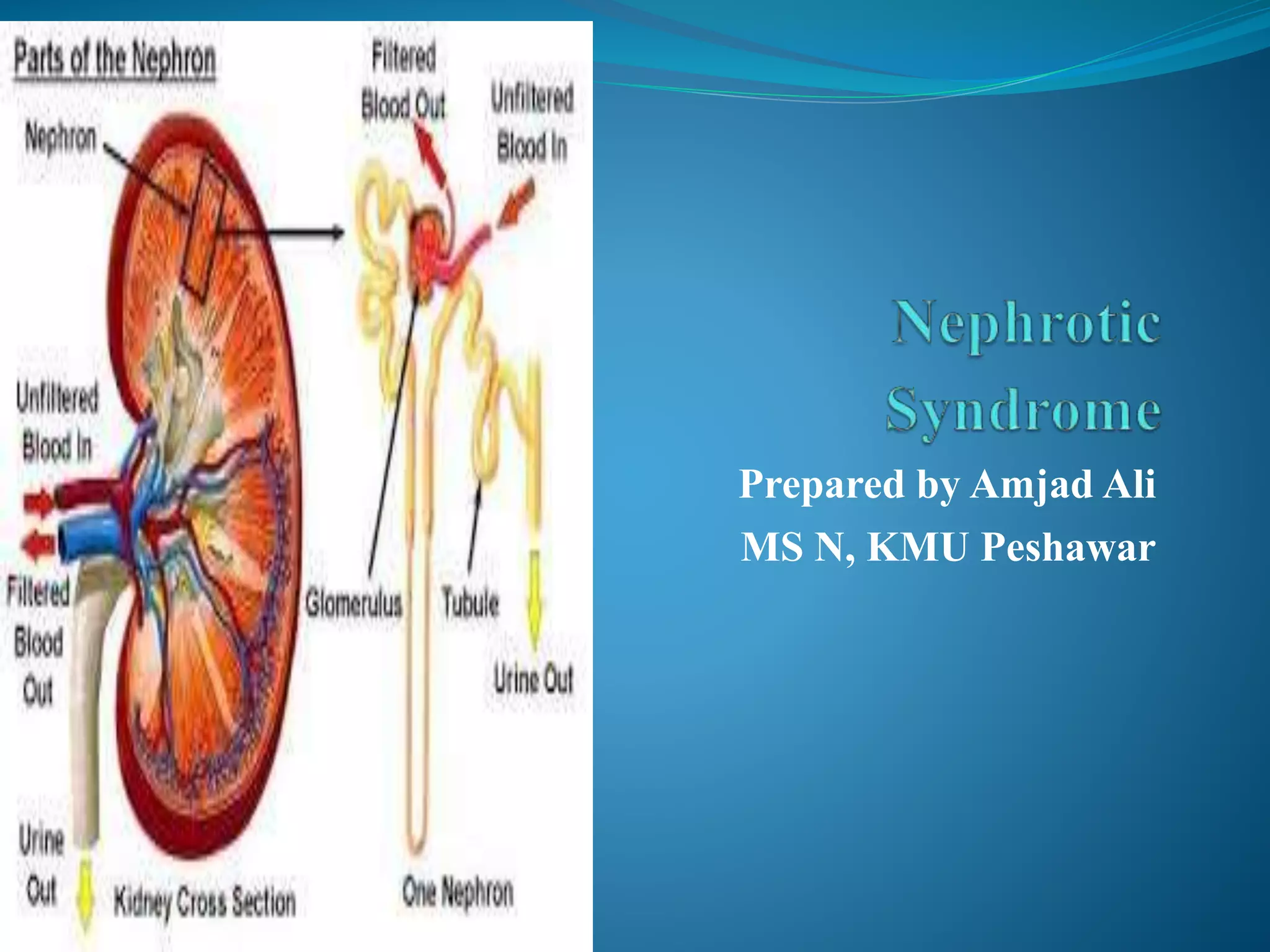
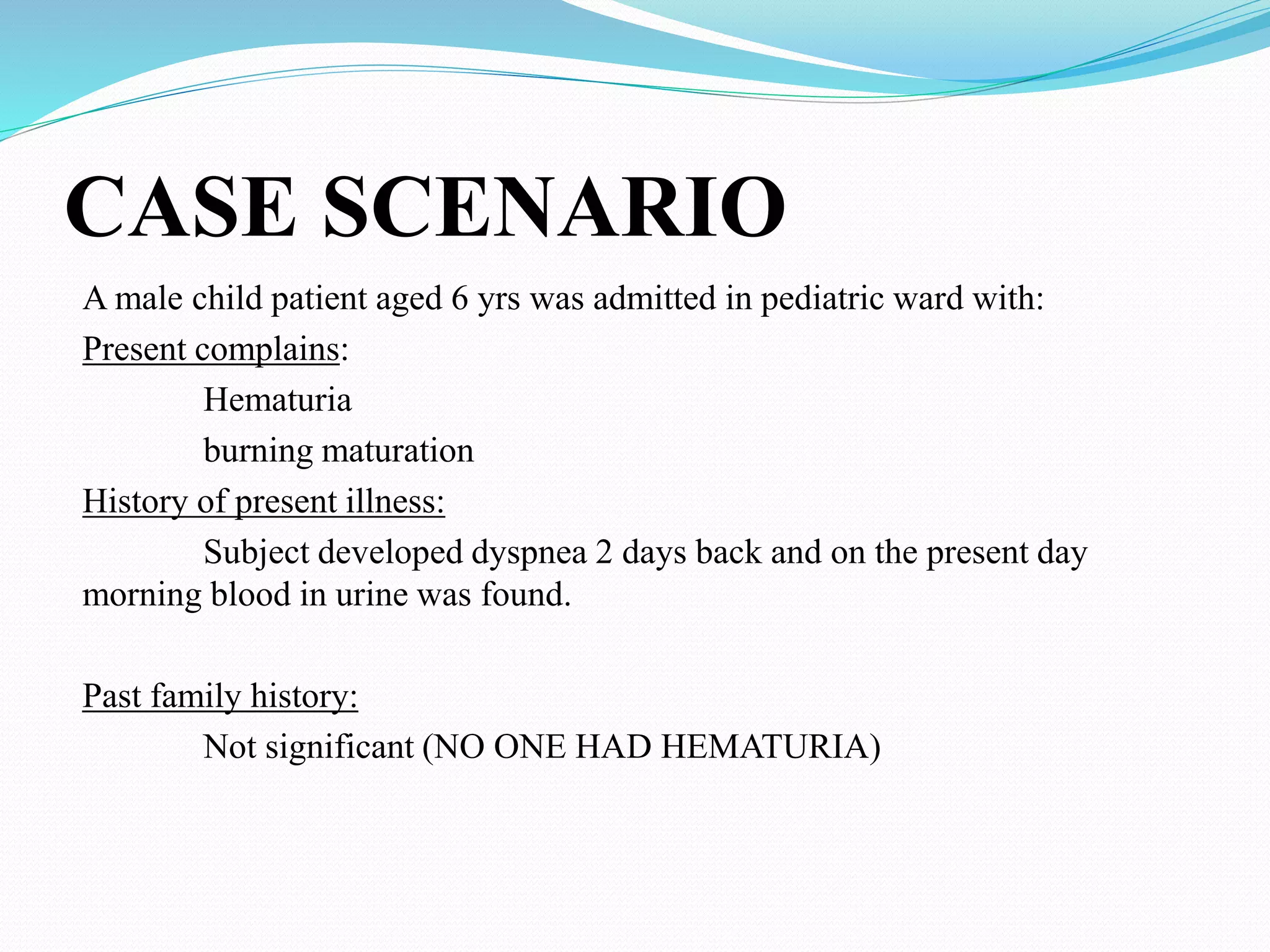
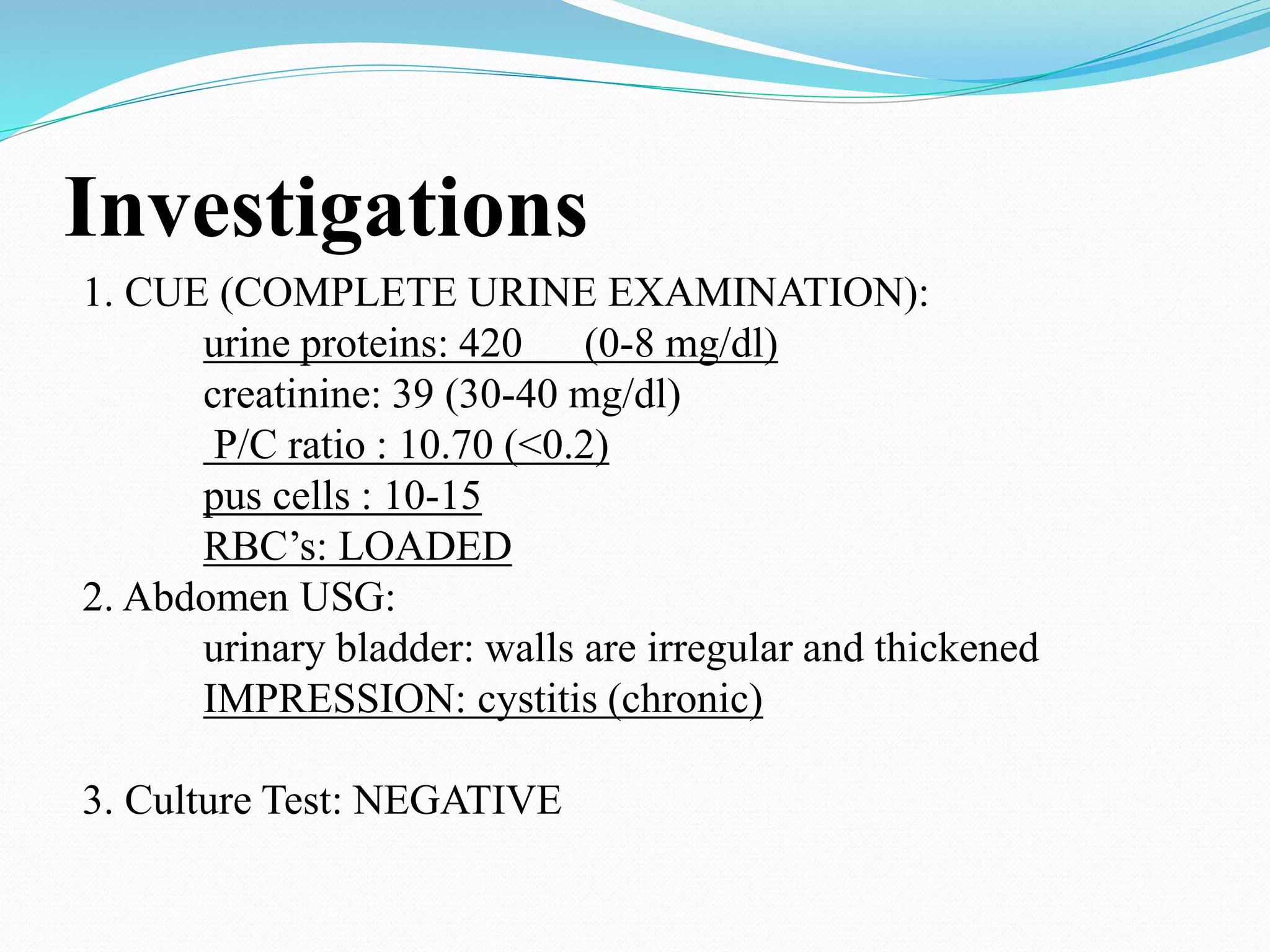
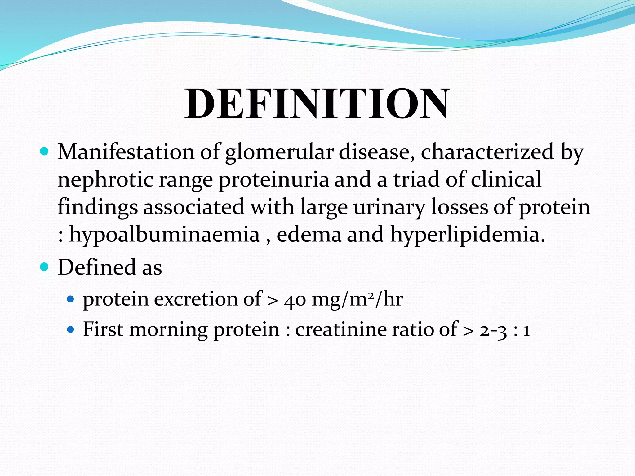
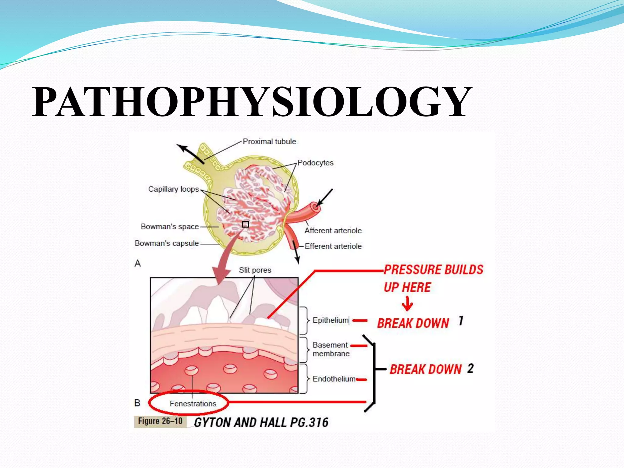
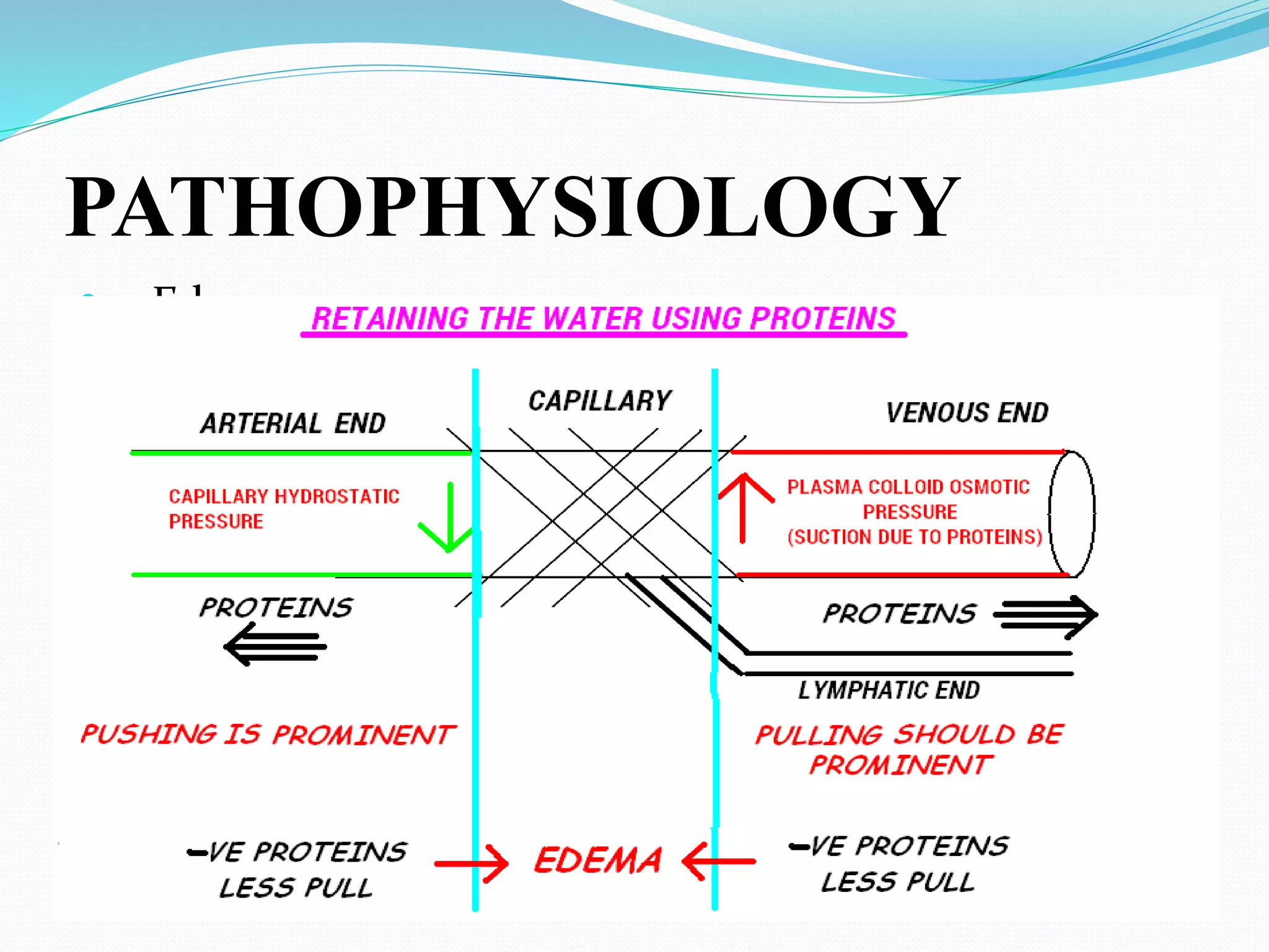
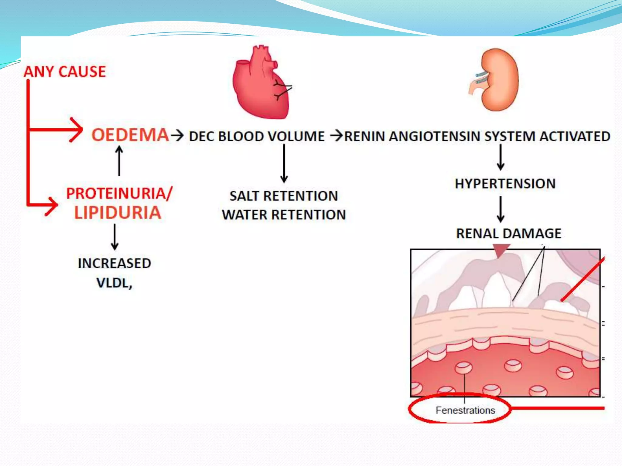
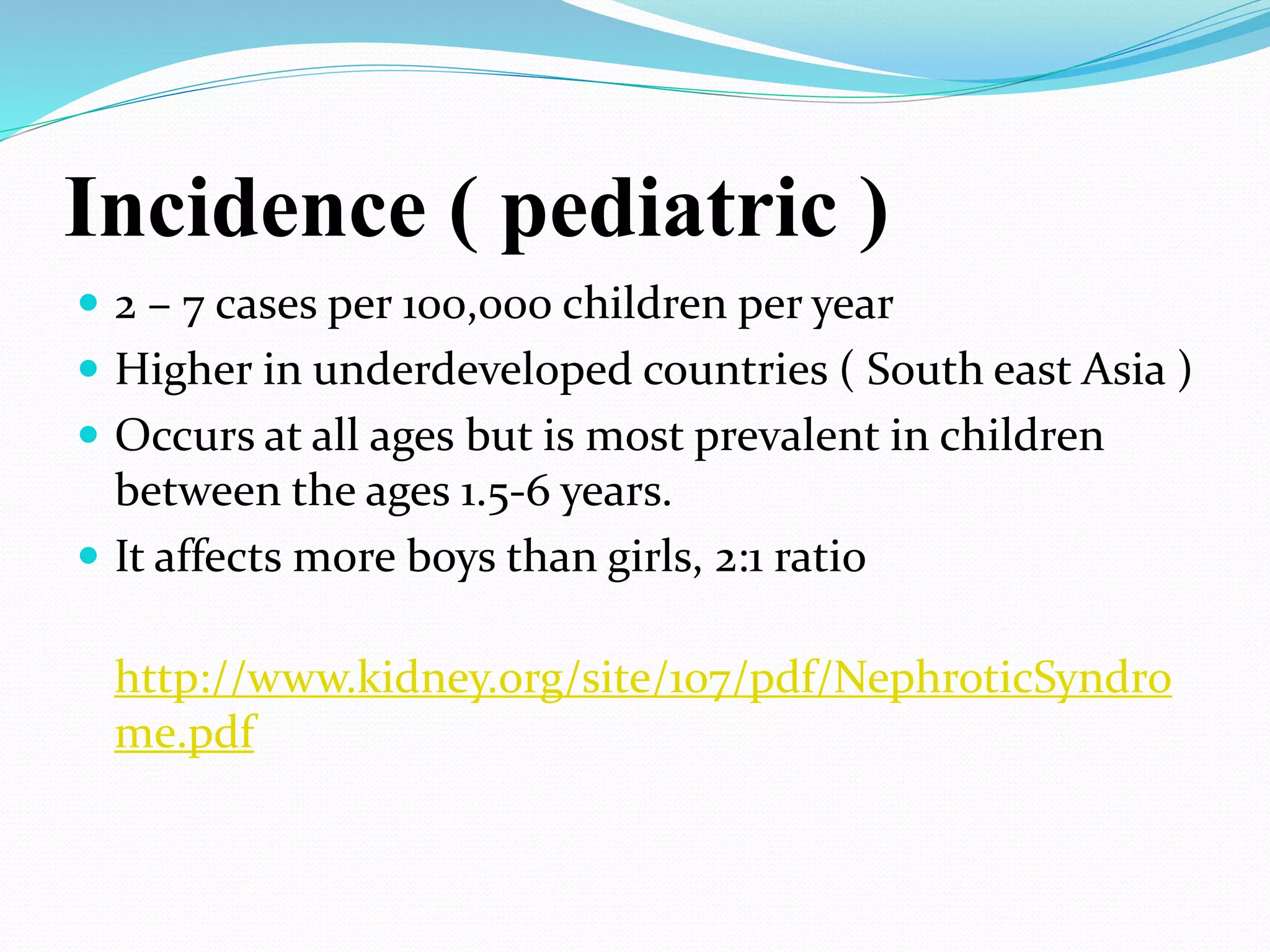
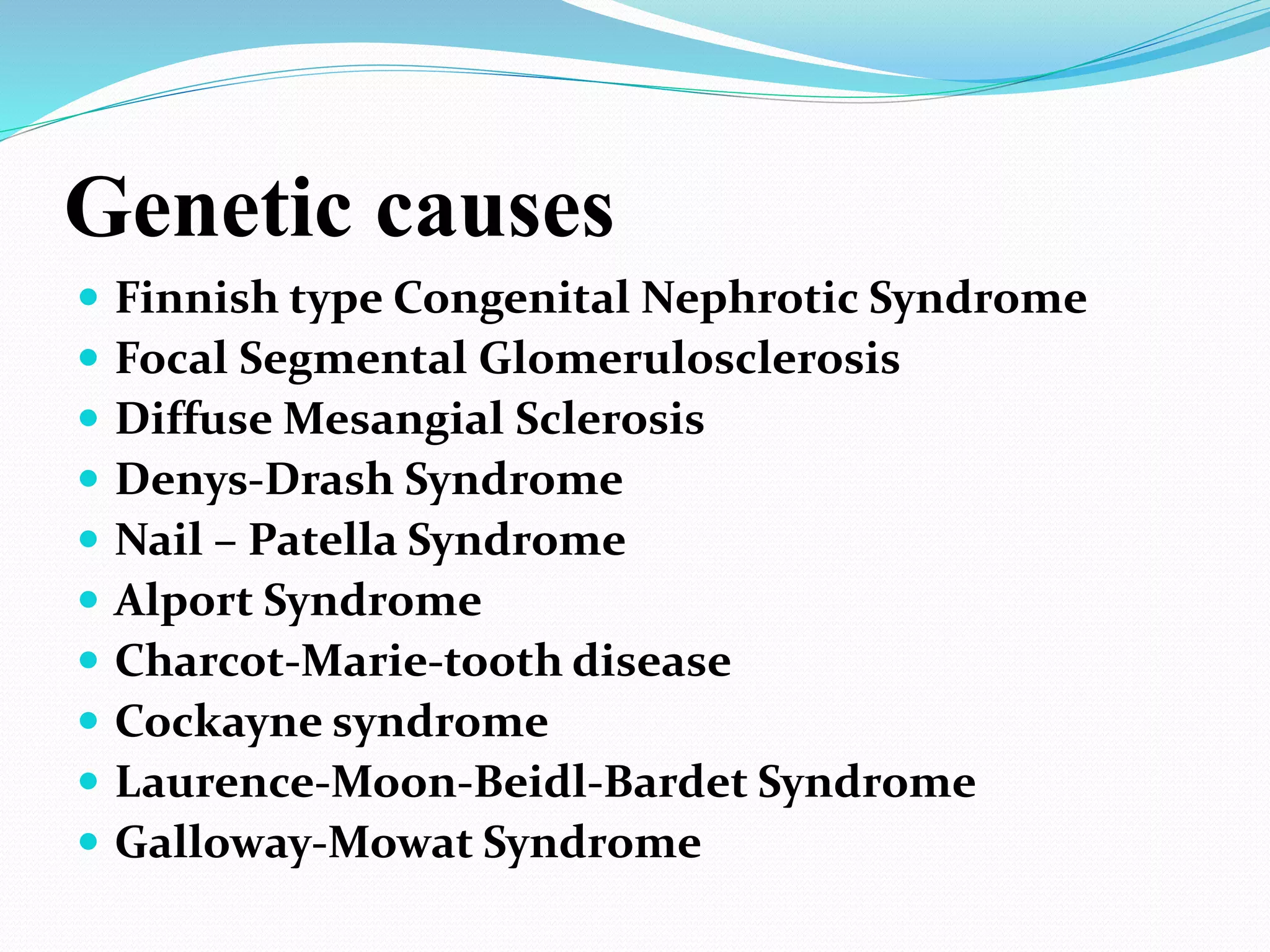
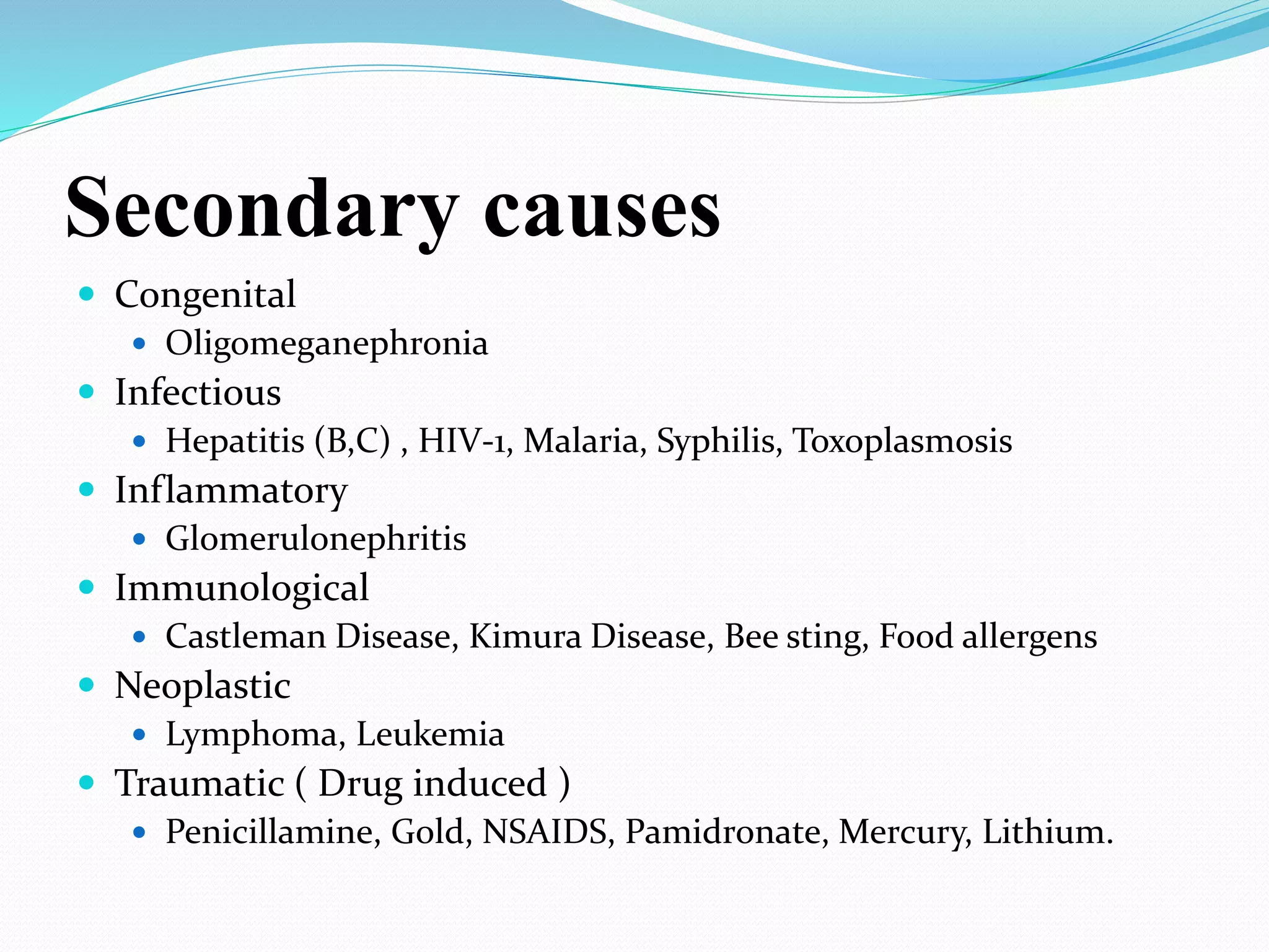
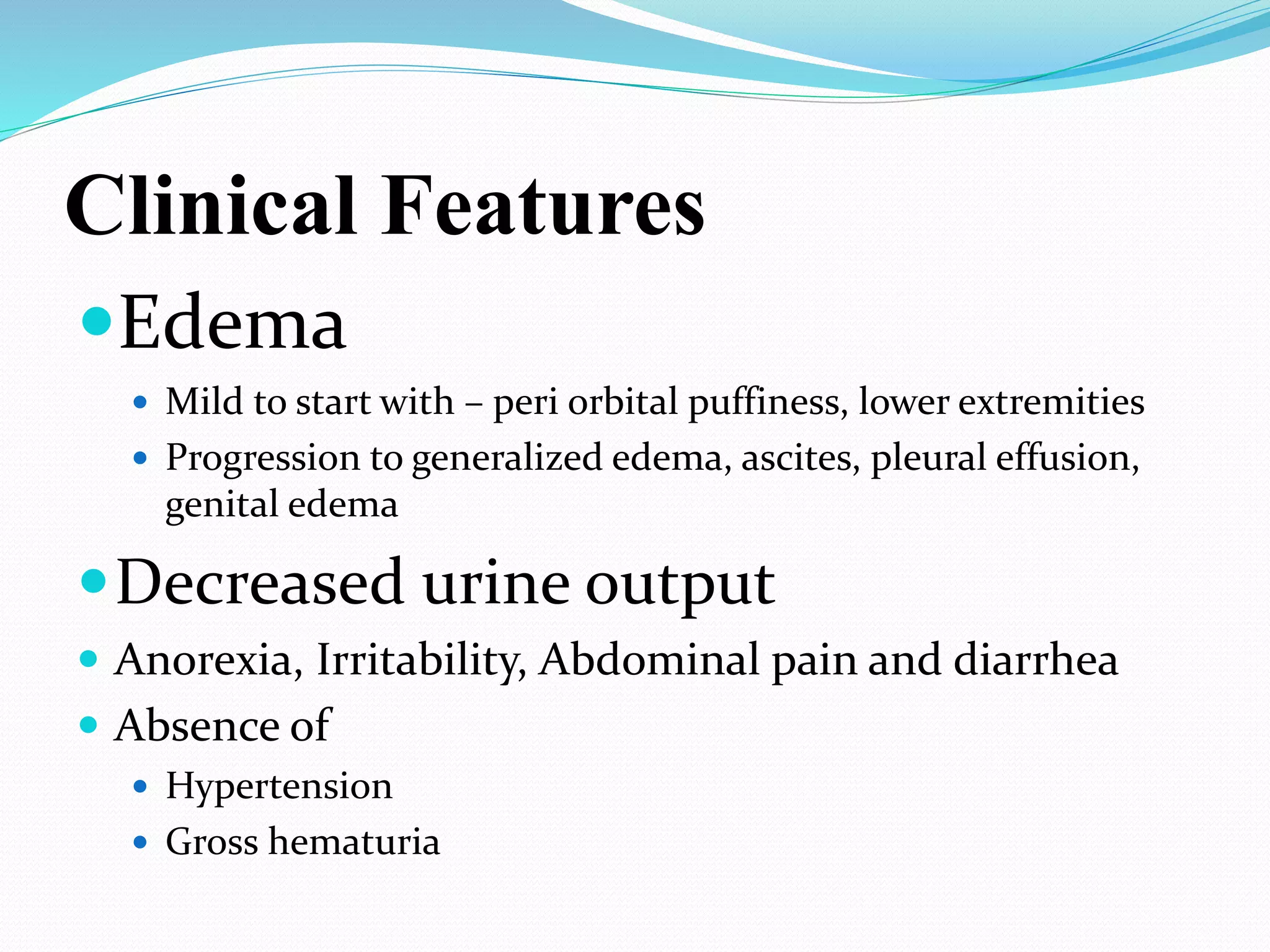
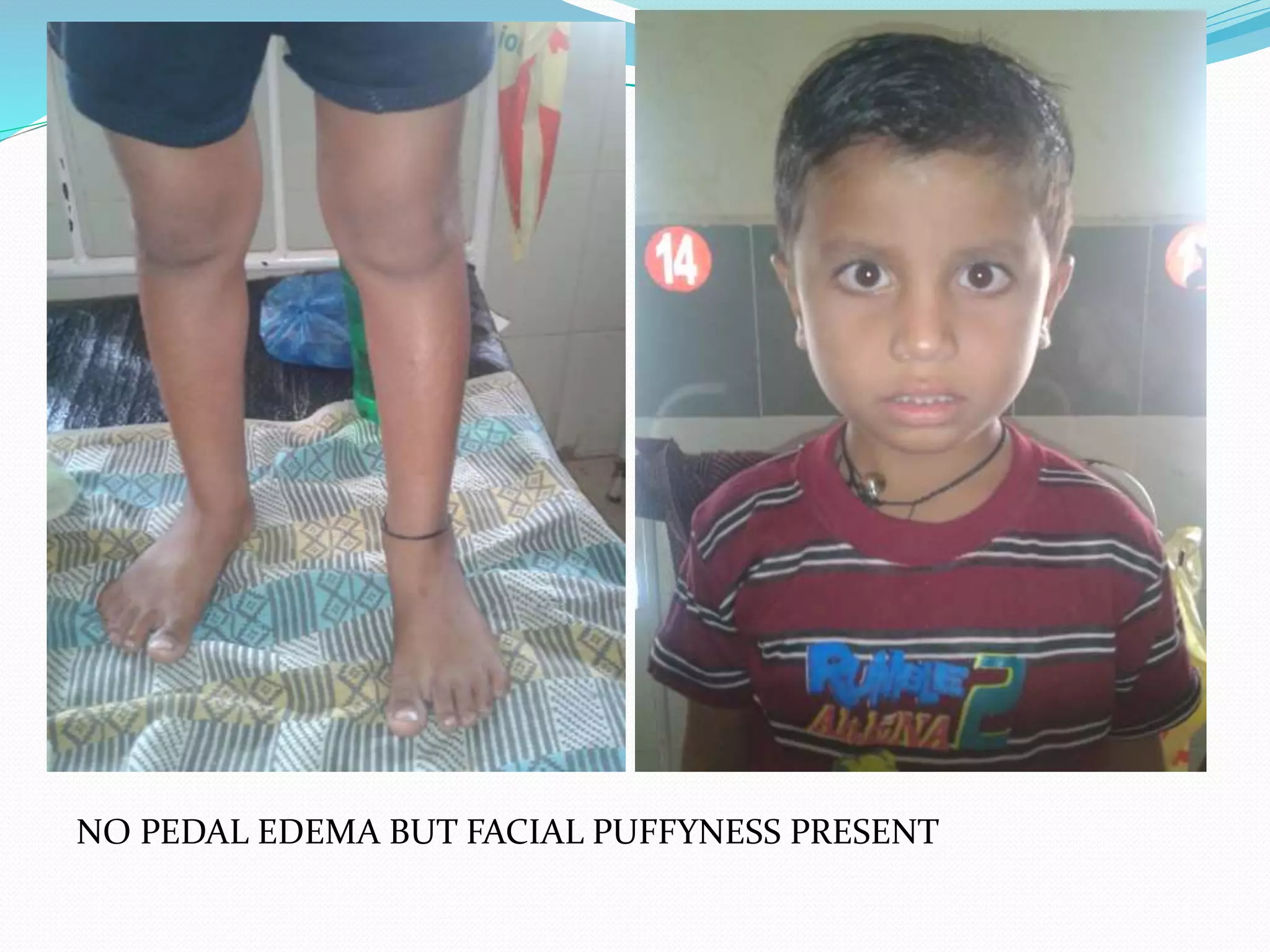
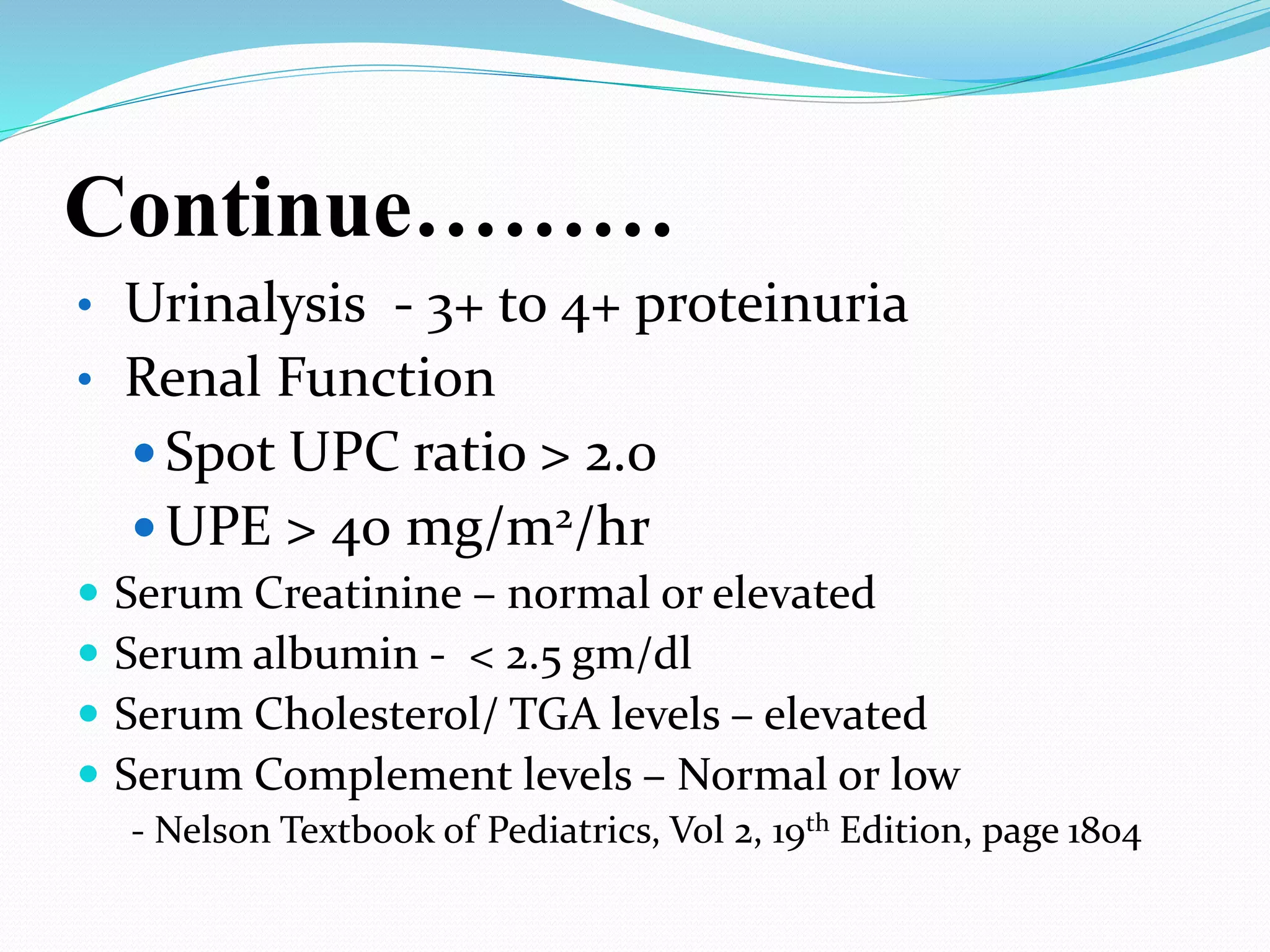
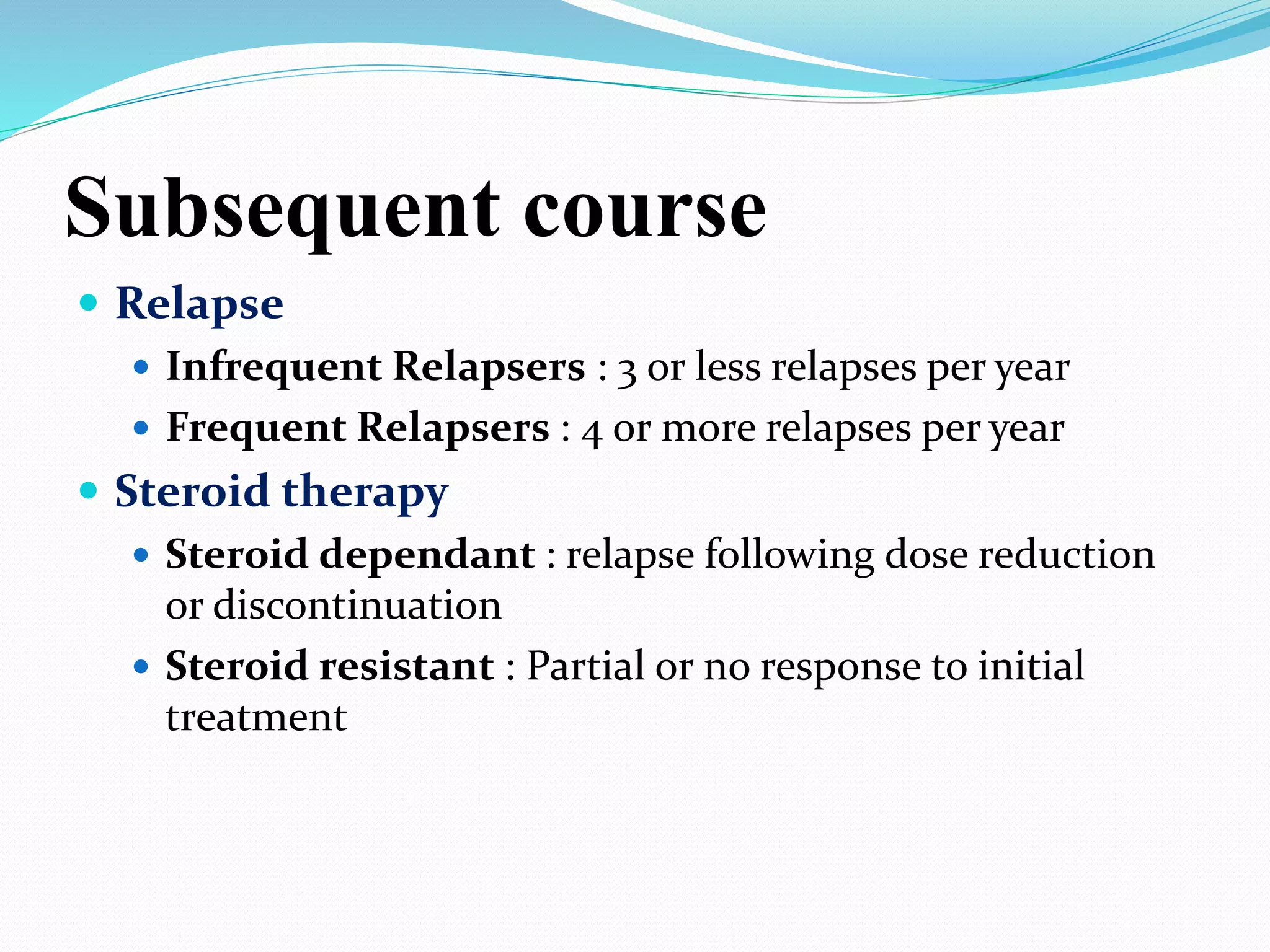
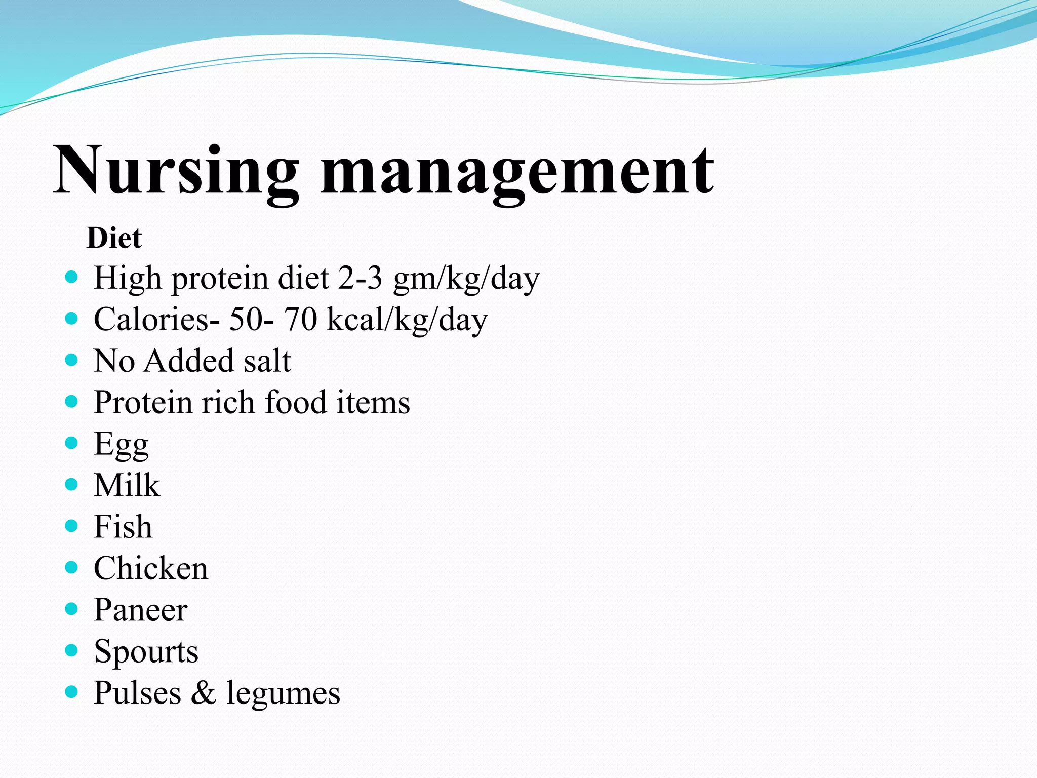
Prepared by Amjad Ali
MS N, KMU Peshawar
CASE SCENARIO
A male child patient aged 6 yrs was admitted in pediatric ward with:
Present complains:
Hematuria
burning maturation
History of present illness:
Subject developed dyspnea 2 days back and on the present day
morning blood in urine was found.
Past family history:
Not significant (NO ONE HAD HEMATURIA)
Investigations
1. CUE (COMPLETE URINE EXAMINATION):
urine proteins: 420 (0-8 mg/dl)
creatinine: 39 (30-40 mg/dl)
P/C ratio : 10.70 (<0.2)
pus cells : 10-15
RBC’s: LOADED
2. Abdomen USG:
urinary bladder: walls are irregular and thickened
IMPRESSION: cystitis (chronic)
3. Culture Test: NEGATIVE
DEFINITION
 Manifestation of glomerular disease, characterized by
nephrotic range proteinuria and a triad of clinical
findings associated with large urinary losses of protein
: hypoalbuminaemia , edema and hyperlipidemia.
 Defined as
 protein excretion of > 40 mg/m2/hr
 First morning protein : creatinine ratio of > 2-3 : 1
PATHOPHYSIOLOGY
PATHOPHYSIOLOGY
 1. Edema
Incidence ( pediatric )
 2 – 7 cases per 100,000 children per year
 Higher in underdeveloped countries ( South east Asia )
 Occurs at all ages but is most prevalent in children
between the ages 1.5-6 years.
 It affects more boys than girls, 2:1 ratio
http://www.kidney.org/site/107/pdf/NephroticSyndro
me.pdf
Etiology
 Genetic
 Secondary
 Idiopathic or Primary
Genetic causes
 Finnish type Congenital Nephrotic Syndrome
 Focal Segmental Glomerulosclerosis
 Diffuse Mesangial Sclerosis
 Denys-Drash Syndrome
 Nail – Patella Syndrome
 Alport Syndrome
 Charcot-Marie-tooth disease
 Cockayne syndrome
 Laurence-Moon-Beidl-Bardet Syndrome
 Galloway-Mowat Syndrome
Secondary causes
 Congenital
 Oligomeganephronia
 Infectious
 Hepatitis (B,C) , HIV-1, Malaria, Syphilis, Toxoplasmosis
 Inflammatory
 Glomerulonephritis
 Immunological
 Castleman Disease, Kimura Disease, Bee sting, Food allergens
 Neoplastic
 Lymphoma, Leukemia
 Traumatic ( Drug induced )
 Penicillamine, Gold, NSAIDS, Pamidronate, Mercury, Lithium.
Idiopathic
 Minimal Change disease ( >80 % )
 Mesangial proliferation
 Focal segmental Glomerulosclerosis
 Membranous Nephropathy
 Membranoproliferative glomerulonephritis
Clinical Features
Edema
 Mild to start with – peri orbital puffiness, lower extremities
 Progression to generalized edema, ascites, pleural effusion,
genital edema
Decreased urine output
 Anorexia, Irritability, Abdominal pain and diarrhea
 Absence of
 Hypertension
 Gross hematuria
NO PEDAL EDEMA BUT FACIAL PUFFYNESS PRESENT
DIFFERENTIALS
 Protein losing enteropathy
 Hepatic failure
 Heart failure
 Acute/Chronic Glomerulonephritis
 Protein Malnutrition
Lab Investigations
 Urine Examination
 Complete Blood Count & Blood picture
 Renal parameters :
 Spot Urine Protein : Creatinine ratio
 Urinary protein excretion
 protein selectivity ratio
 Liver Function Test
 Renal Biopsy ???
Continue………
• Urinalysis - 3+ to 4+ proteinuria
• Renal Function
 Spot UPC ratio > 2.0
 UPE > 40 mg/m2/hr
 Serum Creatinine – normal or elevated
 Serum albumin - < 2.5 gm/dl
 Serum Cholesterol/ TGA levels – elevated
 Serum Complement levels – Normal or low
- Nelson Textbook of Pediatrics, Vol 2, 19th Edition, page 1804
Additional Tests
 C3 and antistreptolysin
 Chest X ray and tuberculin test
 Hepatitis B surface antigen
Indications for Biopsy
 Age below 12 months
 Gross or persistent microscopic hematuria
 Low blood C3
 Hypertension
 Impaired renal Function
 Failure of steroid therapy
Management
(Initial Episode)
 High protein diet
 Salt moderation
 Treatment of infections
 If significant edema – diuretics Aldosterone
antagonist ( Fursemide, spironolactone )
 Corticosteroid therapy with Prednisolone or
prednisone
 ( 2mg/kg per day for 6 weeks followed by
1.5 mg/kg single morning dose on alternate days for
6 weeks )
Subsequent course
 Relapse
 Infrequent Relapsers : 3 or less relapses per year
 Frequent Relapsers : 4 or more relapses per year
 Steroid therapy
 Steroid dependant : relapse following dose reduction
or discontinuation
 Steroid resistant : Partial or no response to initial
treatment
Management of Relapse
 Parent Education
 Symptomatic therapy for infections in case of low
grade proteinuria
 Persistent proteinuria ( 3 - 4+ ) –
 Prednisolone
( 2mg/kg/day until protein is negative for 3 days )
1.5 mg/kg on alternate days for 4 weeks )
Ghai Essential Pediatrics,8th edition, page
479.
Complications
 Edema
 Infections
 Thrombotic complications
 Hypovolaemia and Acute renal Failure
 Steroid Toxicity
Nursing management
 Goals of nursing management
 Reducing edema
 Nutrition
 Administering medications
 Skin care
 Infection prevention
 Promoting psychosocial growth
 Parental teaching
Nursing management
Assessment
 Age, height and weight
 Vital signs
 Past hospital admissions
 Immunization status
 Edema
 Urinary output
 Presence of infections
 Dietary pattern
 Malnourishment
Nursing management
 Monitoring of Vital signs
 Intake output
 Urinary protein
 Diet
 Weight
 Abdominal girth
Nursing management
Diet
 High protein diet 2-3 gm/kg/day
 Calories- 50- 70 kcal/kg/day
 No Added salt
 Protein rich food items
 Egg
 Milk
 Fish
 Chicken
 Paneer
 Spourts
 Pulses & legumes
FLUID MANAGEMENT
 Strict intake output monitoring
 Hypovolemic symptoms
 persistent tachycardia
 Hypotension
 abdominal pain
 Capillary refill >2sec urine Na <10mmol/l being
indicative of severe hypovolaemia
 Previous day output + insensible water loss
 Urine output monitoring
Nursing management
 Immunization.
 Live vaccines should not be given to immunosuppressed
children.
 killed vaccines are safe to administer
 However live vaccine can be considered if the child is not
on steroids for minimum of 03 months.
 Recommended -varicella, pneumococcal vaccines during
remission.
References
 Lau, Keith, et al. "Steroid Responsive Nephrotic Syndrome
in IgA Nephropathy with FSGS." The
 Internet Journal of Nephrology 4 (2008): n. pag. Print.
 "Pediatric Nephrotic Syndrome." Pediatric Nephrotic
Syndrome. N.p., n.d. Web.
<http://emedicine.medscape.com>.
 USA. NIH. NIDDK. Childhood Nephrotic Syndrome. N.p.:
n.p., 2008. Print.
 Trachtman, Howard. “Common Diseases: Minimal Change
Nephrotic Syndrome.” Nephrology
 Self Assessment Program 11 (2012) 19-20. Print.
 Trachtman, Howard. “Common Diseases: Focal Segmental
Glomerulosclerosis.” Nephrology
Presentation on nephrotic syndrome

Presentation on nephrotic syndrome

  • 1.
    Prepared by AmjadAli MS N, KMU Peshawar
  • 2.
    CASE SCENARIO A malechild patient aged 6 yrs was admitted in pediatric ward with: Present complains: Hematuria burning maturation History of present illness: Subject developed dyspnea 2 days back and on the present day morning blood in urine was found. Past family history: Not significant (NO ONE HAD HEMATURIA)
  • 3.
    Investigations 1. CUE (COMPLETEURINE EXAMINATION): urine proteins: 420 (0-8 mg/dl) creatinine: 39 (30-40 mg/dl) P/C ratio : 10.70 (<0.2) pus cells : 10-15 RBC’s: LOADED 2. Abdomen USG: urinary bladder: walls are irregular and thickened IMPRESSION: cystitis (chronic) 3. Culture Test: NEGATIVE
  • 4.
    DEFINITION  Manifestation ofglomerular disease, characterized by nephrotic range proteinuria and a triad of clinical findings associated with large urinary losses of protein : hypoalbuminaemia , edema and hyperlipidemia.  Defined as  protein excretion of > 40 mg/m2/hr  First morning protein : creatinine ratio of > 2-3 : 1
  • 5.
  • 6.
  • 8.
    Incidence ( pediatric)  2 – 7 cases per 100,000 children per year  Higher in underdeveloped countries ( South east Asia )  Occurs at all ages but is most prevalent in children between the ages 1.5-6 years.  It affects more boys than girls, 2:1 ratio http://www.kidney.org/site/107/pdf/NephroticSyndro me.pdf
  • 9.
  • 10.
    Genetic causes  Finnishtype Congenital Nephrotic Syndrome  Focal Segmental Glomerulosclerosis  Diffuse Mesangial Sclerosis  Denys-Drash Syndrome  Nail – Patella Syndrome  Alport Syndrome  Charcot-Marie-tooth disease  Cockayne syndrome  Laurence-Moon-Beidl-Bardet Syndrome  Galloway-Mowat Syndrome
  • 11.
    Secondary causes  Congenital Oligomeganephronia  Infectious  Hepatitis (B,C) , HIV-1, Malaria, Syphilis, Toxoplasmosis  Inflammatory  Glomerulonephritis  Immunological  Castleman Disease, Kimura Disease, Bee sting, Food allergens  Neoplastic  Lymphoma, Leukemia  Traumatic ( Drug induced )  Penicillamine, Gold, NSAIDS, Pamidronate, Mercury, Lithium.
  • 12.
    Idiopathic  Minimal Changedisease ( >80 % )  Mesangial proliferation  Focal segmental Glomerulosclerosis  Membranous Nephropathy  Membranoproliferative glomerulonephritis
  • 13.
    Clinical Features Edema  Mildto start with – peri orbital puffiness, lower extremities  Progression to generalized edema, ascites, pleural effusion, genital edema Decreased urine output  Anorexia, Irritability, Abdominal pain and diarrhea  Absence of  Hypertension  Gross hematuria
  • 14.
    NO PEDAL EDEMABUT FACIAL PUFFYNESS PRESENT
  • 15.
    DIFFERENTIALS  Protein losingenteropathy  Hepatic failure  Heart failure  Acute/Chronic Glomerulonephritis  Protein Malnutrition
  • 16.
    Lab Investigations  UrineExamination  Complete Blood Count & Blood picture  Renal parameters :  Spot Urine Protein : Creatinine ratio  Urinary protein excretion  protein selectivity ratio  Liver Function Test  Renal Biopsy ???
  • 17.
    Continue……… • Urinalysis -3+ to 4+ proteinuria • Renal Function  Spot UPC ratio > 2.0  UPE > 40 mg/m2/hr  Serum Creatinine – normal or elevated  Serum albumin - < 2.5 gm/dl  Serum Cholesterol/ TGA levels – elevated  Serum Complement levels – Normal or low - Nelson Textbook of Pediatrics, Vol 2, 19th Edition, page 1804
  • 18.
    Additional Tests  C3and antistreptolysin  Chest X ray and tuberculin test  Hepatitis B surface antigen Indications for Biopsy  Age below 12 months  Gross or persistent microscopic hematuria  Low blood C3  Hypertension  Impaired renal Function  Failure of steroid therapy
  • 19.
    Management (Initial Episode)  Highprotein diet  Salt moderation  Treatment of infections  If significant edema – diuretics Aldosterone antagonist ( Fursemide, spironolactone )  Corticosteroid therapy with Prednisolone or prednisone  ( 2mg/kg per day for 6 weeks followed by 1.5 mg/kg single morning dose on alternate days for 6 weeks )
  • 20.
    Subsequent course  Relapse Infrequent Relapsers : 3 or less relapses per year  Frequent Relapsers : 4 or more relapses per year  Steroid therapy  Steroid dependant : relapse following dose reduction or discontinuation  Steroid resistant : Partial or no response to initial treatment
  • 21.
    Management of Relapse Parent Education  Symptomatic therapy for infections in case of low grade proteinuria  Persistent proteinuria ( 3 - 4+ ) –  Prednisolone ( 2mg/kg/day until protein is negative for 3 days ) 1.5 mg/kg on alternate days for 4 weeks ) Ghai Essential Pediatrics,8th edition, page 479.
  • 22.
    Complications  Edema  Infections Thrombotic complications  Hypovolaemia and Acute renal Failure  Steroid Toxicity
  • 23.
    Nursing management  Goalsof nursing management  Reducing edema  Nutrition  Administering medications  Skin care  Infection prevention  Promoting psychosocial growth  Parental teaching
  • 24.
    Nursing management Assessment  Age,height and weight  Vital signs  Past hospital admissions  Immunization status  Edema  Urinary output  Presence of infections  Dietary pattern  Malnourishment
  • 25.
    Nursing management  Monitoringof Vital signs  Intake output  Urinary protein  Diet  Weight  Abdominal girth
  • 26.
    Nursing management Diet  Highprotein diet 2-3 gm/kg/day  Calories- 50- 70 kcal/kg/day  No Added salt  Protein rich food items  Egg  Milk  Fish  Chicken  Paneer  Spourts  Pulses & legumes
  • 27.
    FLUID MANAGEMENT  Strictintake output monitoring  Hypovolemic symptoms  persistent tachycardia  Hypotension  abdominal pain  Capillary refill >2sec urine Na <10mmol/l being indicative of severe hypovolaemia  Previous day output + insensible water loss  Urine output monitoring
  • 28.
    Nursing management  Immunization. Live vaccines should not be given to immunosuppressed children.  killed vaccines are safe to administer  However live vaccine can be considered if the child is not on steroids for minimum of 03 months.  Recommended -varicella, pneumococcal vaccines during remission.
  • 29.
    References  Lau, Keith,et al. "Steroid Responsive Nephrotic Syndrome in IgA Nephropathy with FSGS." The  Internet Journal of Nephrology 4 (2008): n. pag. Print.  "Pediatric Nephrotic Syndrome." Pediatric Nephrotic Syndrome. N.p., n.d. Web. <http://emedicine.medscape.com>.  USA. NIH. NIDDK. Childhood Nephrotic Syndrome. N.p.: n.p., 2008. Print.  Trachtman, Howard. “Common Diseases: Minimal Change Nephrotic Syndrome.” Nephrology  Self Assessment Program 11 (2012) 19-20. Print.  Trachtman, Howard. “Common Diseases: Focal Segmental Glomerulosclerosis.” Nephrology