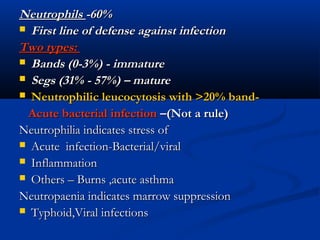
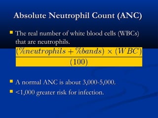
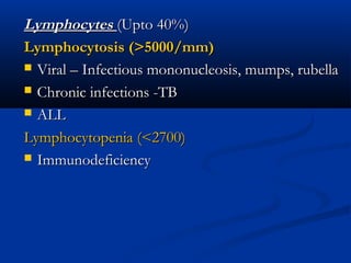
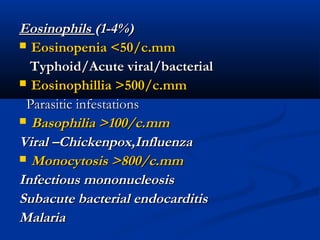
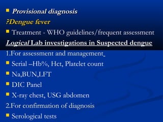
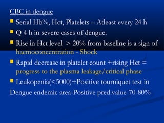
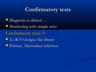
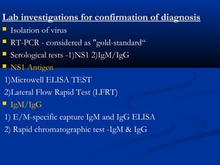
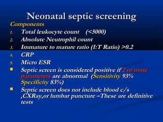
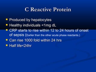
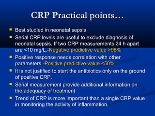
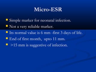
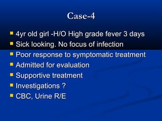
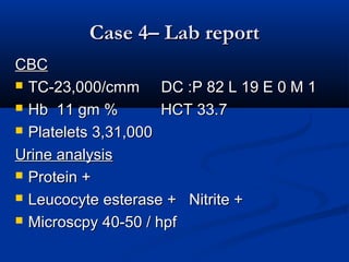
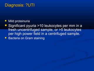
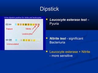
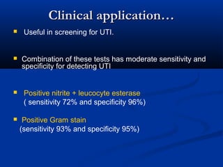
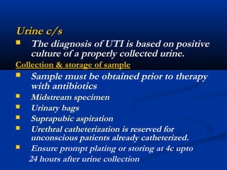
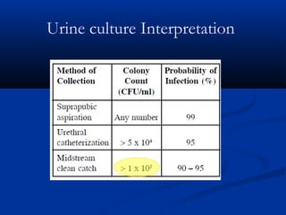
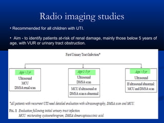
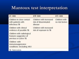
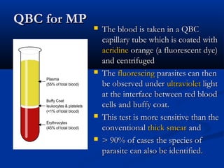
Common Lab investigationsCommon Lab investigations
in Pediatric Practicein Pediatric Practice
Dr.S.Srinivas. DCHDr.S.Srinivas. DCH
1.CBC – Few practical points1.CBC – Few practical points
 Commonest lab test in clinical practiceCommonest lab test in clinical practice
 Results are not specific to a particular disease
 Requires interpretation based on patients clinical condition
 Peripheral smear -most important but negleted part of CBC
 All three cell lines and subtypes of WBC need properAll three cell lines and subtypes of WBC need proper
evaluationevaluation
1.1. WBC count and DCWBC count and DC
2.2. RBC count ,Hb% and IndicesRBC count ,Hb% and Indices
3.3. Platelet count and IndicesPlatelet count and Indices
LeucocytesLeucocytes
Neutrophils : 60% (>70%-Abnormal)
Lymphocytes : Upto 40%
WBCWBC
 Normal range varies with ageNormal range varies with age
Increased WBCIncreased WBC
Acute stress of various causesAcute stress of various causes
 Infection, tissue necrosisInfection, tissue necrosis
 bone marrow malignancies,bone marrow malignancies,
inflammationinflammation
Decreased WBCDecreased WBC
 InfectionsInfections
 conditions or medications thatconditions or medications that
suppress or weaken the immunesuppress or weaken the immune
system or bone marrowsystem or bone marrow
NeutrophilsNeutrophils -60%-60%
 First line of defense against infectionFirst line of defense against infection
Two types:Two types:
 Bands (0-3%) - immatureBands (0-3%) - immature
 Segs (31% - 57%) – matureSegs (31% - 57%) – mature
 Neutrophilic leucocytosis with >20% band-Neutrophilic leucocytosis with >20% band-
Acute bacterial infectionAcute bacterial infection –(Not a rule)–(Not a rule)
Neutrophilia indicates stress ofNeutrophilia indicates stress of
 Acute infection-Bacterial/viralAcute infection-Bacterial/viral
 InflammationInflammation
 Others – Burns ,acute asthmaOthers – Burns ,acute asthma
Neutropaenia indicates marrow suppressionNeutropaenia indicates marrow suppression
 Typhoid,Viral infectionsTyphoid,Viral infections
Absolute Neutrophil Count (ANC)Absolute Neutrophil Count (ANC)
 The real number of white blood cells (WBCs)The real number of white blood cells (WBCs)
that are neutrophils.that are neutrophils.
 A normal ANC is about 3,000-5,000.A normal ANC is about 3,000-5,000.
 <1,000 greater risk for infection.<1,000 greater risk for infection.
LymphocytesLymphocytes (Upto 40%)(Upto 40%)
Lymphocytosis (>5000/mm)Lymphocytosis (>5000/mm)
 Viral – Infectious mononucleosis, mumps, rubellaViral – Infectious mononucleosis, mumps, rubella
 Chronic infections -TBChronic infections -TB
 ALLALL
Lymphocytopenia (<2700)Lymphocytopenia (<2700)
 ImmunodeficiencyImmunodeficiency
EosinophilsEosinophils (1-4%)(1-4%)
 Eosinopenia <50/c.mmEosinopenia <50/c.mm
Typhoid/Acute viral/bacterialTyphoid/Acute viral/bacterial
 Eosinophillia >500/c.mmEosinophillia >500/c.mm
Parasitic infestationsParasitic infestations
 Basophilia >100/c.mmBasophilia >100/c.mm
Viral –Chickenpox,InfluenzaViral –Chickenpox,Influenza
 Monocytosis >800/c.mmMonocytosis >800/c.mm
Infectious mononucleosisInfectious mononucleosis
Subacute bacterial endocarditisSubacute bacterial endocarditis
MalariaMalaria
Leukocyte abnormalities –limitations…
 TC -low predictive value due to wide range of
normal count (8000 to 20,000/cu.mm).
 Non-specific-infective /non-infective inflammation
 Non specific-bacterial /viral.
 Leukamoid reaction (WBC count > 50,000) should
not be mistaken for malignancy like leukemia.
(Leukocyte alkaline phosphatase increased in LR)
RBC indicesRBC indices
 Hb% - Degree of anemiaHb% - Degree of anemia
 RBC countRBC count
 MCV – Cell size (75-95)MCV – Cell size (75-95)
 RDW (11.5%-14.5%) High =AnisocytosisRDW (11.5%-14.5%) High =Anisocytosis
MCV- Low MCV -Normal MCV-High
RDW-Normal RDW-High Chronic disease
Spherocytosis
RDW-Normal RDW-High
Thal. Trait IDA
Thal .major
Aplasia B12-deficiency
Folate -deficiency
Platelets – Known facts…Platelets – Known facts…
ThrombocytosisThrombocytosis
 Viral infectionsViral infections
 Inflammatory disordersInflammatory disorders
 IDAIDA
 Lab errorLab error
ThrombocytopaeniaThrombocytopaenia
 Viral (including dengue)Viral (including dengue)
 TyphoidTyphoid
 MalariaMalaria
 ITP/Aplastic anemia/LeukemiaITP/Aplastic anemia/Leukemia
 DICDIC
 SepsisSepsis
MPV reflects the average size of plateletsMPV reflects the average size of platelets
Predicts functionally good plateletsPredicts functionally good platelets
 High MPV in a person with a low platelet countHigh MPV in a person with a low platelet count
suggests the bone marrow is producing plateletssuggests the bone marrow is producing platelets
and releasing them into circulation rapidly.and releasing them into circulation rapidly.
 Low MPV with low platelet counts - due to aLow MPV with low platelet counts - due to a
disorder affecting production of platelet by thedisorder affecting production of platelet by the
bone marrowbone marrow
PDW reflects how uniform the platelets are in size.PDW reflects how uniform the platelets are in size.
 Normal PDW - platelets are mostly same sizeNormal PDW - platelets are mostly same size
 High PDW - platelet size varies in sizeHigh PDW - platelet size varies in size
Neglected part – MPV & RDW
HbHb WBCWBC PP LL EE PltPlt DiseaseDisease
NN ++++++ ++++++ 00 NN Acute bacterial infAcute bacterial inf
NN ++++++ ++++++ NN ++++ Sys.Inflammatory disSys.Inflammatory dis
NN ++++ ++++ 00 N/LN/L Acute viral infAcute viral inf
NN LL ++++ oo LL TyphoidTyphoid
NN +/-+/- ++++ NN N/LN/L Chronic infectionsChronic infections
LowLow +/-+/- ++++ NN N/LN/L MalariaMalaria
LowLow ++++++ ++++
++
NN LowLow A L LA L L
HighHigh +/-+/- ++++ 00 LowLow DengueDengue
CBC in common clinical conditionsCBC in common clinical conditions
AlertsAlerts
 High Hb +Pcv(>20%) –Dengue feverHigh Hb +Pcv(>20%) –Dengue fever
 Low WBC + High Hb - DengueLow WBC + High Hb - Dengue
 High WBC + High lymphocytes – ALLHigh WBC + High lymphocytes – ALL
 Low WBC + Low platelets + low Hb – BoneLow WBC + Low platelets + low Hb – Bone
marrow diseasemarrow disease
 Low lymphocytes <2700 -ImmunodeficiencyLow lymphocytes <2700 -Immunodeficiency
CBC in fever-limitationsCBC in fever-limitations
 CBC done very early during a febrile illness does notCBC done very early during a febrile illness does not
help muchhelp much
 Interpret only in conjunction with the clinical pictureInterpret only in conjunction with the clinical picture
 It should be repeated if fever persists without diagnosisIt should be repeated if fever persists without diagnosis
 Serial counts (eg) DengueSerial counts (eg) Dengue
Case 1Case 1
 5 yr old boy –Acute onset of high grade fever-2days5 yr old boy –Acute onset of high grade fever-2days
 Interfebrile state –SickInterfebrile state –Sick
 Poor response to supportive treatmentPoor response to supportive treatment
 D2 –CBC-D2 –CBC-HbHb 11.5g% HCT-3411.5g% HCT-34 TCTC-7600-7600 DCDC-- PP6262 LL3636 EE 00
 Plt-1,60,000 ,Plt-1,60,000 , Urine R/E –Normal
 D3-C/O Abd.pain,headache,2 episodes of vomitingD3-C/O Abd.pain,headache,2 episodes of vomiting
 Sick looking,Temp – 104 F ,Moderate dehydrationSick looking,Temp – 104 F ,Moderate dehydration
 CRFT-3sec ,Low pulse volumeCRFT-3sec ,Low pulse volume
 Flushing-Flushing-white island in the red seawhite island in the red sea
 Admitted –Supportive treatment –Rpt CBC -Admitted –Supportive treatment –Rpt CBC -Hb-13.4g%
HCT 41.4 TC 3100 DC P38 L 61E0 Plt- 95,000
 Provisional diagnosisProvisional diagnosis
?Dengue fever?Dengue fever
 Treatment - WHO guidelines/frequent assessment
Logical Lab investigations in Suspected dengue
1.For assessment and management
 Serial –Hb%, Hct, Platelet count
 Na,BUN,LFT
 DIC Panel
 X-ray chest, USG abdomen
2.For confirmation of diagnosis
 Serological tests
CBC in dengue
 Serial Hb%, Hct, Platelets – Atleast every 24 h
 Q 4 h in severe cases of dengue.
 Rise in Hct level > 20% from baseline is a sign of
haemoconcentration - Shock
 Rapid decrease in platelet count +rising Hct =
progress to the plasma leakage/critical phase
 Leukopenia(<5000)+Positive tourniquet test in
Dengue endemic area-Positive pred.value-70-80%
Confirmatory testsConfirmatory tests
 Diagnosis is clinical …Diagnosis is clinical …
 Monitoring with simple testsMonitoring with simple tests
Confirmatory tests ??Confirmatory tests ??
 To R/O dengue like illnessTo R/O dengue like illness
 Primary /Secondary infectionPrimary /Secondary infection
Lab investigations for confirmation of diagnosis
 Isolation of virus
 RT-PCRRT-PCR - considered as "gold-standard“
 Serological tests -1)NS1 2)IgM/IgGSerological tests -1)NS1 2)IgM/IgG
 NS1 AntigenNS1 Antigen
1)Microwell ELISA TEST
2)Lateral Flow Rapid Test (LFRT)
 IgM/IgG
1) E/M-specific capture IgM and IgG ELISA
2) Rapid chromatographic test -IgM & IgG
 NS1 antigen –Non structural proteinNS1 antigen –Non structural protein
 High concentration in the acute-phase of primary and
secondary dengue virus infections
 Up to 9 days after the onset of illness
NS1 testNS1 test ELISAELISA RAPID TESTRAPID TEST
Time requiredTime required 2 hr2 hr 15-30 min15-30 min
Cost/availabilityCost/availability Costly / +Costly / + Cheap / -Cheap / -
SpecificitySpecificity 100%100% 100%100%
SensitivitySensitivity 82%82% 72%72%
Primary/secondaryPrimary/secondary -- --
Basic immunologyBasic immunology
InterpretationInterpretation
Case - 2Case - 2
 4hr old term male baby with maternal H/O4hr old term male baby with maternal H/O
PROMPROM
 O/EO/E
C/C/A –GoodC/C/A –Good
Clinical exam - NormalClinical exam - Normal
 Neonatal septic screeningNeonatal septic screening
2.Neonatal septic screening2.Neonatal septic screening
IndicationsIndications
1. Suspected sepsis
2. Preterm <34wks / wt <1800gm
3. PROM/Maternal fever/UTI
4. Respiratory difficulties
5. Temperature instability/fever
Neonatal septic screeningNeonatal septic screening
ComponentsComponents
1.1. Total leukocyte count (<5000)Total leukocyte count (<5000)
2.2. Absolute Neutrophil countAbsolute Neutrophil count
3.3. Immature to mature ratio (I:T Ratio) >0.2Immature to mature ratio (I:T Ratio) >0.2
4.4. CRPCRP
5.5. Micro ESRMicro ESR
 Septic screen is considered positive ifSeptic screen is considered positive if 2 or more2 or more
parametersparameters are abnormal (are abnormal (SensitivitySensitivity 93%93%
SpecificitySpecificity 83%)83%)
 Septic screen does not include blood c/sSeptic screen does not include blood c/s
,CXRay,or lumbar puncture –These are definitive,CXRay,or lumbar puncture –These are definitive
teststests
Sepsis screening – limitationsSepsis screening – limitations
 Interpret sepsis screen taking into considerationInterpret sepsis screen taking into consideration
the risk factors/gestation of infant/clinicalthe risk factors/gestation of infant/clinical
status/timing of screenstatus/timing of screen
 Taken isolation none of the tests are definiteTaken isolation none of the tests are definite
indicators of sepsisindicators of sepsis
 Do not use septic screen as a substitute forDo not use septic screen as a substitute for
blood cultureblood culture
 Do not use SC to decide duration of antibiotic.Do not use SC to decide duration of antibiotic.
C Reactive ProteinC Reactive Protein
 Produced by hepatocytesProduced by hepatocytes
 Healthy individuals <1mg dLHealthy individuals <1mg dL
 CRP starts to rise within 12 to 24 hours of onset
of sepsis (Earlier than the other acute phase reactants.)
 Can rise 1000 fold within 24 hrsCan rise 1000 fold within 24 hrs
 Half life<24hrHalf life<24hr
CRP Practical points…CRP Practical points…
 Best studied in neonatal sepsisBest studied in neonatal sepsis
 Serial CRP levels are useful to exclude diagnosis of
neonatal sepsis. If two CRP measurements 24 h apart
are <10 mg/L.--Negative predictive value >98%Negative predictive value >98%
 Positive response needs correlation with otherPositive response needs correlation with other
parametersparameters -Positive predictive value <50%-Positive predictive value <50%
 It is not justified to start the antibiotics only on the groundIt is not justified to start the antibiotics only on the ground
of positive CRP.of positive CRP.
 Serial measurement provide additional information onSerial measurement provide additional information on
the adequacy of treatmentthe adequacy of treatment
 Trend of CRP is more important than a single CRP value
in monitoring the activity of inflammation.
Micro-ESR
 Simple marker for neonatal infection.
 Not a very reliable marker.
 Its normal value is 6 mm -first 3 days of life.
 End of first month, upto 11 mm.
 >15 mm is suggestive of infection.
Procalcitonin (PCT)
 Procalcitonin is a reliable marker of late
onset sepsis in newborns with a sensitivity
specificity of almost 100%.
Case 3Case 3
 7yr old boy – fever 6days7yr old boy – fever 6days
 Continuous - high grade - rising trendContinuous - high grade - rising trend
 D1-3 : No other localizing symptoms/signsD1-3 : No other localizing symptoms/signs
 D 4-5 –Vague abd pain ,loose stoolsD 4-5 –Vague abd pain ,loose stools
 D6 -Toxic ,104 F, Tongue - coatedD6 -Toxic ,104 F, Tongue - coated
 P/A-Liver 2cms /span 8cms,Spleen 1cm /P/A-Liver 2cms /span 8cms,Spleen 1cm /
softsoft
 Provisional diagnosisProvisional diagnosis
 Enteric feverEnteric fever
 CBC,Urine R/E, Blood c/sCBC,Urine R/E, Blood c/s
 Urine R/E –NormalUrine R/E –Normal
 CBC-TC-3400/cmm.CBC-TC-3400/cmm.
 DC-P 60 L 38 E 0 M 2DC-P 60 L 38 E 0 M 2
 Hb 11.6 HCT 33.8Hb 11.6 HCT 33.8
 Platelets 142000Platelets 142000
 Clinical +Clinical +low leukocyte count with
eosinopenia points to possible
Enteric fever
 Blood culture -gold standard
 Sensitivity –highest in the first week of the
illness
 Reduces with advancing illness.
 Overall sensitivity - 50 %
 Drops with prior antibiotic therapy
 Bone marrow culture is a highly sensitive
even in late stages of the illness and with
prior antibiotic therapy.
Blood c/s in practiceBlood c/s in practice
 Gold standard for bacteremiasGold standard for bacteremias
IndicationsIndications
 Age <3mon with suspected infection
 Suspected Enteric fever
 FUO
 Febrile nutropenia/ Immuno compromised child
 Hospital acquired infection
Practical points
 Cleaning -70% Alcohol / 2% chlorhexidine
 Universal precautions
 Amount of blood – 1-5ml
 Blood : Media – 1:5
 Preferably before antibiotics
Blood culture typesBlood culture types
 ConventionalConventional
LimitationsLimitations
1. Labor-intensive
2. Time consuming
3. Sub culture - each sample manually after 12,24,48 hr
 Automated blood culture systemsAutomated blood culture systems
1.1. Bacterial growth is detected by microbial productionBacterial growth is detected by microbial production
of COof CO22
2. BACTEC / BacT-alert / MGIT / BACTEC +
Automated culture
AdvantagesAdvantages
 Improved recovery of organismImproved recovery of organism
 Rapid isolationRapid isolation
 Better identificationBetter identification
 Accurate susceptibilityAccurate susceptibility
LimitationLimitation
 Non availability
 Cost
BETTER
MANAGEMENT
Widal testWidal test
Widal test
 Most widely used serological test
 Order this test only after 5-7 days of fever
 Tube method is better than slide method
 Both H and O antibodies of 1 in 160 dilution
should be taken as cut off value of diagnosis.
 H antibodies once positive can remain positive
for long time.
Serum Widal –Limitations….
 Timeline - Send only after first week of fever
 Poor sensitivity- false negative 30%
 Previous antibiotic treatmentPrevious antibiotic treatment
 Poor specificity - endemicity & anamnestic reasons
 Method : slide test – Not reliable
 VaccinationVaccination
whole cell vaccine-High baseline anti-o &anti-Hwhole cell vaccine-High baseline anti-o &anti-H
(Vi-polysaccharide vaccine does not interfere )
 Inter laboratory variations
Typhidot test
 A dot enzyme immunoassay that detects specific
IgM and IgG antibodies - 50KD outer membrane
protein antigen of S. typhi.
 Limitation: False positive results in endemic areas.
-Persistence of high IgG levels
 Typhidot-M® - only IgM is detected. IgG is
inactivated
 Typhidot-M® -sensitivity (>93%)
Molecular methods
PCR for Typhoid
 Superior sensitivity than culture
 The turnaround time < 24 hours
Limitations
 Clinical utility - inadequately evaluated
 False positive –Vaccination , Old infections
 Costly
Tubex test:
 Tubex test is simple one-step test taking 2minutes
 It detects only IgM and not IgG
 Positive result -Suggests recent Salmonella
infection
IgM dipstick test
 Based on the binding of S. typhi-specific IgM
antibodies to S. typhi lipopolysaccharide (LPS)
antigen.
 It can be done in serum or whole blood and
requires incubation for 3 hours
Case-4Case-4
 4yr old girl -H/O High grade fever 3 days4yr old girl -H/O High grade fever 3 days
 Sick looking. No focus of infectionSick looking. No focus of infection
 Poor response to symptomatic treatmentPoor response to symptomatic treatment
 Admitted for evaluationAdmitted for evaluation
 Supportive treatmentSupportive treatment
 Investigations ?Investigations ?
 CBC, Urine R/ECBC, Urine R/E
Case 4– Lab reportCase 4– Lab report
CBCCBC
 TC-23,000/cmm DC :P 82 L 19 E 0 M 1TC-23,000/cmm DC :P 82 L 19 E 0 M 1
 Hb 11 gm % HCT 33.7Hb 11 gm % HCT 33.7
 Platelets 3,31,000Platelets 3,31,000
Urine analysisUrine analysis
 Protein +Protein +
 Leucocyte esterase + Nitrite +Leucocyte esterase + Nitrite +
 Microscpy 40-50 / hpfMicroscpy 40-50 / hpf
Diagnosis: ?UTIDiagnosis: ?UTI
 Mild proteinuria
 Significant pyuria >10 leukocytes per mm in a
fresh uncentrifuged sample, or >5 leukocytes
per high power field in a centrifuged sample.
 Bacteria on Gram staining
Dipstick
 Leucocyte esterase test –
Pyuria
 Nitrite test - significant
Bacteriuria
 Leucocyte esterase + Nitrite
- more sensitive
Clinical application…Clinical application…
 Useful in screening for UTI.
 Combination of these tests has moderate sensitivity and
specificity for detecting UTI
 Positive nitrite + leucocyte esterase
( sensitivity 72% and specificity 96%)
 Positive Gram stain
(sensitivity 93% and specificity 95%)
Urine c/sUrine c/s
 The diagnosis of UTI is based on positive
culture of a properly collected urine.
Collection & storage of sampleCollection & storage of sample
 Sample must be obtained prior to therapy
with antibiotics
 Midstream specimen
 Urinary bags
 Suprapubic aspiration
 Urethral catheterization is reserved for
unconscious patients already catheterized.
 Ensure prompt plating or storing at 4c upto
24 hours after urine collection
Urine culture Interpretation
Radio imaging studies
• Recommended for all children with UTI.
• Aim - to identify patients at-risk of renal damage, mainly those below 5 years of
age, with VUR or urinary tract obstruction.
USG in UTIUSG in UTI
 USG to be done in all cases of UTIUSG to be done in all cases of UTI
To L/FTo L/F
1.1. Posterior urethral valvePosterior urethral valve
2.2. Post void residual urinePost void residual urine
3.3. Bladder hypertrophyBladder hypertrophy
4.4. HydronephrosisHydronephrosis
5.5. Pelvicalyceal anomaliesPelvicalyceal anomalies
MCU ScanMCU Scan
 It will show anatomy of the bladder and theIt will show anatomy of the bladder and the
ureters.ureters.
 It is basically for bladder and VUR issuesIt is basically for bladder and VUR issues
A. The MCU showing a
dilated posterior
urethra, mildly irregular
appearance of the
edge of the bladder and
bilateral vesicoureteric
reflux into dilated
tortuous ureters.
B. Hydronephrosis
with 'clubbing' of
the calyces.
DMSA ScanDMSA Scan
 Gold standard test for detecting renal corticalGold standard test for detecting renal cortical
scarsscars
 Can be done during acute phase of UTICan be done during acute phase of UTI
Case 5: Fever with rashCase 5: Fever with rash
 3 yr old girl with fever – 4 days ,3 yr old girl with fever – 4 days ,
 Skin rash – 2 daysSkin rash – 2 days
 Fever-moderate-highFever-moderate-high
 Rash increased over 2 days and lead to blackRash increased over 2 days and lead to black
patches over one ear lobe and one toepatches over one ear lobe and one toe
O/EO/E
 Sick child with high feverSick child with high fever
 Maculonodular rashMaculonodular rash
 Gangrenous patches-Ear lobe, one toeGangrenous patches-Ear lobe, one toe
Clinical diagnosis…Clinical diagnosis…
 Rickettsial diseaseRickettsial disease
 InvestigationInvestigation
 No rapid laboratory tests are available toNo rapid laboratory tests are available to
diagnose rickettsial diseases early in thediagnose rickettsial diseases early in the
course of illness.course of illness.
 Gold standard –(Gold standard –(IFA )Immuno fluorescence assayIFA )Immuno fluorescence assay
 Weile Felix - Useful and cheapest available tool forWeile Felix - Useful and cheapest available tool for
the laboratory diagnosis of Rickettsial diseases inthe laboratory diagnosis of Rickettsial diseases in
IndiaIndia
 ELISA / RT-PCR /ELISA / RT-PCR / dot blot immunoassaydot blot immunoassay
 Weil-FelixWeil-Felix is a nonspecific agglutination testis a nonspecific agglutination test
 It detects anti-rickettsial antibodies in patient’sIt detects anti-rickettsial antibodies in patient’s
serum.serum.
 If the  titer is greater than or equal to 1:320 orIf the  titer is greater than or equal to 1:320 or
4-fold rise from baseline -4-fold rise from baseline - POSITIVEPOSITIVE
 sensitivity(33%) and specificity(46%)sensitivity(33%) and specificity(46%)
IFA -Immunofluorescence assayIFA -Immunofluorescence assay
 Gold standard for diagnosisGold standard for diagnosis
 IFA can detect IgG or IgM antibodies.
 Blood samples taken early (7-10days) and late
(14-21days) in the disease are the preferred
specimens for evaluation.
 Rising titres of IgM is indicative of a current
infection
Case 6Case 6
 5yr old boy – Frequent cough cold & fever – 1yr5yr old boy – Frequent cough cold & fever – 1yr
 6 episodes /yr6 episodes /yr
 Each episode -2-4days of fever Cough&cold-5-10 dEach episode -2-4days of fever Cough&cold-5-10 d
 No hospitalizations/No family h/o AtopyNo hospitalizations/No family h/o Atopy
 Clinical exam - NormalClinical exam - Normal
 Inv-Hb 11g% TC 14300 P35 L62 E3 ESR 42 mmInv-Hb 11g% TC 14300 P35 L62 E3 ESR 42 mm
 Mx test- 10 mm PositiveMx test- 10 mm Positive..
 Rx –ATT – Referred for 2Rx –ATT – Referred for 2nd Opinionnd Opinion
 No h/o contact , X – Ray chest –NormalNo h/o contact , X – Ray chest –Normal
DiagnosisDiagnosis
 Recurrent viral respiratory infectionRecurrent viral respiratory infection
Tuberculosis diagnosisTuberculosis diagnosis
 Golden triadGolden triad
(Clinical features +Abnormal chest X-ray +Mx)(Clinical features +Abnormal chest X-ray +Mx)
++ contact history/close exposure with an adult havingcontact history/close exposure with an adult having
active TB in the last 2 yearsactive TB in the last 2 years
 Demonstration of AFBDemonstration of AFB - Confirmatory diagnosis- Confirmatory diagnosis
AFB in smear/cultureAFB in smear/culture
 Isolation of MTBIsolation of MTB –Clinical specimen/histopathology–Clinical specimen/histopathology
Mantoux testMantoux test
 ID - PPD-2TU- 2-4 inch belowID - PPD-2TU- 2-4 inch below
elbowelbow
 Transverse diameter of theTransverse diameter of the
induration (not erythema)induration (not erythema)
measured- 48 -72 hours in mmmeasured- 48 -72 hours in mm
 Positive >10mm-InfectionPositive >10mm-Infection
 Reaction could be due to
1. Infection with tubercle bacilli
2. Cross sensitivity to
environmental mycobacteria
3. BCG-induced sensitivity
Mantoux test interpretationMantoux test interpretation
TB diagnosis- Newer methods
1. Direct molecular methods
2. Automated liquid culture methods
Automated liquid culture
BACTEC TB 460
 Sensitive, specific and rapid culture method
 Respiratory /non-respiratory specimens.
 200 viable M. tuberculosis bacilli could be detected in
12-13days
Mycobacterial growth indicator tube(MGIT) 960 TB:
 Fluorescent technology - based on oxygen quenching
with a fluorescent dye.
 Result - 7-10 days.
Direct molecular methods
 Do not require growth of the bacteria,
 Possible to detect M.tuberculosis complex within
3-5 hours.
 Nucleic Acid Amplification assay tests
1. The Gen-Probe AMPLIFIEDTM
Mycobacterium tuberculosis Direct (MTD) Test.
2. The Roche AMPLICOR® Mycobacterium
tuberculosis (MTB) Test.
Limitation
 Results of NAA tests are preliminary; the
mycobacterial culture is needed for species
identification, confirmation and for drug-
susceptibility testing.
 A positive result may be misleading as the NAA
test can amplify DNA from both viable and
non-viable organisms
Case 7Case 7
 5 yr old boy with fever 5days5 yr old boy with fever 5days
 High grade, intermittent, Chills & rigors +High grade, intermittent, Chills & rigors +
 Headache ,Vomiting +Headache ,Vomiting +
 O/E Temp 103 FO/E Temp 103 F
 P/A Spleen measuring about 3 cm below costal marginP/A Spleen measuring about 3 cm below costal margin
– firm , non-tender– firm , non-tender
 No hepatomegalyNo hepatomegaly
DiagnosisDiagnosis
MalariaMalaria
Lab investigationsLab investigations
 Lab: Hb 11.2 TC 13500 P72 L26 M2Lab: Hb 11.2 TC 13500 P72 L26 M2
 MP smear – Plasmodiam vivax ring ,gametocytesMP smear – Plasmodiam vivax ring ,gametocytes
MalariaMalaria
Diagnostic techniques
1. Blood smear examination- gold standard
2. Quantitative buffy coat technique (QBC)
3. RDTs
4. Fluorescent microscopy
Blood smear for malarial parasiteBlood smear for malarial parasite

Thick blood smears are most useful for
detecting the presence of parasites,
because they examine a larger sample of
blood.
Thin blood smears helps doctors
discover what species of malaria is
causing the infection.
Disadvantages of MicroscoprDisadvantages of Microscopr
 Time consuming >60minTime consuming >60min
 Labour intensiveLabour intensive
 Skilled lab technicianSkilled lab technician
 It cannot detect parasites sequestered deep inIt cannot detect parasites sequestered deep in
the vascular compartmentthe vascular compartment
QBC for MPQBC for MP
 The blood is taken in a QBCThe blood is taken in a QBC
capillary tube which is coated with capillary tube which is coated with 
acridineacridine orangeorange (a fluorescent dye) (a fluorescent dye)
and centrifugedand centrifuged
 TheThe fluorescingfluorescing parasites can then parasites can then
be observed under be observed under ultravioletultraviolet light light
at the interface between red bloodat the interface between red blood
cells and buffy coat.cells and buffy coat.
 This test is more sensitive than theThis test is more sensitive than the
conventional conventional thick smearthick smear and and
 > 90% of cases the species of> 90% of cases the species of
parasite can also be identified.parasite can also be identified.
Rapid tests for malaria
 Immunochromatographic tests based on the
‘Dipstick’ format
 Detects plasmodium specific antigens in blood
sample.
 Common RDTs available Parasight ,OptiMAL.
Targeted antigens
 Histidine rich protein II of P. Falciparum (pfHRP-II)
 Plasmodium aldolase- produced by all plasmodium
species.
 Plasmodium lactate dehydrogenase(pLDH), is
produced by all four species of plasmodia.
 Antibodies produced against pLDH either specific for
P.falciparum or P.vivax alone or a pan specific antibody
which reacts with all the four species of plasmodium.
Commercially available kit can detect falciparum and
vivax but cannot differentiate ovale and malariae.
Advantages
 Rapid tests are easy to use with minimal training
 Results are available within minutes.
 They can diagnose falciparum infection even
when the parasite is deeply sequestered.
Disadvantages
 More expensive than microscopy
 Limitations in species identification.
 Persistent positivity even after effective
treatment. HRPII remains positive for 4 weeks
and pLDH remains positive for1 week.
 No prognostic value- they can not quantify
parasites.

Common lab investigations in Paediatric Office Practice

  • 1.
    Common Lab investigationsCommonLab investigations in Pediatric Practicein Pediatric Practice Dr.S.Srinivas. DCHDr.S.Srinivas. DCH
  • 2.
    1.CBC – Fewpractical points1.CBC – Few practical points  Commonest lab test in clinical practiceCommonest lab test in clinical practice  Results are not specific to a particular disease  Requires interpretation based on patients clinical condition  Peripheral smear -most important but negleted part of CBC  All three cell lines and subtypes of WBC need properAll three cell lines and subtypes of WBC need proper evaluationevaluation 1.1. WBC count and DCWBC count and DC 2.2. RBC count ,Hb% and IndicesRBC count ,Hb% and Indices 3.3. Platelet count and IndicesPlatelet count and Indices
  • 3.
    LeucocytesLeucocytes Neutrophils : 60%(>70%-Abnormal) Lymphocytes : Upto 40%
  • 4.
    WBCWBC  Normal rangevaries with ageNormal range varies with age
  • 5.
    Increased WBCIncreased WBC Acutestress of various causesAcute stress of various causes  Infection, tissue necrosisInfection, tissue necrosis  bone marrow malignancies,bone marrow malignancies, inflammationinflammation Decreased WBCDecreased WBC  InfectionsInfections  conditions or medications thatconditions or medications that suppress or weaken the immunesuppress or weaken the immune system or bone marrowsystem or bone marrow
  • 6.
    NeutrophilsNeutrophils -60%-60%  Firstline of defense against infectionFirst line of defense against infection Two types:Two types:  Bands (0-3%) - immatureBands (0-3%) - immature  Segs (31% - 57%) – matureSegs (31% - 57%) – mature  Neutrophilic leucocytosis with >20% band-Neutrophilic leucocytosis with >20% band- Acute bacterial infectionAcute bacterial infection –(Not a rule)–(Not a rule) Neutrophilia indicates stress ofNeutrophilia indicates stress of  Acute infection-Bacterial/viralAcute infection-Bacterial/viral  InflammationInflammation  Others – Burns ,acute asthmaOthers – Burns ,acute asthma Neutropaenia indicates marrow suppressionNeutropaenia indicates marrow suppression  Typhoid,Viral infectionsTyphoid,Viral infections
  • 7.
    Absolute Neutrophil Count(ANC)Absolute Neutrophil Count (ANC)  The real number of white blood cells (WBCs)The real number of white blood cells (WBCs) that are neutrophils.that are neutrophils.  A normal ANC is about 3,000-5,000.A normal ANC is about 3,000-5,000.  <1,000 greater risk for infection.<1,000 greater risk for infection.
  • 8.
    LymphocytesLymphocytes (Upto 40%)(Upto40%) Lymphocytosis (>5000/mm)Lymphocytosis (>5000/mm)  Viral – Infectious mononucleosis, mumps, rubellaViral – Infectious mononucleosis, mumps, rubella  Chronic infections -TBChronic infections -TB  ALLALL Lymphocytopenia (<2700)Lymphocytopenia (<2700)  ImmunodeficiencyImmunodeficiency
  • 9.
    EosinophilsEosinophils (1-4%)(1-4%)  Eosinopenia<50/c.mmEosinopenia <50/c.mm Typhoid/Acute viral/bacterialTyphoid/Acute viral/bacterial  Eosinophillia >500/c.mmEosinophillia >500/c.mm Parasitic infestationsParasitic infestations  Basophilia >100/c.mmBasophilia >100/c.mm Viral –Chickenpox,InfluenzaViral –Chickenpox,Influenza  Monocytosis >800/c.mmMonocytosis >800/c.mm Infectious mononucleosisInfectious mononucleosis Subacute bacterial endocarditisSubacute bacterial endocarditis MalariaMalaria
  • 10.
    Leukocyte abnormalities –limitations… TC -low predictive value due to wide range of normal count (8000 to 20,000/cu.mm).  Non-specific-infective /non-infective inflammation  Non specific-bacterial /viral.  Leukamoid reaction (WBC count > 50,000) should not be mistaken for malignancy like leukemia. (Leukocyte alkaline phosphatase increased in LR)
  • 11.
    RBC indicesRBC indices Hb% - Degree of anemiaHb% - Degree of anemia  RBC countRBC count  MCV – Cell size (75-95)MCV – Cell size (75-95)  RDW (11.5%-14.5%) High =AnisocytosisRDW (11.5%-14.5%) High =Anisocytosis MCV- Low MCV -Normal MCV-High RDW-Normal RDW-High Chronic disease Spherocytosis RDW-Normal RDW-High Thal. Trait IDA Thal .major Aplasia B12-deficiency Folate -deficiency
  • 12.
    Platelets – Knownfacts…Platelets – Known facts… ThrombocytosisThrombocytosis  Viral infectionsViral infections  Inflammatory disordersInflammatory disorders  IDAIDA  Lab errorLab error ThrombocytopaeniaThrombocytopaenia  Viral (including dengue)Viral (including dengue)  TyphoidTyphoid  MalariaMalaria  ITP/Aplastic anemia/LeukemiaITP/Aplastic anemia/Leukemia  DICDIC  SepsisSepsis
  • 13.
    MPV reflects theaverage size of plateletsMPV reflects the average size of platelets Predicts functionally good plateletsPredicts functionally good platelets  High MPV in a person with a low platelet countHigh MPV in a person with a low platelet count suggests the bone marrow is producing plateletssuggests the bone marrow is producing platelets and releasing them into circulation rapidly.and releasing them into circulation rapidly.  Low MPV with low platelet counts - due to aLow MPV with low platelet counts - due to a disorder affecting production of platelet by thedisorder affecting production of platelet by the bone marrowbone marrow PDW reflects how uniform the platelets are in size.PDW reflects how uniform the platelets are in size.  Normal PDW - platelets are mostly same sizeNormal PDW - platelets are mostly same size  High PDW - platelet size varies in sizeHigh PDW - platelet size varies in size Neglected part – MPV & RDW
  • 14.
    HbHb WBCWBC PPLL EE PltPlt DiseaseDisease NN ++++++ ++++++ 00 NN Acute bacterial infAcute bacterial inf NN ++++++ ++++++ NN ++++ Sys.Inflammatory disSys.Inflammatory dis NN ++++ ++++ 00 N/LN/L Acute viral infAcute viral inf NN LL ++++ oo LL TyphoidTyphoid NN +/-+/- ++++ NN N/LN/L Chronic infectionsChronic infections LowLow +/-+/- ++++ NN N/LN/L MalariaMalaria LowLow ++++++ ++++ ++ NN LowLow A L LA L L HighHigh +/-+/- ++++ 00 LowLow DengueDengue CBC in common clinical conditionsCBC in common clinical conditions
  • 15.
    AlertsAlerts  High Hb+Pcv(>20%) –Dengue feverHigh Hb +Pcv(>20%) –Dengue fever  Low WBC + High Hb - DengueLow WBC + High Hb - Dengue  High WBC + High lymphocytes – ALLHigh WBC + High lymphocytes – ALL  Low WBC + Low platelets + low Hb – BoneLow WBC + Low platelets + low Hb – Bone marrow diseasemarrow disease  Low lymphocytes <2700 -ImmunodeficiencyLow lymphocytes <2700 -Immunodeficiency
  • 16.
    CBC in fever-limitationsCBCin fever-limitations  CBC done very early during a febrile illness does notCBC done very early during a febrile illness does not help muchhelp much  Interpret only in conjunction with the clinical pictureInterpret only in conjunction with the clinical picture  It should be repeated if fever persists without diagnosisIt should be repeated if fever persists without diagnosis  Serial counts (eg) DengueSerial counts (eg) Dengue
  • 17.
    Case 1Case 1 5 yr old boy –Acute onset of high grade fever-2days5 yr old boy –Acute onset of high grade fever-2days  Interfebrile state –SickInterfebrile state –Sick  Poor response to supportive treatmentPoor response to supportive treatment  D2 –CBC-D2 –CBC-HbHb 11.5g% HCT-3411.5g% HCT-34 TCTC-7600-7600 DCDC-- PP6262 LL3636 EE 00  Plt-1,60,000 ,Plt-1,60,000 , Urine R/E –Normal  D3-C/O Abd.pain,headache,2 episodes of vomitingD3-C/O Abd.pain,headache,2 episodes of vomiting  Sick looking,Temp – 104 F ,Moderate dehydrationSick looking,Temp – 104 F ,Moderate dehydration  CRFT-3sec ,Low pulse volumeCRFT-3sec ,Low pulse volume  Flushing-Flushing-white island in the red seawhite island in the red sea  Admitted –Supportive treatment –Rpt CBC -Admitted –Supportive treatment –Rpt CBC -Hb-13.4g% HCT 41.4 TC 3100 DC P38 L 61E0 Plt- 95,000
  • 18.
     Provisional diagnosisProvisionaldiagnosis ?Dengue fever?Dengue fever  Treatment - WHO guidelines/frequent assessment Logical Lab investigations in Suspected dengue 1.For assessment and management  Serial –Hb%, Hct, Platelet count  Na,BUN,LFT  DIC Panel  X-ray chest, USG abdomen 2.For confirmation of diagnosis  Serological tests
  • 19.
    CBC in dengue Serial Hb%, Hct, Platelets – Atleast every 24 h  Q 4 h in severe cases of dengue.  Rise in Hct level > 20% from baseline is a sign of haemoconcentration - Shock  Rapid decrease in platelet count +rising Hct = progress to the plasma leakage/critical phase  Leukopenia(<5000)+Positive tourniquet test in Dengue endemic area-Positive pred.value-70-80%
  • 20.
    Confirmatory testsConfirmatory tests Diagnosis is clinical …Diagnosis is clinical …  Monitoring with simple testsMonitoring with simple tests Confirmatory tests ??Confirmatory tests ??  To R/O dengue like illnessTo R/O dengue like illness  Primary /Secondary infectionPrimary /Secondary infection
  • 21.
    Lab investigations forconfirmation of diagnosis  Isolation of virus  RT-PCRRT-PCR - considered as "gold-standard“  Serological tests -1)NS1 2)IgM/IgGSerological tests -1)NS1 2)IgM/IgG  NS1 AntigenNS1 Antigen 1)Microwell ELISA TEST 2)Lateral Flow Rapid Test (LFRT)  IgM/IgG 1) E/M-specific capture IgM and IgG ELISA 2) Rapid chromatographic test -IgM & IgG
  • 22.
     NS1 antigen–Non structural proteinNS1 antigen –Non structural protein  High concentration in the acute-phase of primary and secondary dengue virus infections  Up to 9 days after the onset of illness NS1 testNS1 test ELISAELISA RAPID TESTRAPID TEST Time requiredTime required 2 hr2 hr 15-30 min15-30 min Cost/availabilityCost/availability Costly / +Costly / + Cheap / -Cheap / - SpecificitySpecificity 100%100% 100%100% SensitivitySensitivity 82%82% 72%72% Primary/secondaryPrimary/secondary -- --
  • 23.
  • 24.
    Case - 2Case- 2  4hr old term male baby with maternal H/O4hr old term male baby with maternal H/O PROMPROM  O/EO/E C/C/A –GoodC/C/A –Good Clinical exam - NormalClinical exam - Normal  Neonatal septic screeningNeonatal septic screening
  • 25.
    2.Neonatal septic screening2.Neonatalseptic screening IndicationsIndications 1. Suspected sepsis 2. Preterm <34wks / wt <1800gm 3. PROM/Maternal fever/UTI 4. Respiratory difficulties 5. Temperature instability/fever
  • 26.
    Neonatal septic screeningNeonatalseptic screening ComponentsComponents 1.1. Total leukocyte count (<5000)Total leukocyte count (<5000) 2.2. Absolute Neutrophil countAbsolute Neutrophil count 3.3. Immature to mature ratio (I:T Ratio) >0.2Immature to mature ratio (I:T Ratio) >0.2 4.4. CRPCRP 5.5. Micro ESRMicro ESR  Septic screen is considered positive ifSeptic screen is considered positive if 2 or more2 or more parametersparameters are abnormal (are abnormal (SensitivitySensitivity 93%93% SpecificitySpecificity 83%)83%)  Septic screen does not include blood c/sSeptic screen does not include blood c/s ,CXRay,or lumbar puncture –These are definitive,CXRay,or lumbar puncture –These are definitive teststests
  • 27.
    Sepsis screening –limitationsSepsis screening – limitations  Interpret sepsis screen taking into considerationInterpret sepsis screen taking into consideration the risk factors/gestation of infant/clinicalthe risk factors/gestation of infant/clinical status/timing of screenstatus/timing of screen  Taken isolation none of the tests are definiteTaken isolation none of the tests are definite indicators of sepsisindicators of sepsis  Do not use septic screen as a substitute forDo not use septic screen as a substitute for blood cultureblood culture  Do not use SC to decide duration of antibiotic.Do not use SC to decide duration of antibiotic.
  • 28.
    C Reactive ProteinCReactive Protein  Produced by hepatocytesProduced by hepatocytes  Healthy individuals <1mg dLHealthy individuals <1mg dL  CRP starts to rise within 12 to 24 hours of onset of sepsis (Earlier than the other acute phase reactants.)  Can rise 1000 fold within 24 hrsCan rise 1000 fold within 24 hrs  Half life<24hrHalf life<24hr
  • 29.
    CRP Practical points…CRPPractical points…  Best studied in neonatal sepsisBest studied in neonatal sepsis  Serial CRP levels are useful to exclude diagnosis of neonatal sepsis. If two CRP measurements 24 h apart are <10 mg/L.--Negative predictive value >98%Negative predictive value >98%  Positive response needs correlation with otherPositive response needs correlation with other parametersparameters -Positive predictive value <50%-Positive predictive value <50%  It is not justified to start the antibiotics only on the groundIt is not justified to start the antibiotics only on the ground of positive CRP.of positive CRP.  Serial measurement provide additional information onSerial measurement provide additional information on the adequacy of treatmentthe adequacy of treatment  Trend of CRP is more important than a single CRP value in monitoring the activity of inflammation.
  • 30.
    Micro-ESR  Simple markerfor neonatal infection.  Not a very reliable marker.  Its normal value is 6 mm -first 3 days of life.  End of first month, upto 11 mm.  >15 mm is suggestive of infection.
  • 31.
    Procalcitonin (PCT)  Procalcitoninis a reliable marker of late onset sepsis in newborns with a sensitivity specificity of almost 100%.
  • 32.
    Case 3Case 3 7yr old boy – fever 6days7yr old boy – fever 6days  Continuous - high grade - rising trendContinuous - high grade - rising trend  D1-3 : No other localizing symptoms/signsD1-3 : No other localizing symptoms/signs  D 4-5 –Vague abd pain ,loose stoolsD 4-5 –Vague abd pain ,loose stools  D6 -Toxic ,104 F, Tongue - coatedD6 -Toxic ,104 F, Tongue - coated  P/A-Liver 2cms /span 8cms,Spleen 1cm /P/A-Liver 2cms /span 8cms,Spleen 1cm / softsoft  Provisional diagnosisProvisional diagnosis  Enteric feverEnteric fever
  • 33.
     CBC,Urine R/E,Blood c/sCBC,Urine R/E, Blood c/s  Urine R/E –NormalUrine R/E –Normal  CBC-TC-3400/cmm.CBC-TC-3400/cmm.  DC-P 60 L 38 E 0 M 2DC-P 60 L 38 E 0 M 2  Hb 11.6 HCT 33.8Hb 11.6 HCT 33.8  Platelets 142000Platelets 142000  Clinical +Clinical +low leukocyte count with eosinopenia points to possible Enteric fever
  • 34.
     Blood culture-gold standard  Sensitivity –highest in the first week of the illness  Reduces with advancing illness.  Overall sensitivity - 50 %  Drops with prior antibiotic therapy  Bone marrow culture is a highly sensitive even in late stages of the illness and with prior antibiotic therapy.
  • 35.
    Blood c/s inpracticeBlood c/s in practice  Gold standard for bacteremiasGold standard for bacteremias IndicationsIndications  Age <3mon with suspected infection  Suspected Enteric fever  FUO  Febrile nutropenia/ Immuno compromised child  Hospital acquired infection Practical points  Cleaning -70% Alcohol / 2% chlorhexidine  Universal precautions  Amount of blood – 1-5ml  Blood : Media – 1:5  Preferably before antibiotics
  • 36.
    Blood culture typesBloodculture types  ConventionalConventional LimitationsLimitations 1. Labor-intensive 2. Time consuming 3. Sub culture - each sample manually after 12,24,48 hr  Automated blood culture systemsAutomated blood culture systems 1.1. Bacterial growth is detected by microbial productionBacterial growth is detected by microbial production of COof CO22 2. BACTEC / BacT-alert / MGIT / BACTEC +
  • 37.
    Automated culture AdvantagesAdvantages  Improvedrecovery of organismImproved recovery of organism  Rapid isolationRapid isolation  Better identificationBetter identification  Accurate susceptibilityAccurate susceptibility LimitationLimitation  Non availability  Cost BETTER MANAGEMENT
  • 38.
  • 39.
    Widal test  Mostwidely used serological test  Order this test only after 5-7 days of fever  Tube method is better than slide method  Both H and O antibodies of 1 in 160 dilution should be taken as cut off value of diagnosis.  H antibodies once positive can remain positive for long time.
  • 40.
    Serum Widal –Limitations…. Timeline - Send only after first week of fever  Poor sensitivity- false negative 30%  Previous antibiotic treatmentPrevious antibiotic treatment  Poor specificity - endemicity & anamnestic reasons  Method : slide test – Not reliable  VaccinationVaccination whole cell vaccine-High baseline anti-o &anti-Hwhole cell vaccine-High baseline anti-o &anti-H (Vi-polysaccharide vaccine does not interfere )  Inter laboratory variations
  • 41.
    Typhidot test  Adot enzyme immunoassay that detects specific IgM and IgG antibodies - 50KD outer membrane protein antigen of S. typhi.  Limitation: False positive results in endemic areas. -Persistence of high IgG levels  Typhidot-M® - only IgM is detected. IgG is inactivated  Typhidot-M® -sensitivity (>93%)
  • 42.
    Molecular methods PCR forTyphoid  Superior sensitivity than culture  The turnaround time < 24 hours Limitations  Clinical utility - inadequately evaluated  False positive –Vaccination , Old infections  Costly
  • 43.
    Tubex test:  Tubextest is simple one-step test taking 2minutes  It detects only IgM and not IgG  Positive result -Suggests recent Salmonella infection IgM dipstick test  Based on the binding of S. typhi-specific IgM antibodies to S. typhi lipopolysaccharide (LPS) antigen.  It can be done in serum or whole blood and requires incubation for 3 hours
  • 44.
    Case-4Case-4  4yr oldgirl -H/O High grade fever 3 days4yr old girl -H/O High grade fever 3 days  Sick looking. No focus of infectionSick looking. No focus of infection  Poor response to symptomatic treatmentPoor response to symptomatic treatment  Admitted for evaluationAdmitted for evaluation  Supportive treatmentSupportive treatment  Investigations ?Investigations ?  CBC, Urine R/ECBC, Urine R/E
  • 45.
    Case 4– LabreportCase 4– Lab report CBCCBC  TC-23,000/cmm DC :P 82 L 19 E 0 M 1TC-23,000/cmm DC :P 82 L 19 E 0 M 1  Hb 11 gm % HCT 33.7Hb 11 gm % HCT 33.7  Platelets 3,31,000Platelets 3,31,000 Urine analysisUrine analysis  Protein +Protein +  Leucocyte esterase + Nitrite +Leucocyte esterase + Nitrite +  Microscpy 40-50 / hpfMicroscpy 40-50 / hpf
  • 46.
    Diagnosis: ?UTIDiagnosis: ?UTI Mild proteinuria  Significant pyuria >10 leukocytes per mm in a fresh uncentrifuged sample, or >5 leukocytes per high power field in a centrifuged sample.  Bacteria on Gram staining
  • 47.
    Dipstick  Leucocyte esterasetest – Pyuria  Nitrite test - significant Bacteriuria  Leucocyte esterase + Nitrite - more sensitive
  • 48.
    Clinical application…Clinical application… Useful in screening for UTI.  Combination of these tests has moderate sensitivity and specificity for detecting UTI  Positive nitrite + leucocyte esterase ( sensitivity 72% and specificity 96%)  Positive Gram stain (sensitivity 93% and specificity 95%)
  • 49.
    Urine c/sUrine c/s The diagnosis of UTI is based on positive culture of a properly collected urine. Collection & storage of sampleCollection & storage of sample  Sample must be obtained prior to therapy with antibiotics  Midstream specimen  Urinary bags  Suprapubic aspiration  Urethral catheterization is reserved for unconscious patients already catheterized.  Ensure prompt plating or storing at 4c upto 24 hours after urine collection
  • 50.
  • 51.
    Radio imaging studies •Recommended for all children with UTI. • Aim - to identify patients at-risk of renal damage, mainly those below 5 years of age, with VUR or urinary tract obstruction.
  • 52.
    USG in UTIUSGin UTI  USG to be done in all cases of UTIUSG to be done in all cases of UTI To L/FTo L/F 1.1. Posterior urethral valvePosterior urethral valve 2.2. Post void residual urinePost void residual urine 3.3. Bladder hypertrophyBladder hypertrophy 4.4. HydronephrosisHydronephrosis 5.5. Pelvicalyceal anomaliesPelvicalyceal anomalies
  • 53.
    MCU ScanMCU Scan It will show anatomy of the bladder and theIt will show anatomy of the bladder and the ureters.ureters.  It is basically for bladder and VUR issuesIt is basically for bladder and VUR issues
  • 54.
    A. The MCUshowing a dilated posterior urethra, mildly irregular appearance of the edge of the bladder and bilateral vesicoureteric reflux into dilated tortuous ureters. B. Hydronephrosis with 'clubbing' of the calyces.
  • 55.
    DMSA ScanDMSA Scan Gold standard test for detecting renal corticalGold standard test for detecting renal cortical scarsscars  Can be done during acute phase of UTICan be done during acute phase of UTI
  • 56.
    Case 5: Feverwith rashCase 5: Fever with rash  3 yr old girl with fever – 4 days ,3 yr old girl with fever – 4 days ,  Skin rash – 2 daysSkin rash – 2 days  Fever-moderate-highFever-moderate-high  Rash increased over 2 days and lead to blackRash increased over 2 days and lead to black patches over one ear lobe and one toepatches over one ear lobe and one toe O/EO/E  Sick child with high feverSick child with high fever  Maculonodular rashMaculonodular rash  Gangrenous patches-Ear lobe, one toeGangrenous patches-Ear lobe, one toe
  • 57.
    Clinical diagnosis…Clinical diagnosis… Rickettsial diseaseRickettsial disease  InvestigationInvestigation  No rapid laboratory tests are available toNo rapid laboratory tests are available to diagnose rickettsial diseases early in thediagnose rickettsial diseases early in the course of illness.course of illness.  Gold standard –(Gold standard –(IFA )Immuno fluorescence assayIFA )Immuno fluorescence assay  Weile Felix - Useful and cheapest available tool forWeile Felix - Useful and cheapest available tool for the laboratory diagnosis of Rickettsial diseases inthe laboratory diagnosis of Rickettsial diseases in IndiaIndia  ELISA / RT-PCR /ELISA / RT-PCR / dot blot immunoassaydot blot immunoassay
  • 58.
     Weil-FelixWeil-Felix isa nonspecific agglutination testis a nonspecific agglutination test  It detects anti-rickettsial antibodies in patient’sIt detects anti-rickettsial antibodies in patient’s serum.serum.  If the  titer is greater than or equal to 1:320 orIf the  titer is greater than or equal to 1:320 or 4-fold rise from baseline -4-fold rise from baseline - POSITIVEPOSITIVE  sensitivity(33%) and specificity(46%)sensitivity(33%) and specificity(46%)
  • 59.
    IFA -Immunofluorescence assayIFA-Immunofluorescence assay  Gold standard for diagnosisGold standard for diagnosis  IFA can detect IgG or IgM antibodies.  Blood samples taken early (7-10days) and late (14-21days) in the disease are the preferred specimens for evaluation.  Rising titres of IgM is indicative of a current infection
  • 60.
    Case 6Case 6 5yr old boy – Frequent cough cold & fever – 1yr5yr old boy – Frequent cough cold & fever – 1yr  6 episodes /yr6 episodes /yr  Each episode -2-4days of fever Cough&cold-5-10 dEach episode -2-4days of fever Cough&cold-5-10 d  No hospitalizations/No family h/o AtopyNo hospitalizations/No family h/o Atopy  Clinical exam - NormalClinical exam - Normal  Inv-Hb 11g% TC 14300 P35 L62 E3 ESR 42 mmInv-Hb 11g% TC 14300 P35 L62 E3 ESR 42 mm  Mx test- 10 mm PositiveMx test- 10 mm Positive..  Rx –ATT – Referred for 2Rx –ATT – Referred for 2nd Opinionnd Opinion  No h/o contact , X – Ray chest –NormalNo h/o contact , X – Ray chest –Normal DiagnosisDiagnosis  Recurrent viral respiratory infectionRecurrent viral respiratory infection
  • 61.
    Tuberculosis diagnosisTuberculosis diagnosis Golden triadGolden triad (Clinical features +Abnormal chest X-ray +Mx)(Clinical features +Abnormal chest X-ray +Mx) ++ contact history/close exposure with an adult havingcontact history/close exposure with an adult having active TB in the last 2 yearsactive TB in the last 2 years  Demonstration of AFBDemonstration of AFB - Confirmatory diagnosis- Confirmatory diagnosis AFB in smear/cultureAFB in smear/culture  Isolation of MTBIsolation of MTB –Clinical specimen/histopathology–Clinical specimen/histopathology
  • 62.
    Mantoux testMantoux test ID - PPD-2TU- 2-4 inch belowID - PPD-2TU- 2-4 inch below elbowelbow  Transverse diameter of theTransverse diameter of the induration (not erythema)induration (not erythema) measured- 48 -72 hours in mmmeasured- 48 -72 hours in mm  Positive >10mm-InfectionPositive >10mm-Infection  Reaction could be due to 1. Infection with tubercle bacilli 2. Cross sensitivity to environmental mycobacteria 3. BCG-induced sensitivity
  • 63.
  • 64.
    TB diagnosis- Newermethods 1. Direct molecular methods 2. Automated liquid culture methods
  • 65.
    Automated liquid culture BACTECTB 460  Sensitive, specific and rapid culture method  Respiratory /non-respiratory specimens.  200 viable M. tuberculosis bacilli could be detected in 12-13days Mycobacterial growth indicator tube(MGIT) 960 TB:  Fluorescent technology - based on oxygen quenching with a fluorescent dye.  Result - 7-10 days.
  • 66.
    Direct molecular methods Do not require growth of the bacteria,  Possible to detect M.tuberculosis complex within 3-5 hours.  Nucleic Acid Amplification assay tests 1. The Gen-Probe AMPLIFIEDTM Mycobacterium tuberculosis Direct (MTD) Test. 2. The Roche AMPLICOR® Mycobacterium tuberculosis (MTB) Test.
  • 67.
    Limitation  Results ofNAA tests are preliminary; the mycobacterial culture is needed for species identification, confirmation and for drug- susceptibility testing.  A positive result may be misleading as the NAA test can amplify DNA from both viable and non-viable organisms
  • 68.
    Case 7Case 7 5 yr old boy with fever 5days5 yr old boy with fever 5days  High grade, intermittent, Chills & rigors +High grade, intermittent, Chills & rigors +  Headache ,Vomiting +Headache ,Vomiting +  O/E Temp 103 FO/E Temp 103 F  P/A Spleen measuring about 3 cm below costal marginP/A Spleen measuring about 3 cm below costal margin – firm , non-tender– firm , non-tender  No hepatomegalyNo hepatomegaly DiagnosisDiagnosis MalariaMalaria
  • 69.
    Lab investigationsLab investigations Lab: Hb 11.2 TC 13500 P72 L26 M2Lab: Hb 11.2 TC 13500 P72 L26 M2  MP smear – Plasmodiam vivax ring ,gametocytesMP smear – Plasmodiam vivax ring ,gametocytes
  • 70.
    MalariaMalaria Diagnostic techniques 1. Bloodsmear examination- gold standard 2. Quantitative buffy coat technique (QBC) 3. RDTs 4. Fluorescent microscopy
  • 71.
    Blood smear formalarial parasiteBlood smear for malarial parasite  Thick blood smears are most useful for detecting the presence of parasites, because they examine a larger sample of blood. Thin blood smears helps doctors discover what species of malaria is causing the infection.
  • 72.
    Disadvantages of MicroscoprDisadvantagesof Microscopr  Time consuming >60minTime consuming >60min  Labour intensiveLabour intensive  Skilled lab technicianSkilled lab technician  It cannot detect parasites sequestered deep inIt cannot detect parasites sequestered deep in the vascular compartmentthe vascular compartment
  • 73.
    QBC for MPQBCfor MP  The blood is taken in a QBCThe blood is taken in a QBC capillary tube which is coated with capillary tube which is coated with  acridineacridine orangeorange (a fluorescent dye) (a fluorescent dye) and centrifugedand centrifuged  TheThe fluorescingfluorescing parasites can then parasites can then be observed under be observed under ultravioletultraviolet light light at the interface between red bloodat the interface between red blood cells and buffy coat.cells and buffy coat.  This test is more sensitive than theThis test is more sensitive than the conventional conventional thick smearthick smear and and  > 90% of cases the species of> 90% of cases the species of parasite can also be identified.parasite can also be identified.
  • 74.
    Rapid tests formalaria  Immunochromatographic tests based on the ‘Dipstick’ format  Detects plasmodium specific antigens in blood sample.  Common RDTs available Parasight ,OptiMAL.
  • 75.
    Targeted antigens  Histidinerich protein II of P. Falciparum (pfHRP-II)  Plasmodium aldolase- produced by all plasmodium species.  Plasmodium lactate dehydrogenase(pLDH), is produced by all four species of plasmodia.  Antibodies produced against pLDH either specific for P.falciparum or P.vivax alone or a pan specific antibody which reacts with all the four species of plasmodium. Commercially available kit can detect falciparum and vivax but cannot differentiate ovale and malariae.
  • 76.
    Advantages  Rapid testsare easy to use with minimal training  Results are available within minutes.  They can diagnose falciparum infection even when the parasite is deeply sequestered.
  • 77.
    Disadvantages  More expensivethan microscopy  Limitations in species identification.  Persistent positivity even after effective treatment. HRPII remains positive for 4 weeks and pLDH remains positive for1 week.  No prognostic value- they can not quantify parasites.