URINARY TRACT INFECTION IN CHILDREN
•Download as PPTX, PDF•
27 likes•5,799 views

UTI IN PEDIATRICS, RENAL TRACT INFECTION

Recommended
More Related Content
What's hot
What's hot (20)
Similar to URINARY TRACT INFECTION IN CHILDREN
Similar to URINARY TRACT INFECTION IN CHILDREN (20)
More from Virendra Hindustani
More from Virendra Hindustani (20)
Recently uploaded
Recently uploaded (20)
URINARY TRACT INFECTION IN CHILDREN
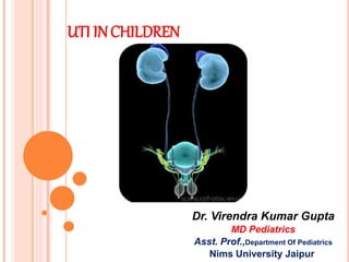
- 1. UTI INCHILDREN Dr. Virendra Kumar Gupta MD Pediatrics Asst. Prof.,Department Of Pediatrics Nims University Jaipur
- 2. DEFINITION: Infection of the urinary tract- Significant number of organisms Presence of symptoms inflammation of urinary tract Recurrent UTI- Recurrence of symptoms Significant bacteriuria Patients who have recovered clinically following treatment Common in girls.
- 3. EPIDEMIOLOGY UTI is a common bacterial infection in infants and children. The risk of having a UTI before the age of 14 yrs -1- 3% in boys - 3-10% in girls . In girls, the first UTI usually occurs by the age of 5 yr, with peaks during infancy and toilet training. In boys, most UTIs occur during the 1st yr of life; more common in uncircumcised boys. During the 1st yr of life, -M : F ratio is 2.8–5.4 : 1. Beyond 1–2 yr, -M : F ratio of 1 : 10.
- 4. Rapid evaluation and treatment of UTI is important to prevent renal parenchymal damage and renal scarring that can cause hypertension and progressive renal damage.
- 5. • Significant bacteriuria: Presence of at least 105 bacteria/ml of urine. • Asymptomatic bacteriuria : Bacteriuria with no symptoms. • Urethritis: infection of anterior urethral tract *dysuria, urgency and frequency of urination. • Cystitis: infection to urinary bladder *dysuria, frequency and urgency, pyuria and haematuria.
- 6. • Acute pyelonephritis: infection of one/both kidneys; sometimes lower tract also. *pyuria, fever, painful micturition • Chronic pyelonephritis: particular type of pathology of kidney; may/may not be due to infection.
- 7. UTI - TERMINOLOGY • Uncomplicated: UTI without underlying renal or neurologic disease. • Complicated: UTI with underlying structural, medical or neurologic disease. • Recurrent : > 3 symptomatic UTIs within 12 months following clinical therapy. • Reinfection: recurrent UTI caused by a different pathogen at any time • Relapse: recurrent UTI caused by same species causing original UTI within 2 wks after therapy.
- 8. CAUSE AND COURSE : G u t f l o r a B a c t e r i a l v i r u l e n c e U r o p a t h o g e n i c s t r a i n C o l o n i s a t i o n o f t h e u r e t h r a a n d t h e p e r i n e u m ( i n f e m a l e s t h e v a g i n a ) M u c o s a b a r r i e r e H o s t I n c r e a s e d a d h e r e n c e i m m u n s t a t u s V U R o b s t r u c t i o n f o r e i g n b o d y p r e v i o u s i n f l a m m a t i o n s c y s t i t i s a k u t e p y e l o n e p h r i t i s h e a l e d u r o s e p s i s s c a r h y p e r t e n s i o n C R F . . .
- 9. ETIOLOGY: Most common infecting pathogen : Escherichia Coli 80% of UTI. Other pathogens: - Staphylococcus & Streptococcus Species Enterobacteria ( Klebsiella, Proteus, pseudomonas) Occasionally Candida albicans
- 10. RISK FACTORS FOR URINARY TRACT INFECTION: Female gender Uncircumcised male Vesicoureteral reflux Toilet training Voiding dysfunction Obstructive uropathy Urethral instrumentation Wiping from back to front in females Bubble bath? Tight clothing Pinworm infestation Constipation Bacteria with P fimbriae Anatomic abnormality (labial adhesion) Neuropathic bladder Sexual activity Pregnancy
- 11. CLINICAL MANIFESTATIONS: The 3 Basic forms of UTI 1. Pyelonephritis 2. Cystitis 3. Asymptomatic bacteriuria
- 12. PYELONEPHRITIS Characterized by any or all of the following: abdominal or flank pain fever malaise nausea vomiting occasionally diarrhea. In newborns show nonspecific symptoms: poor feeding, irritability, and weight loss.
- 13. Acute lobar nephronia (acute lobar nephritis): localized renal bacterial infection involving >1 lobe Renal abscess : following a pyelonephritis or may be secondary to a primary bacteremia Perinephric abscesses: may be secondary to contiguous infection in the perirenal area
- 14. CYSTITIS Bladder involvement. Symptoms include dysuria urgency frequency suprapubic pain incontinence malodorous urine. Cystitis does not cause fever and does not result in renal injury.
- 15. ASYMPTOMATIC BACTERIURIA Positive urine culture without any infection It is most common in girls The incidence is 1–2% in preschool and school-age girls and 0.03% in boys. The incidence declines with increasing age.
- 16. DIAGNOSIS: While urinalysis enables a provisional diagnosis of UTI, a specimen must be obtained for culture prior to therapy with antibiotics Based on positive culture of a properly collected specimen of urine.
- 17. Significant pyuria defined as >10 WBC/mm3 in uncentrifuged sample,or >5 WBC/ mm3 in a centrifuged sample. Rapid dipstick based tests detect leukocyte esterase and nitrite useful in screening for UTI. A combination of these tests has moderate sensitivity and specificity for detecting UTI, and is diagnostically as useful as microscopy
- 18. COLLECTION OF SPECIMEN FOR CULTURE Clean-catch midstream Sample used to minimize contamination washing the genitalia with soap and water forced retraction of the prepuce are not advised Suprapubic aspiration And/or Transurethral catheterization In neonates and infants, urine sample is obtained by either above Both techniques are safe and easy to perform.
- 19. The urine specimen should be promptly plated within one hour of collection. If delay is anticipated, the sample can be stored in a refrigerator at 4ºC for up to 12-24 hours Cultures of specimens collected from urine bags have high false positive rates, and are not recommended.
- 20. A urine culture repeat in case contamination is suspected: mixed growth of two or more pathogens, or growth of organisms that normally constitute the periurethral flora (lacto-bacilli in healthy girls; enterococci in infants and toddlers) strongly suspected but colony counts are equivocal
- 22. With acute renal infection leukocytosis, neutrophilia, and elevated ESR and CRP are common. With a renal abscess the white blood cell count is markedly elevated to >20,000–25,000/mm3. Because sepsis is common in pyelonephritis, particularly in infants and in any child with obstructive uropathy, blood cultures should be considered.
- 23. TREATMENT: The patient’s age, features suggesting toxicity and dehydration, ability to retain oral intake and the likelihood of compliance with medication(s) help in deciding the need for hospitalization. Therapy should be prompt to reduce the morbidity of infection, minimize renal damage and subsequent complications.
- 24. Hospitalized and treated with parenteral antibiotics Choice of antibiotic- local sensitivity patterns A third generation cephalosporin is preferred. Aminoglycoside -in children with normal renal function. Intravenous therapy is given for the first 2-3 days followed by oral antibiotics once the clinical condition improves. Children less than 3 months of age and Those with complicated UTI
- 25. Are treated with oral antibiotics With adequate therapy, there is resolution of fever and reduction of symptoms by 48-72 hours Failure to respond may be due to presence of resistant pathogens, complicating factors or noncompliance; these patients require re-evaluation. Children with simple UTI and Those above 3 months of age
- 26. The duration of therapy -14 days for infants and children with complicated UTI - 7-10 days for uncomplicated UTI. Adolescents with cystitis may be treated with shorter duration of antibiotics, lasting 3 days Following the treatment of the UTI, prophylactic antibiotic therapy is initiated in children below 1 year of age, until appropriate imaging of the urinary tract is completed.
- 27. IMAGING STUDY IN FEBRILE UTI The aim of investigations is to identify patients at high risk of renal damage, chiefly those below one year of age, and those with VUR or urinary tract obstruction. Evaluation includes ultrasonography, DMSA renal scan and micturating cystourethrography (MCU/VCUG) performed USG provides information on kidney size, number and location, presence of hydronephrosis, urinary bladder anomalies and post- void residual urine. DMSA scintigraphy is a sensitive technique for detecting renal parenchymal infection and cortical scarring. MCU / VCUG detects VUR and provides anatomical details regarding the bladder and the urethra.
- 29. Ultrasonography should be done soon after the diagnosis of UTI. The MCU is recommended 2-3 weeks later. The DMSA scan is carried out 2-3 months after treatment.
- 30. PREVENTION OF RECURRENT UTI General Measures: Adequate fluid intake and frequent voiding Constipation should be avoided In children with VUR who are toilet trained, regular and volitional low pressure voiding with complete bladder emptying is encouraged. Double voiding ensures emptying of the bladder of post void residual urine. Circumcision reduces the risk of recurrent UTI in infant boys, and might therefore have benefits in patients with high grade reflux.
- 31. ANTIBIOTIC PROPHYLAXIS Long-term, low dose, antibacterial prophylaxis is used to prevent recurrent, febrile UTI. The antibiotic used should be effective, non-toxic with few side effects and should not alter the growth of commensals or induce bacterial resistance .
- 32. Antibiotic prophylaxis is recommended for patients with UTI below 1-yr of age, while awaiting imaging studies VUR Frequent febrile UTI (3 or more episodes in a year) even if the urinary tract is normal.
- 33. 1- VUR (it might be a complication of UTI, or primary causing UTI. 2- Scarring. Might lead to HTN, if multiple it might lead to renal insufficiency. 3- HTN 4- Renal insufficiency. Normal DMSA Acute Pyelonephritis Scarring VUR Acute: no irregular borders and no scarring and no change in size In chronic: irregular borders and scarring.
- 34. VESICOURETERIC REFLUX VUR is a bladder valve defect that allows urine to reflux from the bladder through one or both ureters and up to the Kidneys. •Febrile urinary tract infection (UTI) is the defining Symptom.
- 35. VUR is seen in 40-50% infants and 30-50% children with UTI, and resolves with age. Its severity is graded using the International Study Classification from grade I to V, based on the appearance of the urinary tract on MCU. The presence of moderate to severe VUR, particularly if bilateral, is an important risk factor for pyelonephritis and renal scarring, with subsequent risk of hypertension, albuminuria and progressive kidney disease. The risk of scarring is highest in the first year of life
- 36. VUR GRADES
- 38. SCREENING OF SIBLINGS AND OFFSPRING: Reflux is inherited in an autosomal dominant manner with incomplete penetrance; 27% siblings and 35% offspring of patients show VUR. Ultrasonography is recommended to screen for the presence of reflux. Further imaging is required if ultrasonography is abnormal THANKS