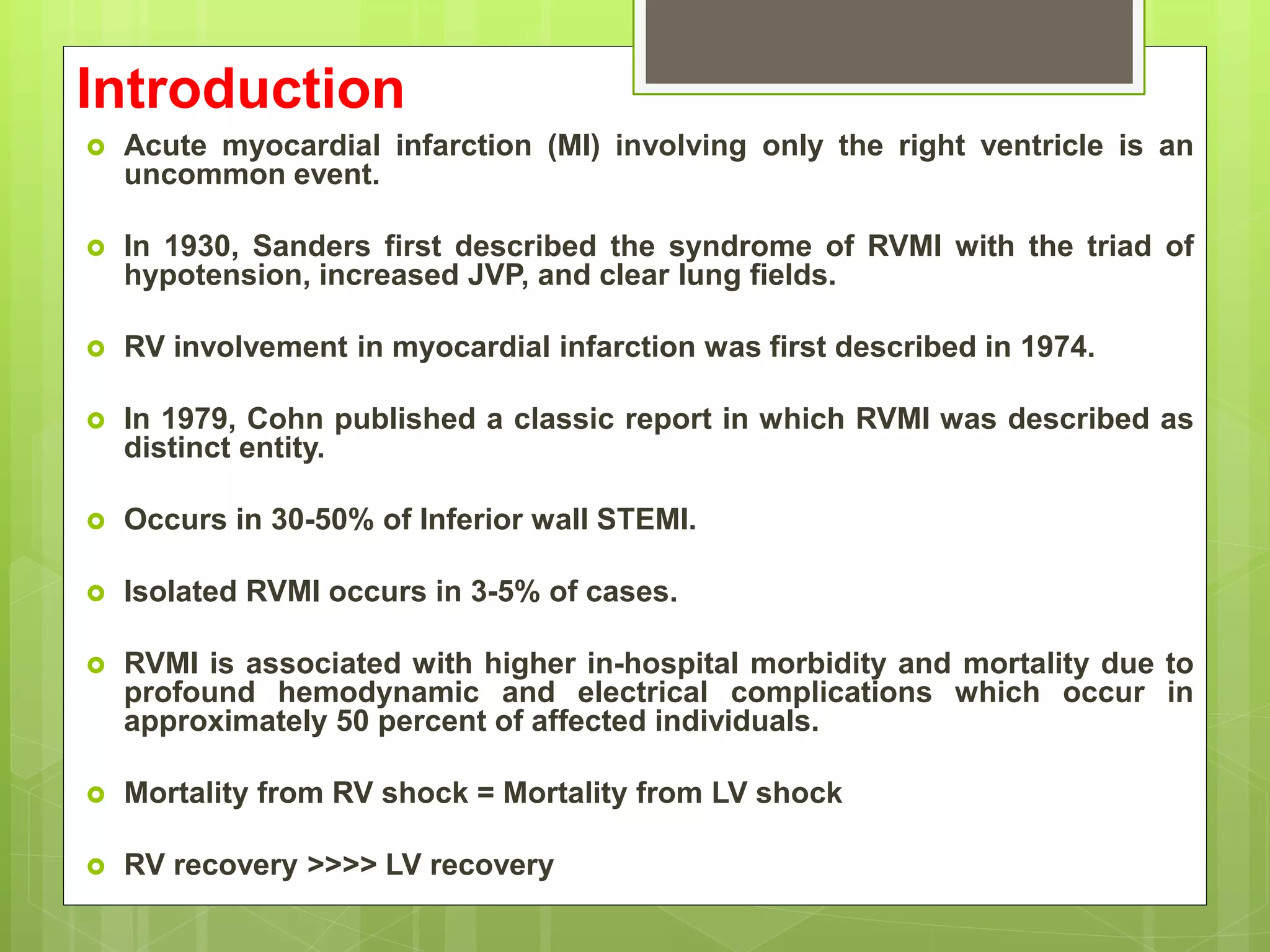
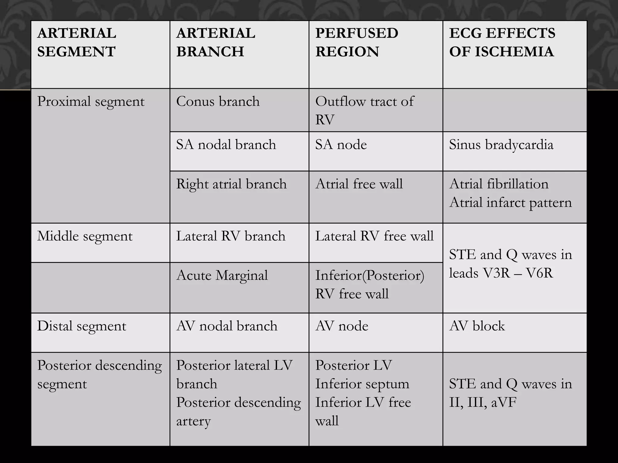
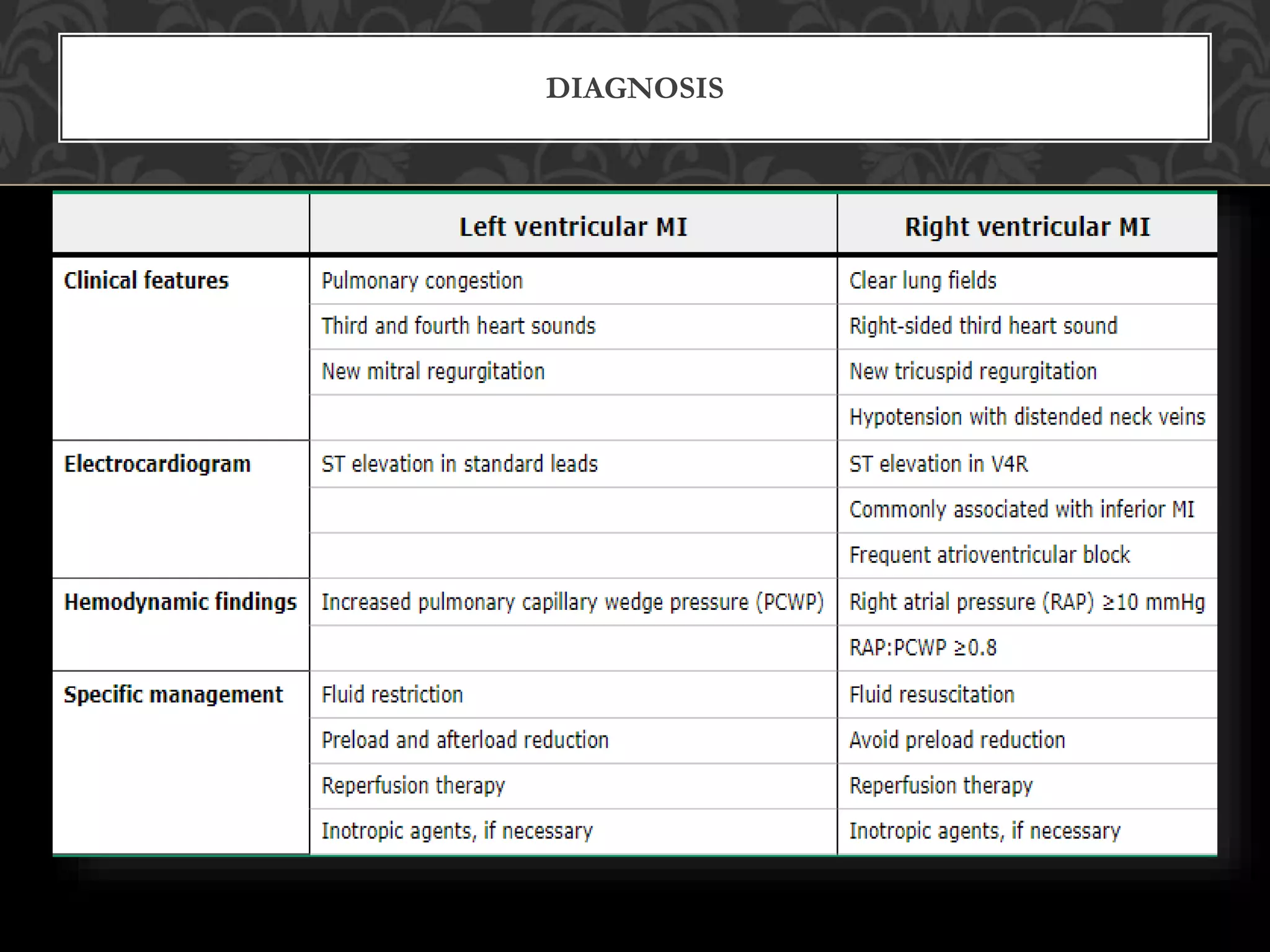
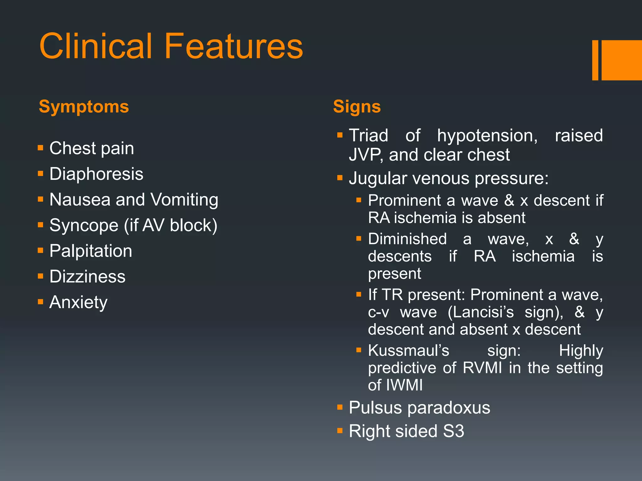
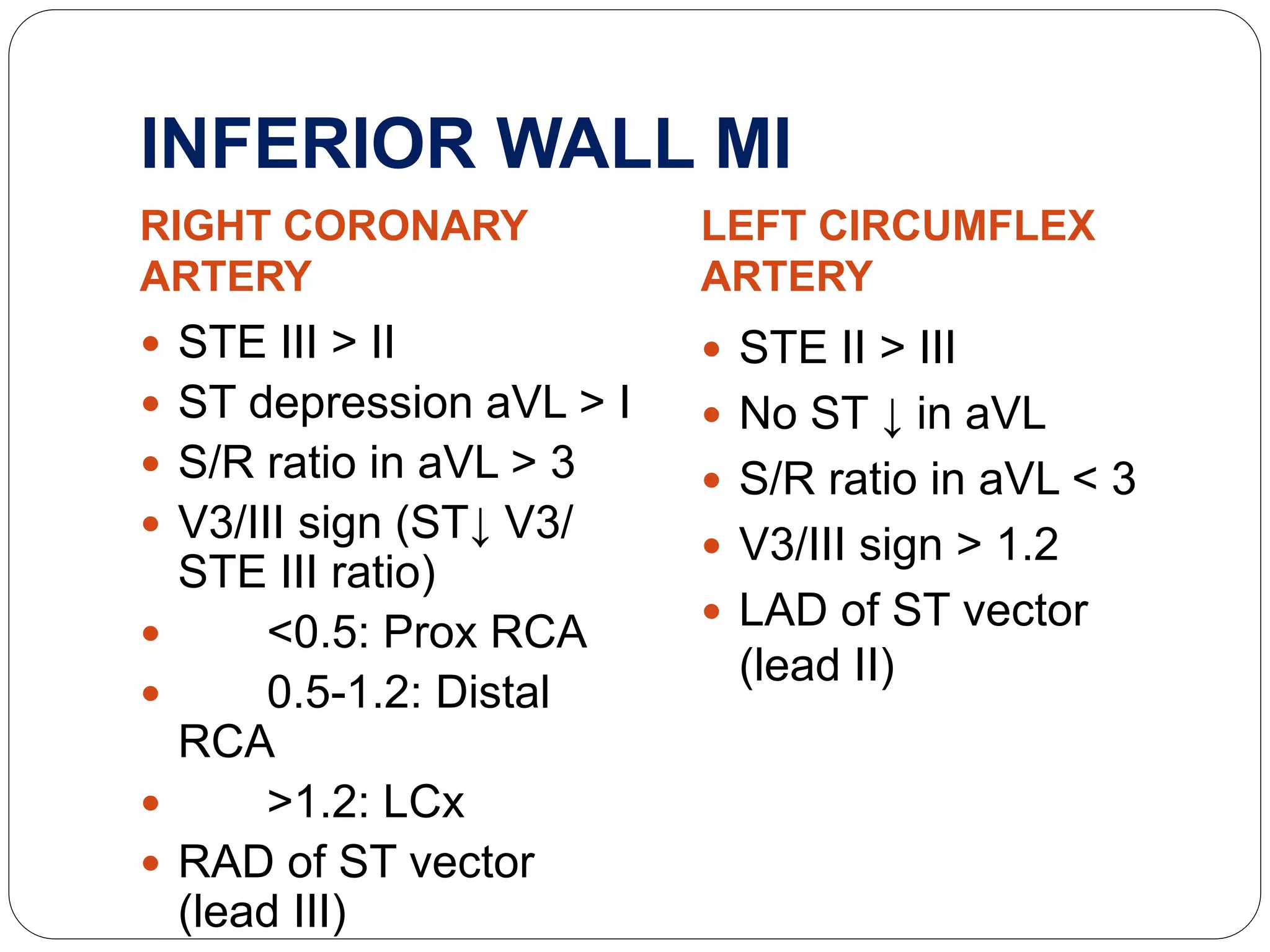
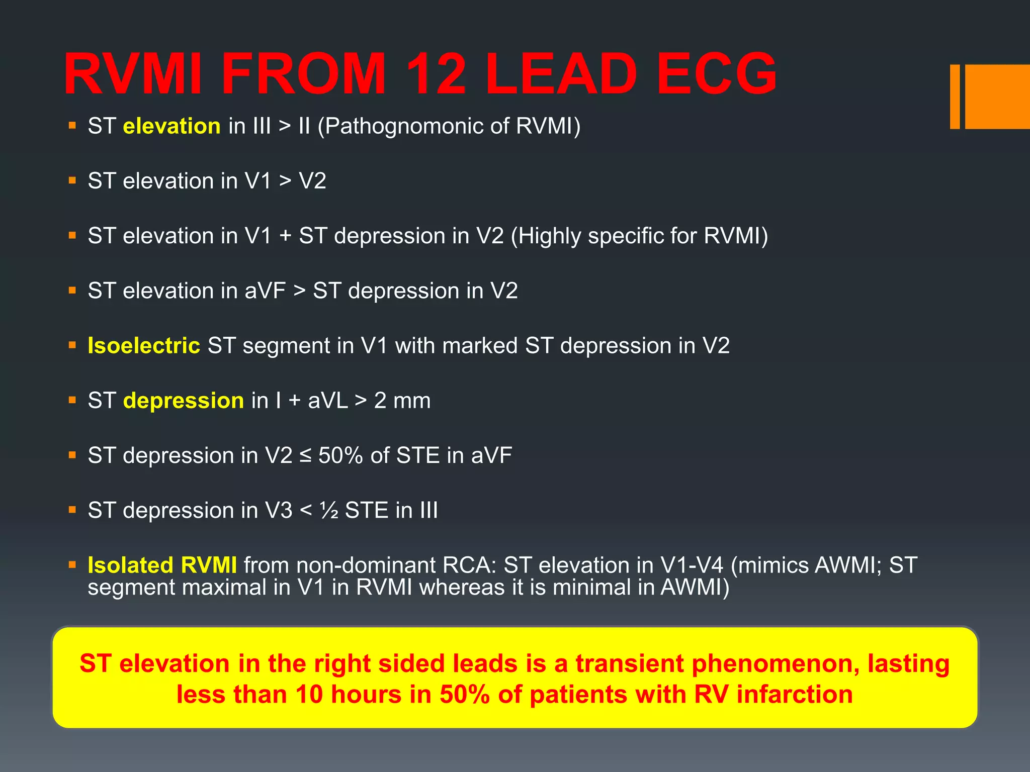
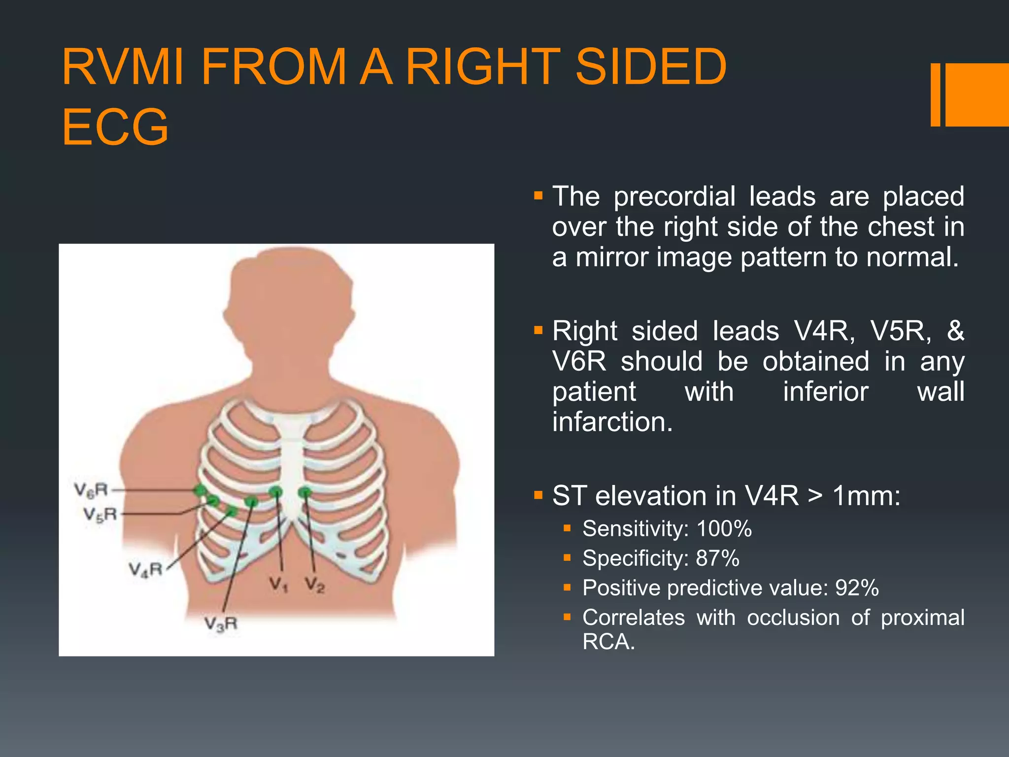
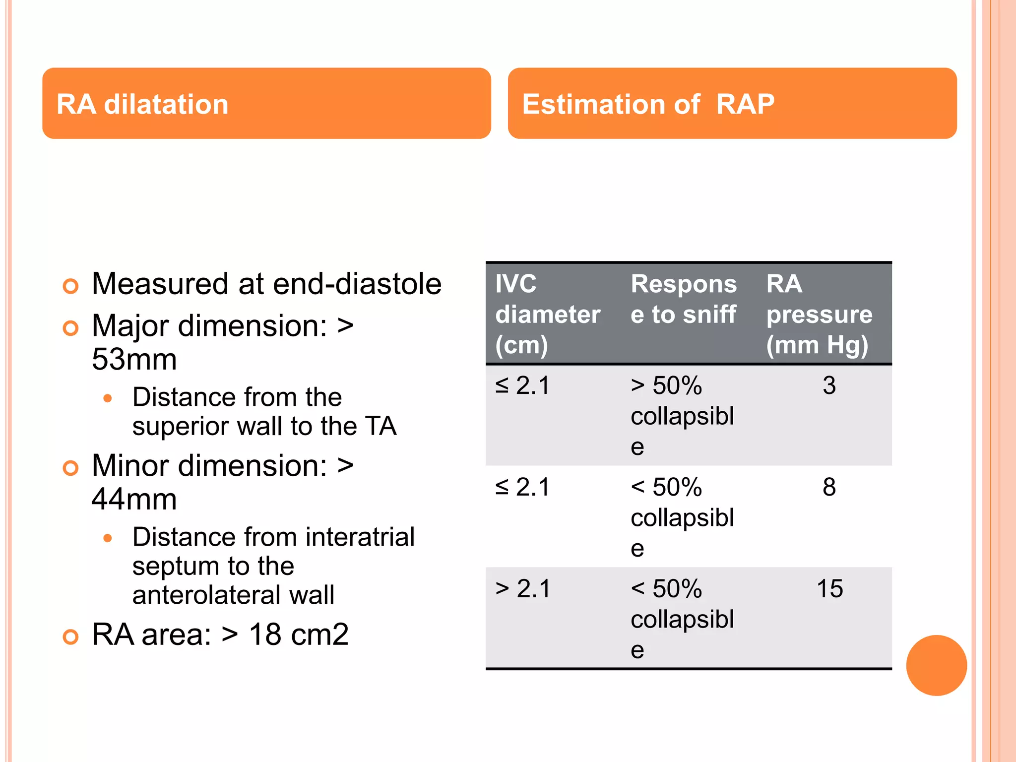
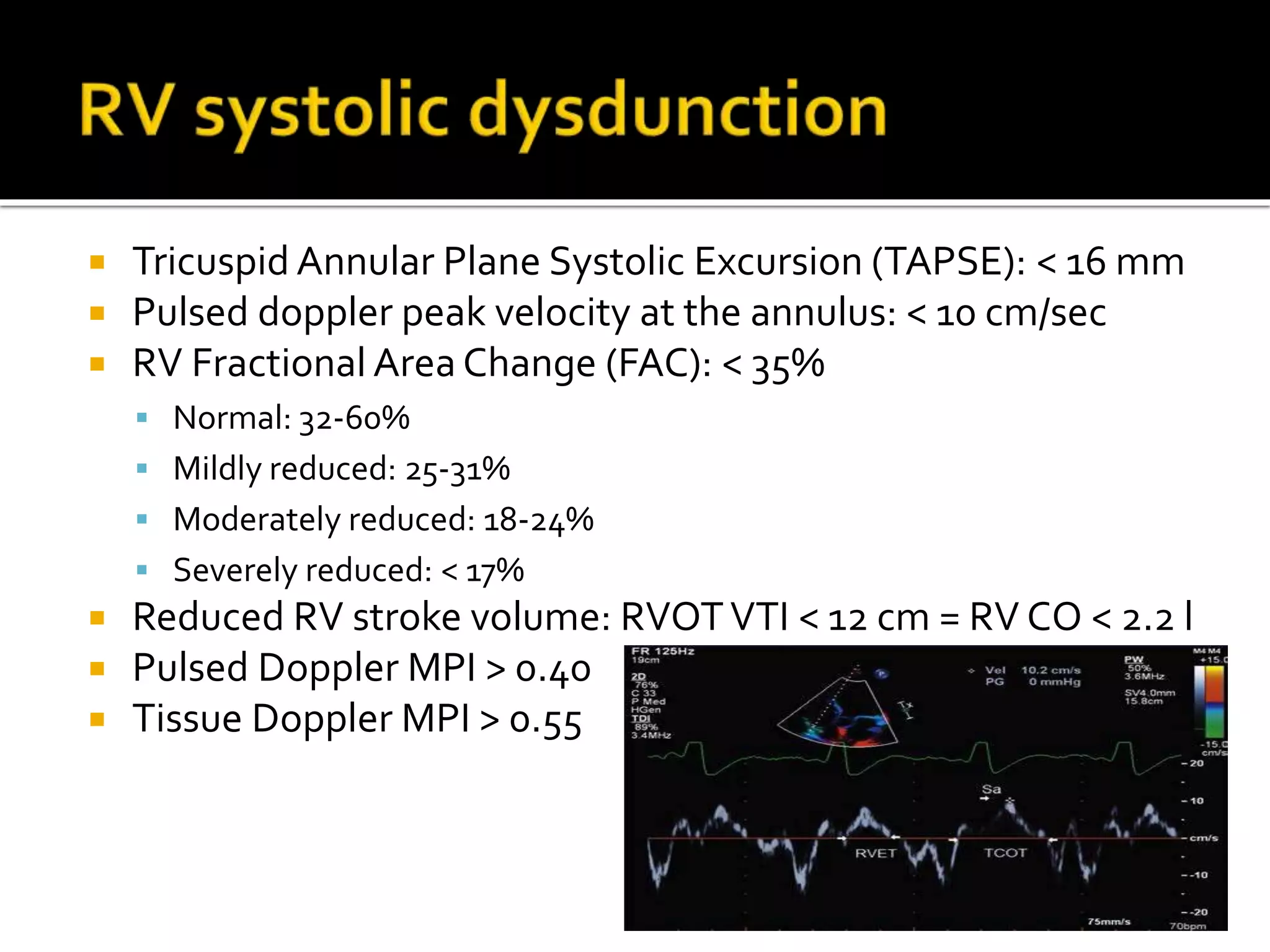
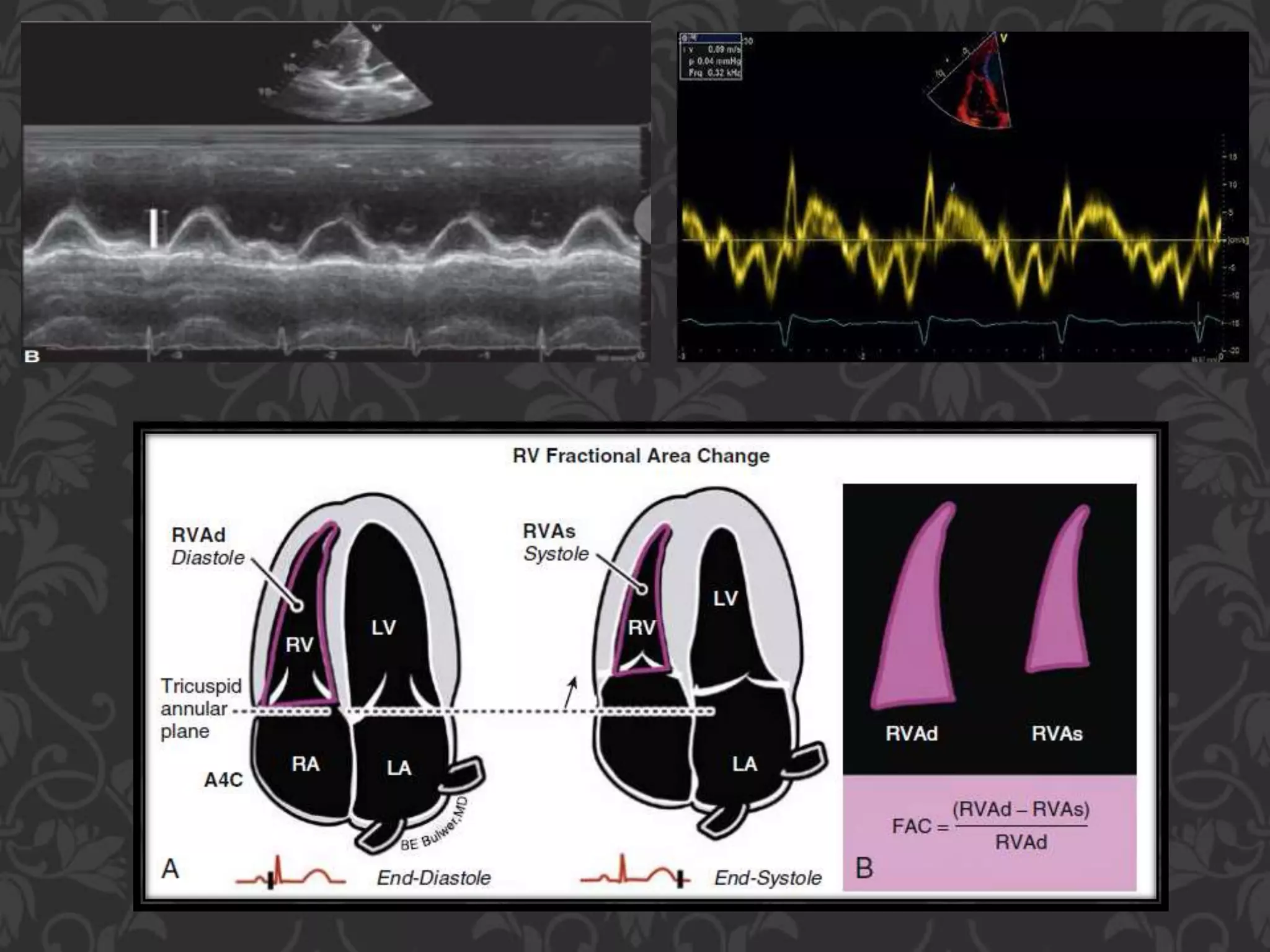
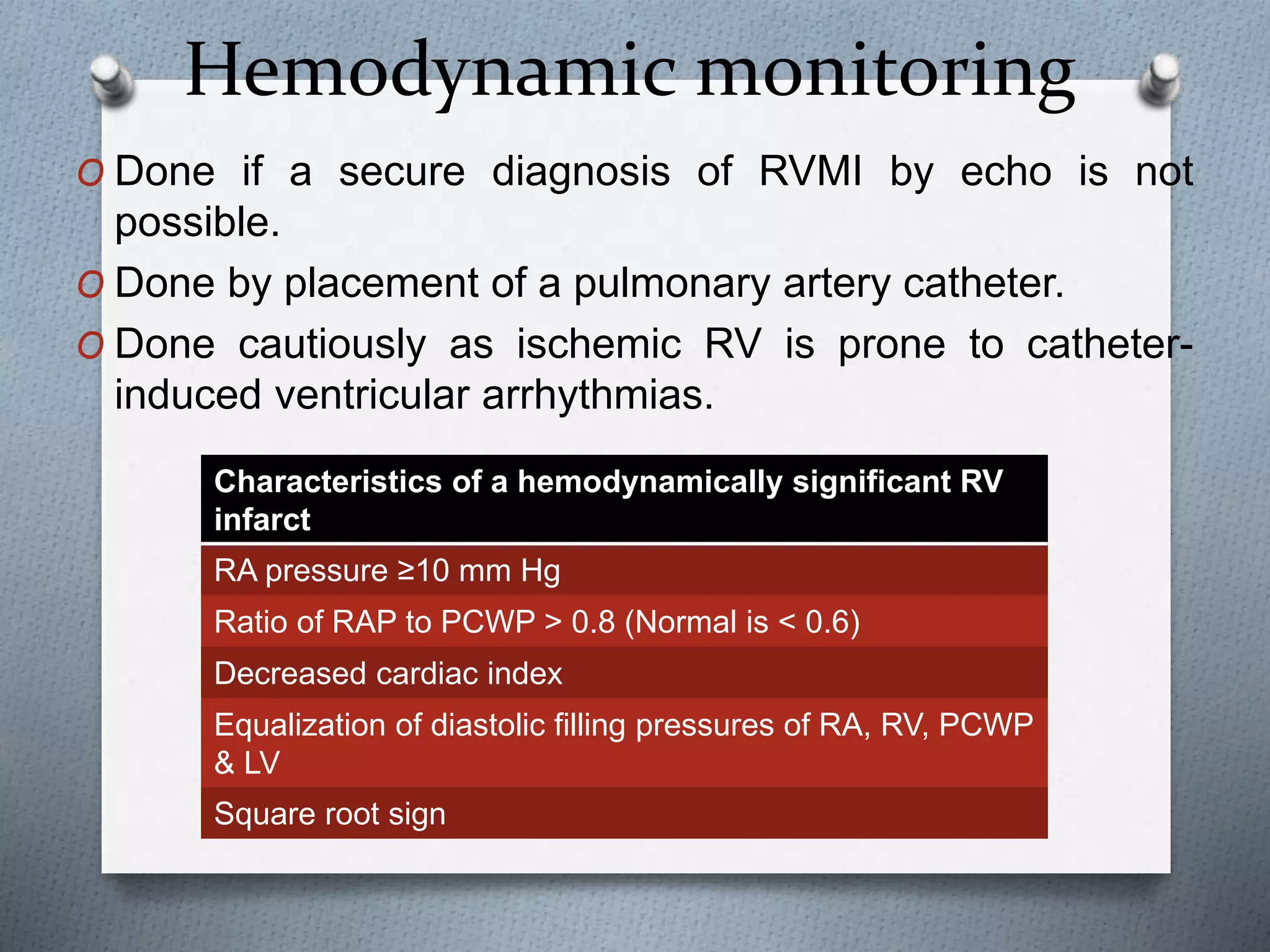
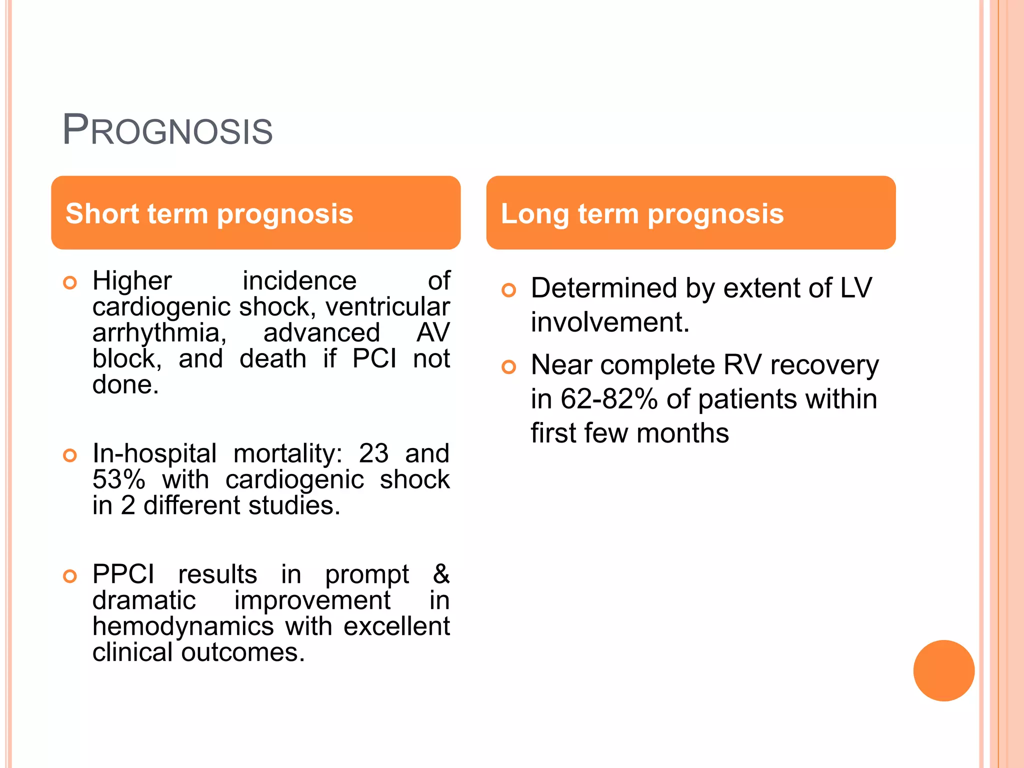
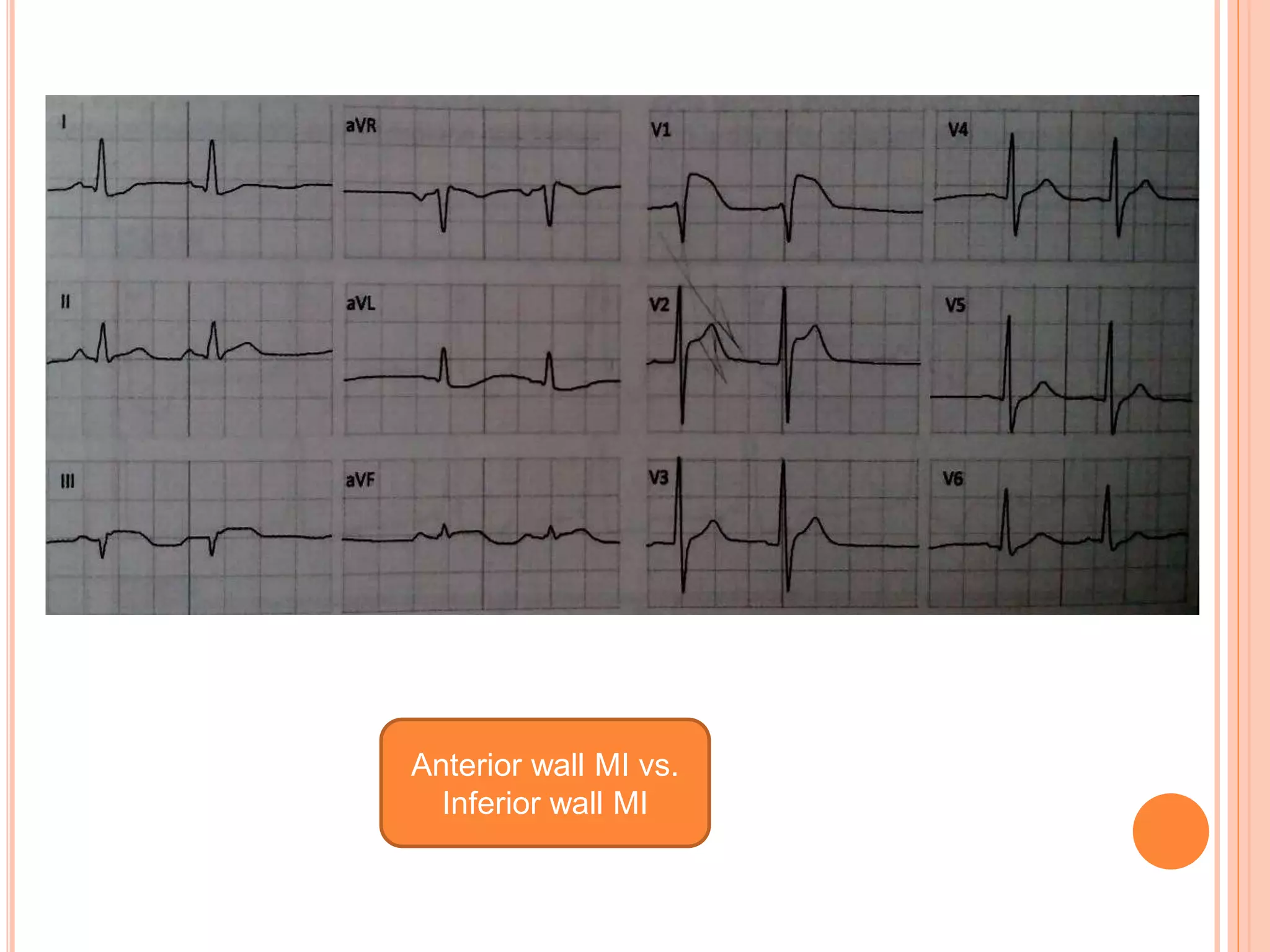
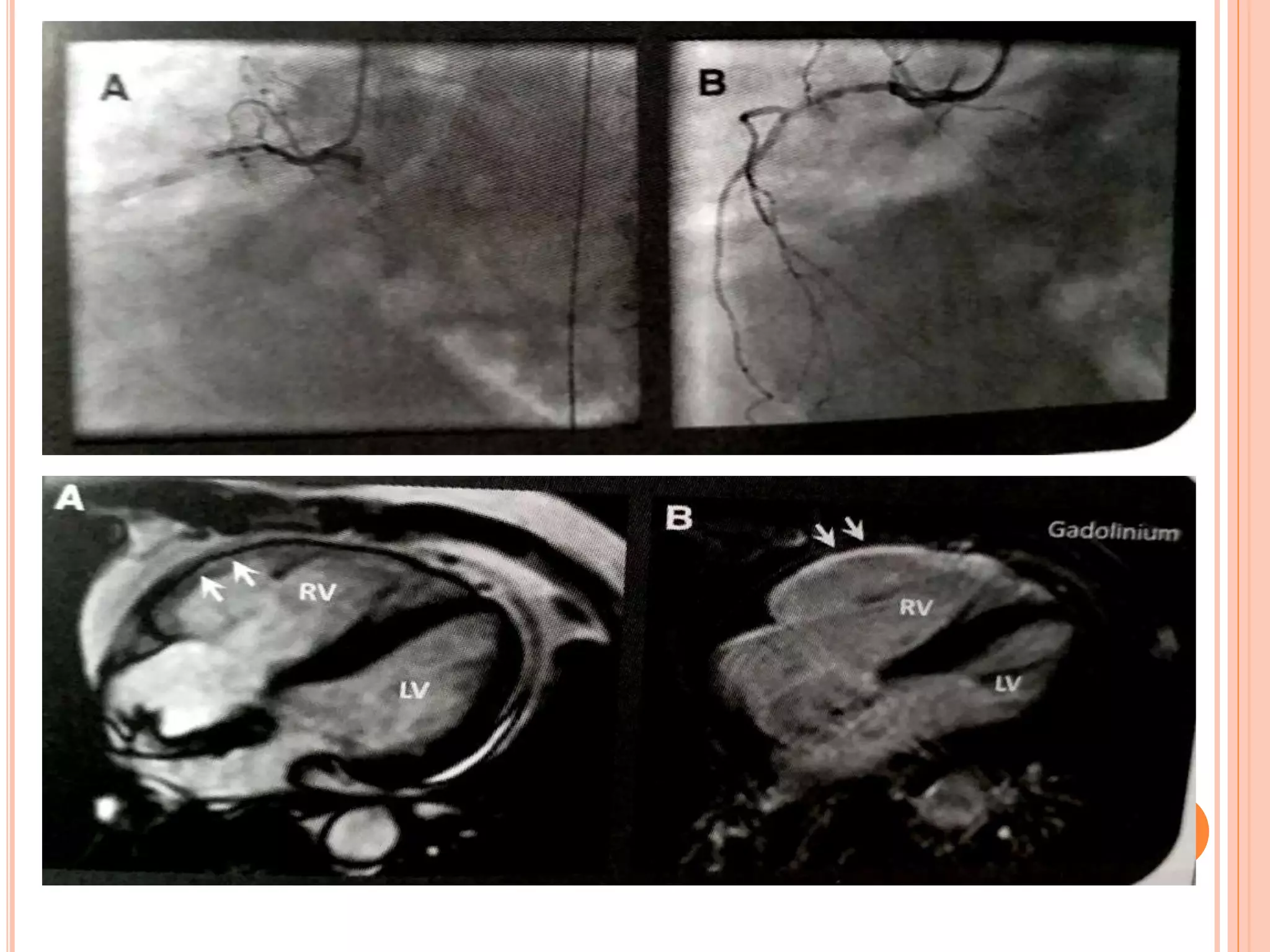
This document discusses right ventricular myocardial infarction (RVMI). Some key points:
1. RVMI occurs in 30-50% of inferior wall ST-elevation MIs and 3-5% of MIs are isolated RVMI.
2. RVMI is associated with higher morbidity and mortality than LV MI due to hemodynamic and electrical complications in around 50% of cases.
3. Diagnosis of RVMI can be made based on ECG findings of ST elevation in leads V1-V3 and III>II, and echo findings of RV dilation, hypokinesis of the RV free wall, and elevated right-sided pressures.
4. Management of RVMI focuses on optimizing RV
Right ventricular infarction (RVI) is rare but can occur alongside inferior wall myocardial infarction. It carries a higher mortality risk than inferior MI alone. RVI results from occlusion of the right coronary artery, with clinical features including hypotension, clear lung fields, and elevated jugular venous pressure. Diagnosis involves ECG showing ST elevations in right-sided leads and echocardiogram demonstrating right ventricular dysfunction. Treatment aims to support right ventricular preload and restore atrioventricular synchrony using inotropic support when needed. Combined therapies including inhaled nitric oxide and IABP may benefit patients with cardiogenic shock.
This document provides an overview of echocardiographic assessment of mitral regurgitation. It describes the anatomy of the mitral valve including the leaflets, annulus, chordae, and papillary muscles. It discusses Carpentier's functional classification system for describing the mechanism of mitral valve dysfunction. Methods for assessing severity are covered, including color flow imaging, continuous wave Doppler, vena contracta width, proximal isovelocity surface area, and volumetric assessment. Key points are made about evaluating jet direction, duration, and velocity in context of blood pressure. The importance of assessing left ventricular and left atrial size and function is also highlighted.
This document discusses cardiac tamponade, which occurs when fluid rapidly accumulates in the pericardial sac, putting pressure on the heart and reducing cardiac function. Key points include:
- Pericardial effusion puts pressure on the heart, causing symptoms like chest pain and shortness of breath.
- Cardiac tamponade occurs when a rapid accumulation of fluid in the pericardial sac severely compresses the heart.
- Echocardiography is useful for diagnosing tamponade by showing findings like pericardial effusion, right ventricular collapse, and reduced respiratory variation in blood flow velocities.
- Tamponade is a medical emergency treated initially with medications and peric
preop TEE assessment of atrial septal defect is very important for making decision for device closure, properly assessed adequate rims of ASD will reduce risk of device embolization to almost nil.
This document provides an overview of echocardiography in pericardial diseases. It begins with an introduction to pericardial anatomy and pathophysiology. It then discusses various pericardial diseases that can be evaluated by echocardiography, including acute pericarditis, recurrent pericarditis, pericardial effusions, cardiac tamponade, and constrictive pericarditis. For each condition, it describes the echocardiographic findings and techniques used to evaluate the condition. It emphasizes that echocardiography is usually the initial imaging test of choice but that CT or CMR may be needed in some complex cases.
This document summarizes the evaluation of aortic valve stenosis using echocardiography. It describes the normal aortic valve anatomy and various types of aortic valve stenosis including calcific, bicuspid, rheumatic, and supravalvular or subvalvular stenosis. Doppler echocardiography is used to evaluate aortic valve stenosis severity based on valve area, mean gradient, and peak jet velocity. Stress echocardiography with dobutamine can help distinguish true severe from pseudo-severe low-flow, low-gradient aortic stenosis.
The document discusses constrictive pericarditis, providing details on:
1) The pathology of constrictive pericarditis which involves thickening and scarring of the pericardium leading to loss of elasticity.
2) The pathophysiology of constrictive pericarditis where the inelastic pericardium constrains cardiac filling and prevents adaptation to volume changes.
3) Key diagnostic features of constrictive pericarditis seen on echocardiogram include septal bounce, rapid early diastolic mitral inflow, and increased mitral annular velocities that rise with inspiration.
Echo assessment of lv systolic function and swmaFuad Farooq
This document discusses various techniques for assessing left ventricular systolic function using echocardiography, including:
- Visual assessment of endocardial motion and wall thickening to evaluate global and regional function
- Quantitative measures like fractional shortening, ejection fraction, and volumes
- Tissue Doppler imaging of mitral annular velocities
- Tissue tracking and strain imaging to evaluate timing and extent of myocardial contraction
- Wall motion scoring to characterize regional abnormalities
This document discusses development of percutaneous mitral valve repair techniques and clinical trials. It provides background on chronic mitral regurgitation (MR) and the limitations of medical and surgical treatment. Percutaneous mitral valve repair offers benefits over surgery like reduced morbidity and shorter recovery. The document describes the four main percutaneous repair methods and focuses on the MitraClip edge-to-edge leaflet repair system, including patient selection criteria, procedure steps, and clinical trial results demonstrating safety and effectiveness for treating MR.
Evaluation of prosthetic valve function and clinical utility.Ramachandra Barik
Many of the prosthesis-related complications can be prevented or their impact minimized through optimal prosthesis selection in the individual patient and careful medical management and follow-up after implantation.
This document provides an overview of cardiac resynchronization therapy (CRT). It discusses how conduction delays can lead to electromechanical dyssynchrony and impair the heart's function. CRT aims to improve this synchrony and thereby improve systolic and diastolic function. The document outlines different types of dyssynchrony and methods to assess it, including echocardiography. Current guidelines recommend CRT for symptomatic heart failure patients with low ejection fraction and wide QRS duration. The implantation procedure involves placing right atrial/ventricular leads and a left ventricular lead via the coronary sinus.
1) Transthoracic and transesophageal echocardiography are important modalities for assessing atrial septal defects (ASDs). TTE can identify RV volume overload and septal flattening, while TEE precisely measures defect size and evaluates rim morphology.
2) The four main types of ASDs - ostium secundum, ostium primum, sinus venosus, and coronary sinus defects - have distinguishing echo features. Doppler can demonstrate shunt direction and magnitude.
3) Echocardiography guides percutaneous ASD closure by assessing defect and rim anatomy, device sizing, and post-procedure result. Understanding echo features is key to ensuring procedure success.
Mitral valve anatomy - ppt by kunwar sidharthkunwar sidharth
The mitral valve, also known as the bicuspid valve or left atrioventricular valve, lies between the left atrium and left ventricle. It is a dual flap valve made up of the mitral annulus, two leaflets (anterior and posterior), chordae tendineae connecting the leaflets to the papillary muscles of the left ventricle, and the left ventricular wall. The mitral valve apparatus works to ensure unidirectional blood flow from the left atrium to the left ventricle during diastole and prevent backflow during systole.
Ventricular septal defects (VSDs) are openings in the wall separating the ventricles of the heart. There are four main types classified by location: membranous, muscular, supracristal, and inlet VSDs. Echocardiography is useful for diagnosing VSDs and assessing their characteristics like location, size, and impact on cardiac function. VSDs range from small and asymptomatic to large defects causing heart failure or pulmonary hypertension. Surgical or catheter-based closure may be required for large VSDs.
This document discusses techniques for localizing the site of origin of ventricular tachycardia based on electrocardiogram characteristics. It describes that right ventricular outflow tract tachycardias typically present with left bundle branch block morphology while left ventricular sites may present with either right or left bundle branch block depending on exit site. Specific leads are discussed that can provide clues about anterior vs posterior, septal vs free wall origin within the outflow tracts. Other areas like fascicles, papillary muscles and mitral/tricuspid annuli are also summarized.
This document discusses several normal anatomic variants that can be mistaken for pathologies in echocardiographic imaging of the heart structures. It describes variants commonly seen in the right atrium, left atrium, left ventricle, aortic valve, and right ventricle which include things like Eustachian valves, Chiari networks, prominent trabeculations, moderator bands, and false tendons. It emphasizes that recognizing these normal variants is important to avoid misdiagnosis and unnecessary treatment based on imaging findings alone.
HCM – Presentation, Hemodynamics and InterventionAnkur Gupta
This document describes a case of a 50-year-old female presenting with symptoms of breathlessness, angina, and presyncope. Echocardiography revealed asymmetric septal hypertrophy and systolic anterior motion of the mitral valve, consistent with hypertrophic obstructive cardiomyopathy (HOCM). The document then provides detailed background information on HOCM, including definitions, pathophysiology, clinical presentation, diagnostic testing, and treatment options such as beta-blockers, septal ablation, and disqualification from competitive sports in severe cases.
Stent thrombosis is a rare but serious complication of percutaneous coronary intervention (PCI) with mortality rates between 25-40%. It is classified based on timing (acute, subacute, late, very late) and etiology (primary, secondary). Risk factors include premature discontinuation of dual antiplatelet therapy, smoking, diabetes, chronic kidney disease, acute coronary syndrome, and high platelet reactivity. Strategies to minimize stent thrombosis involve careful patient selection, optimal stent deployment, adherence to potent dual antiplatelet regimens, and treatment involving emergent thrombectomy with escalated antiplatelet therapy.
The mitral valve lies between the left atrium and left ventricle. Mitral stenosis is usually caused by rheumatic fever which causes scarring of the mitral valve leaflets and commissures. In early mitral stenosis, the leaflets can open but have restricted motion. Over time, the leaflets become thickened and rigid, reducing valve opening. This causes symptoms like dyspnea and pulmonary hypertension. On examination, findings may include an irregular pulse from atrial fibrillation, elevated jugular venous pressure, accentuated S1, and a diastolic murmur. Severe mitral stenosis can lead to right heart failure and complications like hemoptysis.
This document discusses fractional flow reserve (FFR), a technique used during coronary catheterization to measure pressure differences across a coronary stenosis and determine if it is causing myocardial ischemia. An FFR value below 0.75 is considered functionally significant while a value above 0.80 rules out ischemia. FFR is useful for evaluating single-vessel disease, left main stenosis, tandem lesions, diffuse disease, grafts, and ostial lesions. Limitations include inability to assess plaque morphology.
The document defines no-reflow as inadequate myocardial perfusion through a coronary circulation segment without mechanical vessel obstruction. No-reflow occurs in 30% of patients after reperfusion for myocardial infarction and is associated with worse outcomes. It results from microvascular obstruction from distal embolization, ischemic injury, and reperfusion injury. Diagnosis involves assessing TIMI flow, myocardial blush grade, and imaging techniques. Prevention focuses on reducing embolization using thrombectomy or filters while treatment involves vasodilators like adenosine, verapamil, and glycoprotein IIb/IIIa inhibitors.
A lecture on the echocardiographic evaluation of hypertrophic cardiomyopathy. Starts with an overview of the topic then a systematic approach to diagnosis and then a differential diagnosis followed by take-home messages and conclusion.
determining the suitability of the mitral valve for repair most likely in patients with mitral regurgitation due to myxomatous degeneration and is least likely in patients with regurgitation due to endocarditis most likely with posterior prolapse or flail, whereas ileaflet involvement and isolated anterior leaflet prolapse reduce the likelihood of successful repair substantially.
This document summarizes dobutamine stress echocardiography (DSE). Key points include:
- DSE uses the drug dobutamine to simulate exercise and increase heart rate, contractility, and myocardial oxygen demand to detect ischemia.
- It is useful for evaluating ischemia, viability, and valvular dysfunction in patients unable to exercise.
- The document reviews the DSE protocol, interpretation of wall motion abnormalities, indications, side effects, and applications for assessing ischemic heart disease, viability, valvular stenosis like mitral and aortic stenosis, and pulmonary hypertension.
This document discusses left atrial thrombus in patients with rheumatic mitral stenosis. It finds that 26-33% of patients with severe mitral stenosis have left atrial thrombi, which are associated with a higher risk of embolic events. The document classifies different types of left atrial thrombi and examines determinants of thrombus formation like atrial fibrillation, left atrial size, and severity of mitral stenosis. It recommends anticoagulation to reduce thromboembolic risk, noting studies have found anticoagulation facilitates thrombus resolution.
This document discusses the use of echocardiography in evaluating various types of cardiomyopathies. It provides echocardiographic features of dilated cardiomyopathy including dilated chambers, normal wall thickness, and complications like mitral regurgitation. Hypertrophic cardiomyopathy features include unexplained hypertrophy, diastolic dysfunction, and left ventricular outflow tract obstruction. Restrictive cardiomyopathies show hypertrophy, enlarged atria, restricted filling, and elevated pressures. Left ventricular non-compaction and arrhythmogenic right ventricular cardiomyopathy also have distinct echocardiographic characteristics described.
Echocardiographic Evaluation of LV Diastolic FunctionJunhao Koh
The document discusses methods for evaluating left ventricular diastolic function using echocardiography. It describes the four phases of diastole, parameters used to assess diastolic function including mitral inflow patterns, mitral annular tissue Doppler, pulmonary vein flow, left atrial size and the Tei index. Grades of diastolic dysfunction and approaches from ASE/EAE and Mayo Clinic are summarized. Continuous wave Doppler of aortic regurgitation is also presented as a noninvasive method to evaluate left ventricular relaxation.
This document discusses the echocardiographic evaluation of mitral valve prolapse (MVP). It describes the use of M-mode, 2D, and 3D echocardiography to diagnose MVP and assess mitral regurgitation severity. Measurement of the vena contracta and use of the proximal isovelocity surface area method are emphasized for accurate regurgitant quantification. Surgical indications and repair techniques are also summarized.
LOCALIZATION OF CULPRIT ARTERY IN MI.pptxAnayaAnaya14
- The document discusses the coronary artery anatomy and blood supply of the heart. The right coronary artery supplies the right atrium and ventricle as well as parts of the left ventricle. The left main coronary artery branches into the left anterior descending artery and left circumflex artery.
- Electrocardiogram patterns for different types of myocardial infarction are described based on the affected coronary artery. ST segment elevation in different leads helps localize the site of infarction. The hyperacute, evolved and chronic phases of myocardial infarction and the associated ECG changes are also summarized.
Ventricular tachycardia can occur in structurally normal hearts. It is classified based on origin, morphology, response to exercise and drugs. Non-life threatening VT is often monomorphic and originates from sites like outflow tracts and fascicles. Outflow tract VT commonly originates from the right ventricular outflow tract. Other sites include the left ventricular outflow tract and aortic cusps. Treatment includes medications, ablation, and implantable cardioverter-defibrillators for more severe cases. Life-threatening VT is often polymorphic and associated with genetic ion channel disorders like long QT syndrome.
This document reviews features of electrocardiograms (ECGs) that can help localize the occluded vessel in acute myocardial infarction. The ST segment changes seen on ECGs form the basis for diagnosing the location and severity of the infarction. Specific patterns of ST elevation or depression in different leads can indicate occlusion of the left anterior descending coronary artery, right coronary artery, or circumflex artery. Factors like the extent of ST elevation and resolution of changes with reperfusion also provide prognostic information. Localizing the culprit vessel guides treatment decisions and allows assessment of the affected cardiac region.
This document provides information on ECG localization of myocardial infarction. It discusses the branches of the coronary arteries and their blood supply territories. It describes concepts like injury vector, ST elevation and depression cut-offs, and ECG changes seen in different types of MI such as anterior wall MI, inferior wall MI, right ventricular MI, and posterior wall MI. It also discusses ECG patterns that can indicate high-risk coronary artery disease such as left main occlusion. Overall, the document is a guide for using the ECG to localize the site and artery of myocardial infarction.
CT plays an important role in evaluating various congenital heart diseases. It can be used to confirm diagnoses, assess anatomy in detail, evaluate shunts and obstructions, and help determine the best treatment approach. The document discusses how CT can evaluate conditions like atrial and ventricular septal defects, patent ductus arteriosus, transposition of the great arteries, tetralogy of Fallot, total anomalous pulmonary venous return, pulmonary vein anomalies, aortic stenosis, coarctation of the aorta, and more. CT provides detailed anatomical information to complement other imaging modalities in the assessment of complex congenital heart disease.
1. Ventricular septal defects (VSDs) are one of the most common congenital heart defects, accounting for 20-30% of cases in India.
2. The natural history and progression of a VSD depends on factors like its size, location, and the development of pulmonary hypertension.
3. Small VSDs have over a 50% chance of spontaneous closure by age 5, while larger defects often require surgical intervention. Without treatment, complications can include congestive heart failure, pulmonary vascular disease, bacterial endocarditis, and aortic regurgitation.
Left ventricular angiography is used to assess global and regional left ventricular function and anatomy. It involves inserting a catheter into the left ventricle and injecting contrast dye to visualize the ventricle on x-ray imaging. The procedure provides key information on mitral valve function, ventricular shape and wall motion abnormalities, and congenital defects like VSD. LV volumes and ejection fraction are calculated from the images to quantify function. Regional wall motion is graded and correlated to coronary artery territories. Characteristic appearances are seen in conditions like cardiomyopathy, mitral regurgitation, and septal defects. Potential complications include arrhythmias and endocardial injury.
Aortic regurgitation is a type of heart valve disease in which the valve does not close properly as a result some of the blood is back flow from the Aorta into the left ventricle during diastole .Regurgitation is seen in echo Guidance by using Parasternal long axis view AND Parasternal short axis view ,apical 5 chamber view and apical 3 chamber view it is the best seen in color flow imaging .The severity of this is calculated and evaluated by VC,PISA,EROA,PHT,CW DOPPLER, REGURGITANT VOLUME ,REGURGITANT FRACTION this echo approach use to all Healthcare workers and the health care students it is easily understandable .I hope that u will support me and stimulate me to do more presentation related on this category Love u all for the supports Thank u guys
CULPRIT ARTERY LOCALISATION IN STEMI _ DR BIJILESH.ppsxAnayaAnaya14
1) Careful analysis of the surface ECG is highly useful in localizing the culprit vessel in STEMI patients and providing immediate prognostic information.
2) The ECG can help determine whether an aggressive reperfusion strategy is needed by identifying the blocked artery.
3) Different ECG patterns in leads and precordial leads can indicate whether the left anterior descending artery, right coronary artery, or circumflex artery is blocked, as well as the location of blockage along those arteries. Localizing the blocked vessel helps guide reperfusion treatment.
The document summarizes coronary artery anatomy. It describes the origins and branches of the right coronary artery and left coronary artery. The right coronary artery typically arises from the right coronary sinus and supplies the right ventricle. The left main coronary artery bifurcates into the left anterior descending artery and circumflex artery. It also discusses common anatomical variations such as anomalous origins, fistulas, and intrinsic abnormalities like stenosis.
Pulmonary atresia with intact ventricular septum (PAIVS) is a congenital heart defect where the pulmonary valve is blocked, preventing blood flow from the right ventricle to the lungs. It occurs in 1-3% of congenital heart diseases. Surgical interventions for PAIVS have improved, with 5-year survival rates now around 80%. Treatment depends on factors like the size of the tricuspid valve and whether the coronary arteries depend on blood flow from the right ventricle. Options include biventricular repair, univentricular repair, or transplantation.
Definition of LVA Centerline analysis of RWMA on LV angio in 30º RAO shows hypocontractile segments moving more than 2 standard deviations out of normal range.
1. The document discusses electrocardiographic (ECG) interpretation including determining cardiac rate and rhythm, identifying conduction disturbances, myocardial ischemia or infarction, and other abnormalities.
2. It provides details on properly placing ECG leads and determining the cardiac axis. Common rhythms, conduction blocks, hypertrophy, and other ECG findings are explained.
3. A mnemonic device, RRAHIM, is presented to guide the systematic interpretation of an ECG, covering rate, rhythm, axis, hypertrophy, ischemia/infarction, and other findings.
This document discusses hemodynamic principles and waveform analysis from cardiac catheterization. It provides an overview of normal and abnormal pressure waveforms from the right atrium, pulmonary artery, left ventricle, and aorta. Key points include how respiration and different disease states can impact pressure tracings. The document also outlines the components and uses of the Swan-Ganz catheter for measuring pressures in the right heart and pulmonary circulation.
This document provides information about aortic stenosis, including its definition, location, morphology, etiology, types, stages, symptoms, complications, risk factors, diagnostic testing, and management. Aortic stenosis is a narrowing of the aortic valve that reduces or blocks blood flow from the heart to the rest of the body. It is most commonly caused by calcification of the aortic valve and affects older adults. Diagnosis involves echocardiography to evaluate valve anatomy and hemodynamics. Treatment depends on symptom severity and may include medications, lifestyle changes, surgical valve replacement, or transcatheter aortic valve replacement.
Atrial septal defect (ASD), ventricular septal defect (VSD), patent ductus arteriosus (PDA), and tetralogy of Fallot (TOF) are four common types of congenital heart disease. ASD is a hole in the atrial septum that allows blood to flow from the left to the right atrium. VSD is a hole in the ventricular septum that allows blood to flow between the ventricles. PDA is a persistent opening between the aorta and pulmonary artery that normally closes after birth. TOF involves four abnormalities that reduce pulmonary blood flow.
Double outlet right ventricle (DORV) is a heart defect where both the aorta and pulmonary artery arise completely or primarily from the right ventricle. It can cause varying degrees of cyanosis and congestive heart failure depending on pulmonary pressures and associated defects. Echocardiography is important for assessing the relationship of the great vessels to the ventricles, presence of a ventricular septal defect, and other structural issues to determine appropriate surgical repair. Management may involve biventricular repair in the neonatal period or staged palliation depending on the specific anatomy and physiology in each case.
Right Ventricle Anatomy, Physiology & ECHO Assessment by Dr. Vaibhav Yawalka...vaibhavyawalkar
This document provides an overview of right ventricle anatomy, physiology, and echocardiographic assessment. It describes the irregular shape and trabeculated structure of the right ventricle. The physiology section covers the RV's adaptation to volume overload through distensibility and compliance. Echocardiographic assessment techniques are outlined, including measurements of RV dimensions, fractional area change, TAPSE, tissue Doppler imaging, and the TEI index. The document provides a detailed but technical summary of right ventricular structure and function.
This case report describes a rare case of aneurysm of the sinus of Valsalva (ASOV) that dissected into and ruptured within the interventricular septum, creating a communication with the left ventricle. Echocardiography revealed a large ASOV originating from the right coronary cusp that burrowed into and dissected nearly the entire interventricular septum, with a small perforation allowing flow into the left ventricle. The patient underwent surgical repair with patch closure of the ASOV mouth and thrombosed septal cavity. This is a very rare occurrence, as septal dissection is an uncommon complication of ASOV, and left ventricular communication is even r
This document provides guidance on managing common issues in cardiology consultations, including bleeding risks with antithrombotic medications, acute coronary syndrome management in chronic kidney disease patients, anticoagulation in pregnancy with mechanical heart valves, and timing of stopping antiplatelet therapies before surgery. It discusses evaluation and treatment of bleeding events of varying severity caused by anticoagulants or antiplatelet drugs. It also reviews cardiac biomarker interpretation and revascularization approaches in chronic kidney disease.
1) Multiple trials have evaluated various oral antiplatelet agents for reducing mortality in patients with acute coronary syndromes. The ISIS-2 trial showed aspirin reduces mortality in STEMI patients with no increased bleeding risk.
2) Clopidogrel, prasugrel, ticagrelor, and cangrelor are P2Y12 receptor antagonists used in addition to aspirin. They differ in reversibility, activation pathway, onset and offset of action, and drug interactions. Trials have shown these agents reduce ischemic events when added to aspirin.
3) Later trials evaluated higher doses of antiplatelet agents or administering them in alternative ways like crushing tablets. While some strategies led
1) Risk scores help identify patients with NSTE-ACS who are at higher risk of death and recurrent cardiac events in both the short and long term.
2) Studies have found reductions in mortality rates for NSTE-ACS patients in recent years, which is due in part to improvements in pharmacological and invasive coronary strategies.
3) The GRACE risk score is the most widely used and has been shown to accurately predict mortality in NSTE-ACS patients at both 30 days and 1 year. It helps guide clinical decision making regarding treatment strategies.
Principles of diagnosis & management of acute pulmonaryVijay Yadav
This document provides an overview of the principles of diagnosis and management of acute pulmonary embolism (PE). It discusses the epidemiology of PE including incidence and mortality rates. It covers the pre-test clinical assessment, diagnostic testing including D-dimer, chest imaging, echocardiogram and CT pulmonary angiography. It also discusses prognostic factors in echocardiogram and other biomarkers. The document outlines the management of PE including hemodynamic support, anticoagulation, thrombolysis, percutaneous interventions, surgical thrombectomy and inferior vena cava filters. It concludes with recommendations for long-term anticoagulation or secondary prophylaxis based on the condition that caused the PE.
Perioperative myocardial infarction or injury after noncardiac surgeryVijay Yadav
1) Perioperative myocardial injury and infarction are common complications after non-cardiac surgery, occurring in up to 17% of high-risk patients, with many events going unrecognized due to a lack of symptoms.
2) Both symptomatic and asymptomatic perioperative myocardial infarctions are associated with a significant increase in short- and long-term mortality.
3) Routine screening for myocardial injury using high-sensitivity troponin measurements can help identify at-risk patients, and secondary prevention treatments with aspirin, statins, and other medications may provide mortality benefits.
2019 esc guidelines for the management of patientsVijay Yadav
This document discusses guidelines for managing supraventricular tachycardia. It classifies different types of SVT such as atrial tachycardias, AV junctional tachycardias, and AV re-entrant tachycardias. It describes the characteristics, ECG patterns, mechanisms, and treatment recommendations for different SVTs including AV nodal reentrant tachycardia, focal atrial tachycardia, atrial flutter, and AV reciprocating tachycardias. The guidelines provide classifications for acute and chronic therapy for SVTs, with recommendations for electrical or pharmacological cardioversion as well as catheter ablation procedures.
1. Acute viral hepatitis is defined as inflammation of the liver caused by viral infection, resulting in elevated liver enzymes for less than 6 months. The most common causes are hepatitis A, B, C, D, E viruses.
2. The document discusses the pathogenesis, clinical features, investigations, management, and prevention of different viral hepatitis types. Hepatitis A and E are usually self-limiting while hepatitis B, C and D can sometimes lead to chronic liver disease.
3. Supportive care is the main treatment approach for most cases of acute viral hepatitis. Specific antiviral therapy may be used for severe hepatitis B cases to prevent progression to liver failure. Vaccines exist to prevent hepatitis A and B infection
This document provides information about acute liver failure (ALF), including its definition, etiology, pathophysiology, clinical features, investigations, prognosis, and management. ALF is characterized by severe liver injury and encephalopathy within 8 weeks without pre-existing liver disease. Common causes include drugs/toxins, viral hepatitis, and autoimmune conditions. In ALF, liver cells die rapidly, impairing ammonia clearance and coagulation factor production, which can lead to cerebral edema, coagulopathy, and multi-organ failure without transplantation. Prognosis is assessed using tools like KCH or ALFED scores. Management involves supportive care, treating the underlying cause, and considering transplantation for eligible patients.
8 Surprising Reasons To Meditate 40 Minutes A Day That Can Change Your Life.pptxHolistified Wellness
We’re talking about Vedic Meditation, a form of meditation that has been around for at least 5,000 years. Back then, the people who lived in the Indus Valley, now known as India and Pakistan, practised meditation as a fundamental part of daily life. This knowledge that has given us yoga and Ayurveda, was known as Veda, hence the name Vedic. And though there are some written records, the practice has been passed down verbally from generation to generation.
Rasamanikya is a excellent preparation in the field of Rasashastra, it is used in various Kushtha Roga, Shwasa, Vicharchika, Bhagandara, Vatarakta, and Phiranga Roga. In this article Preparation& Comparative analytical profile for both Formulationon i.e Rasamanikya prepared by Kushmanda swarasa & Churnodhaka Shodita Haratala. The study aims to provide insights into the comparative efficacy and analytical aspects of these formulations for enhanced therapeutic outcomes.
Basavarajeeyam is a Sreshta Sangraha grantha (Compiled book ), written by Neelkanta kotturu Basavaraja Virachita. It contains 25 Prakaranas, First 24 Chapters related to Rogas& 25th to Rasadravyas.
TEST BANK For Community Health Nursing A Canadian Perspective, 5th Edition by...Donc Test
TEST BANK For Community Health Nursing A Canadian Perspective, 5th Edition by Stamler, Verified Chapters 1 - 33, Complete Newest Version Community Health Nursing A Canadian Perspective, 5th Edition by Stamler, Verified Chapters 1 - 33, Complete Newest Version Community Health Nursing A Canadian Perspective, 5th Edition by Stamler Community Health Nursing A Canadian Perspective, 5th Edition TEST BANK by Stamler Test Bank For Community Health Nursing A Canadian Perspective, 5th Edition Pdf Chapters Download Test Bank For Community Health Nursing A Canadian Perspective, 5th Edition Pdf Download Stuvia Test Bank For Community Health Nursing A Canadian Perspective, 5th Edition Study Guide Test Bank For Community Health Nursing A Canadian Perspective, 5th Edition Ebook Download Stuvia Test Bank For Community Health Nursing A Canadian Perspective, 5th Edition Questions and Answers Quizlet Test Bank For Community Health Nursing A Canadian Perspective, 5th Edition Studocu Test Bank For Community Health Nursing A Canadian Perspective, 5th Edition Quizlet Test Bank For Community Health Nursing A Canadian Perspective, 5th Edition Stuvia Community Health Nursing A Canadian Perspective, 5th Edition Pdf Chapters Download Community Health Nursing A Canadian Perspective, 5th Edition Pdf Download Course Hero Community Health Nursing A Canadian Perspective, 5th Edition Answers Quizlet Community Health Nursing A Canadian Perspective, 5th Edition Ebook Download Course hero Community Health Nursing A Canadian Perspective, 5th Edition Questions and Answers Community Health Nursing A Canadian Perspective, 5th Edition Studocu Community Health Nursing A Canadian Perspective, 5th Edition Quizlet Community Health Nursing A Canadian Perspective, 5th Edition Stuvia Community Health Nursing A Canadian Perspective, 5th Edition Test Bank Pdf Chapters Download Community Health Nursing A Canadian Perspective, 5th Edition Test Bank Pdf Download Stuvia Community Health Nursing A Canadian Perspective, 5th Edition Test Bank Study Guide Questions and Answers Community Health Nursing A Canadian Perspective, 5th Edition Test Bank Ebook Download Stuvia Community Health Nursing A Canadian Perspective, 5th Edition Test Bank Questions Quizlet Community Health Nursing A Canadian Perspective, 5th Edition Test Bank Studocu Community Health Nursing A Canadian Perspective, 5th Edition Test Bank Quizlet Community Health Nursing A Canadian Perspective, 5th Edition Test Bank Stuvia
Local Advanced Lung Cancer: Artificial Intelligence, Synergetics, Complex Sys...Oleg Kshivets
Overall life span (LS) was 1671.7±1721.6 days and cumulative 5YS reached 62.4%, 10 years – 50.4%, 20 years – 44.6%. 94 LCP lived more than 5 years without cancer (LS=2958.6±1723.6 days), 22 – more than 10 years (LS=5571±1841.8 days). 67 LCP died because of LC (LS=471.9±344 days). AT significantly improved 5YS (68% vs. 53.7%) (P=0.028 by log-rank test). Cox modeling displayed that 5YS of LCP significantly depended on: N0-N12, T3-4, blood cell circuit, cell ratio factors (ratio between cancer cells-CC and blood cells subpopulations), LC cell dynamics, recalcification time, heparin tolerance, prothrombin index, protein, AT, procedure type (P=0.000-0.031). Neural networks, genetic algorithm selection and bootstrap simulation revealed relationships between 5YS and N0-12 (rank=1), thrombocytes/CC (rank=2), segmented neutrophils/CC (3), eosinophils/CC (4), erythrocytes/CC (5), healthy cells/CC (6), lymphocytes/CC (7), stick neutrophils/CC (8), leucocytes/CC (9), monocytes/CC (10). Correct prediction of 5YS was 100% by neural networks computing (error=0.000; area under ROC curve=1.0).
Integrating Ayurveda into Parkinson’s Management: A Holistic ApproachAyurveda ForAll
Explore the benefits of combining Ayurveda with conventional Parkinson's treatments. Learn how a holistic approach can manage symptoms, enhance well-being, and balance body energies. Discover the steps to safely integrate Ayurvedic practices into your Parkinson’s care plan, including expert guidance on diet, herbal remedies, and lifestyle modifications.
TEST BANK For Basic and Clinical Pharmacology, 14th Edition by Bertram G. Kat...rightmanforbloodline
TEST BANK For Basic and Clinical Pharmacology, 14th Edition by Bertram G. Katzung, Verified Chapters 1 - 66, Complete Newest Version.
Histololgy of Female Reproductive System.pptxAyeshaZaid1
Dive into an in-depth exploration of the histological structure of female reproductive system with this comprehensive lecture. Presented by Dr. Ayesha Irfan, Assistant Professor of Anatomy, this presentation covers the Gross anatomy and functional histology of the female reproductive organs. Ideal for students, educators, and anyone interested in medical science, this lecture provides clear explanations, detailed diagrams, and valuable insights into female reproductive system. Enhance your knowledge and understanding of this essential aspect of human biology.
2. Introduction
Acute myocardial infarction (MI) involving only the right ventricle is an
uncommon event.
In 1930, Sanders first described the syndrome of RVMI with the triad of
hypotension, increased JVP, and clear lung fields.
RV involvement in myocardial infarction was first described in 1974.
In 1979, Cohn published a classic report in which RVMI was described as
distinct entity.
Occurs in 30-50% of Inferior wall STEMI.
Isolated RVMI occurs in 3-5% of cases.
RVMI is associated with higher in-hospital morbidity and mortality due to
profound hemodynamic and electrical complications which occur in
approximately 50 percent of affected individuals.
Mortality from RV shock = Mortality from LV shock
RV recovery >>>> LV recovery
3. RIGHTVENTRICLE
Trabeculated endocardial
surface
Thin walled (< 5mm)
Three papillary muscles
Moderator band
Triangular shaped cavity
Tricuspid atrioventricular
valve with relatively apical
insertion
Crista supraventricularis
separates the tricuspid and
pulmonary valves
LEFTVENTRICLE
Smooth endocardial surface
Thick walled
Two papillary muscles
False tendon
Elliptical shaped cavity
Mitral atrioventricular valve
with relatively basal insertion
The mitral and aortic valves
share fibrous continuity
4. RV PHYSIOLOGY AT A GLANCE
The RV wall is thinner (< 5mm) and more complaint than the LV
wall.
Composed of circumferential fibers in the subepicardium and
longtudional fibers in the subendocardium.
There is inward, longitudinal, and circumferential traction in RV due
to LV contraction.
Longitudinal shortening is the major contributor to the overall RV
performance.
Normal RV contraction occurs as a peristaltic wave directed from
inflow to infundibulum.
RV pumps the same stroke volume as the LV but uses only 25% of
the stroke work because of low resistance of pulmonary
vasculature.
RV is closely connected to LV:
◦ Share a wall (IVS)
◦ RV free wall is attached to the anterior and inferior IVS
◦ Have mutually encircling epicardial fibers
◦ Share the same intrapericardial space
◦ Ventricular Interdependence
5.
6. ARTERIAL
SEGMENT
ARTERIAL
BRANCH
PERFUSED
REGION
ECG EFFECTS
OF ISCHEMIA
Proximal segment Conus branch Outflow tract of
RV
SA nodal branch SA node Sinus bradycardia
Right atrial branch Atrial free wall Atrial fibrillation
Atrial infarct pattern
Middle segment Lateral RV branch Lateral RV free wall
STE and Q waves in
leads V3R – V6RAcute Marginal Inferior(Posterior)
RV free wall
Distal segment AV nodal branch AV node AV block
Posterior descending
segment
Posterior lateral LV
branch
Posterior descending
artery
Posterior LV
Inferior septum
Inferior LV free
wall
STE and Q waves in
II, III, aVF
7. Factors that make the right
ventricle less susceptible to
infarction:
8. LC blood flow is impeded by
extravascular compressive forces
generated by systolic LV contraction.
These forces are so high that LC blood
flow is briefly reversed.
LC blood flow increases to a maximum
early in diastole and then falls
gradually following the decline in aortic
pressure during the remainder of
diastole.
Because of a lower developed pressure
in the RV, there is no systolic inhibition
of RC blood flow.
RC blood flow follows the shape of the
aortic pressure curve and remains
appreciable throughout the entire
cardiac cycle.
9. 2) Reduced myocardial oxygen demand because of smaller
muscle mass and low afteralod.
3) Reduced myocardial oxygen uptake and blood flow
4) Oxygen extraction reserve
5) More extensive collateral flow from left to right coronary
arteries
6) Greater degree of ischemic preconditioning
7) Ability to downregulate the metabolic demand during
coronary hypoperfusion.
10. Pathophysiology
RVI results in reduced RV systolic contraction (RVSP and PP are
decreased)
RVI results in RV diastolic dysfunction (elevated right sided filling
pressures like CVP, RA, and RVEDP).
Reduction in RV output & blood supply to the lungs.
Reduced pulmonary flow decreases pulmonary venous return to LA
and LV. (decreases LV preload and LV filling).
Reduced LV output and systolic BP.
RVI leads to RV dilatation which alters the motion of IVS; i.e. leftward
shift of septum during diastole which further impedes LV filling and
eventually reduces CO.
LV dyssynchrony due to abnormal septal motion and loss of AV
synchrony when there is AV block also leads to decreased CO.
Dilatation of RV enlarges tricuspid annulus which results in functional
TR that further reduces RV output.
When RCA occlusion is proximal to right atrial branch, RA ischemia
occurs that diminishes its contraction and increases RA pressure and
further increases the probability of atrial arrhythmia.
11. Elevated right sided filling pressure in the
presence of normal pulmonary artery and left
sided filling pressure is the hallmark of RVI
RA pressure: 10 mm Hg
PCWP: 1-5 mm Hg
Sensitivity: 73%
Specificity: 100%
13. Symptoms Signs
Clinical Features
Chest pain
Diaphoresis
Nausea and Vomiting
Syncope (if AV block)
Palpitation
Dizziness
Anxiety
Triad of hypotension, raised
JVP, and clear chest
Jugular venous pressure:
Prominent a wave & x descent if
RA ischemia is absent
Diminished a wave, x & y
descents if RA ischemia is
present
If TR present: Prominent a wave,
c-v wave (Lancisi’s sign), & y
descent and absent x descent
Kussmaul’s sign: Highly
predictive of RVMI in the setting
of IWMI
Pulsus paradoxus
Right sided S3
14. INFERIOR WALL MI
RIGHT CORONARY
ARTERY
LEFT CIRCUMFLEX
ARTERY
STE III > II
ST depression aVL > I
S/R ratio in aVL > 3
V3/III sign (ST↓ V3/
STE III ratio)
<0.5: Prox RCA
0.5-1.2: Distal
RCA
>1.2: LCx
RAD of ST vector
(lead III)
STE II > III
No ST ↓ in aVL
S/R ratio in aVL < 3
V3/III sign > 1.2
LAD of ST vector
(lead II)
15. RVMI FROM 12 LEAD ECG
ST elevation in III > II (Pathognomonic of RVMI)
ST elevation in V1 > V2
ST elevation in V1 + ST depression in V2 (Highly specific for RVMI)
ST elevation in aVF > ST depression in V2
Isoelectric ST segment in V1 with marked ST depression in V2
ST depression in I + aVL > 2 mm
ST depression in V2 ≤ 50% of STE in aVF
ST depression in V3 < ½ STE in III
Isolated RVMI from non-dominant RCA: ST elevation in V1-V4 (mimics AWMI; ST
segment maximal in V1 in RVMI whereas it is minimal in AWMI)
ST elevation in the right sided leads is a transient phenomenon, lasting
less than 10 hours in 50% of patients with RV infarction
16. RVMI FROM A RIGHT SIDED
ECG
The precordial leads are placed
over the right side of the chest in
a mirror image pattern to normal.
Right sided leads V4R, V5R, &
V6R should be obtained in any
patient with inferior wall
infarction.
ST elevation in V4R > 1mm:
Sensitivity: 100%
Specificity: 87%
Positive predictive value: 92%
Correlates with occlusion of proximal
RCA.
18. *
*Sensitivity: 82% & Specificity: 93% for detection of RVI.
*The specificity may be decreased by pre-existent pulmonary
diseases (COPD, PE)
*Most specific: RV free wall hypokinesia
*RV dilatation with paradoxical septal motion
*RA dilatation and increased RAP
*RV systolic dysfunction
*Functional tricuspid regurgitation : Hallmark
*Persistent bowing of IAS from right to left (RAP > LAP)
*Patent PFO on saline contrast echo leading to profound
hypoxemia
*Dilated IVC with poor respirophasic variation
19. Qualitative
Quantitative
RV dilatation
In A4CH view
Mildly enlarged:
RV is enlarged but < LV
Moderately enlarged:
RV = LV
Severely enlarged:
RV > LV
Apex of heart comprised of
RV
In RV focused A4CH at end-
diastole
RV basal diameter > 4.2 cm
RV midcavity diameter > 3.5 cm
RV longitudional diameter > 8.6
cm
RVOT PLAX proximal diameter
> 3.5 cm
RVOT PSAX distal diameter >
2.7 cm
20. Measured at end-diastole
Major dimension: >
53mm
Distance from the
superior wall to the TA
Minor dimension: >
44mm
Distance from interatrial
septum to the
anterolateral wall
RA area: > 18 cm2
IVC
diameter
(cm)
Respons
e to sniff
RA
pressure
(mm Hg)
≤ 2.1 > 50%
collapsibl
e
3
≤ 2.1 < 50%
collapsibl
e
8
> 2.1 < 50%
collapsibl
e
15
RA dilatation Estimation of RAP
23. PULSED WAVE
DOPPLER MPI
1) Tricuspid valve inflow &
Pulmonary valve outflow
doppler tracings are
acquired in RV modified
A4CH and PSAX views.
2) Time duration from the
end of A wave to the
onset of E wave is
calculated. (TV closure
to opening time)
3) RVET from pulmonary
doppler tracing is
measured.
4) Isovolumic time =
(RVET – TV A to E
duration)
5) RVMPI = Isovolumic
time/RVET
6) In this case it is (386 –
271)/271 = 0.43
25. OTHER IMAGING STUDIES FOR RVMI
Radionuclide
ventriculography &
99mTC-pyrophosphate
myocardial scintigraphy
are sometimes used.
Standard imaging
technique for detailed
evaluation of RV
structure & function.
RV free wall
myonecrosis is
indicated by late
gadolinium
enhancement.
Nuclear Imaging Cardiac MRI
26. Hemodynamic monitoring
O Done if a secure diagnosis of RVMI by echo is not
possible.
O Done by placement of a pulmonary artery catheter.
O Done cautiously as ischemic RV is prone to catheter-
induced ventricular arrhythmias.
Characteristics of a hemodynamically significant RV
infarct
RA pressure ≥10 mm Hg
Ratio of RAP to PCWP > 0.8 (Normal is < 0.6)
Decreased cardiac index
Equalization of diastolic filling pressures of RA, RV, PCWP
& LV
Square root sign
27. Acute pulmonary embolism
Cardiac tamponade
Constrictive pericarditis
Restrictive cardiomyopathy
Severe pulmonary hypertension
Acute anteroseptal wall MI (STE inV1 andV2
seen with an RV injury pattern)
28. MANAGEMENT
Optimization of RV preload:
• IV Fluid ( Isotonic saline) in patients with hypotension & low/N JVP
• 300-600ml preferably through central line over 10-15 minutes while
serially assessing JVP and BP
• Invasive hemodynamic monitoring with a Swan Gang Catheter
• Target PCWP: not to exceed 20 mm Hg
Avoidance of Nitrates, Diuretics, & Opoids:
• Cause venodilatation and further reduces RV preload
Ionotropic agents:
• Hemodynamic instability (raised RAP & PCWP) despite adequate IVF
• Dopamine is the initial agent of choice (5 – 15 mcg/kg/min)
• Dobutamine @ 5 – 20 mcg/kg/min
• Milrinone
• Levosimenden
29. Coronary reperfusion:
◦ Either PPCI or thrombolysis can preserve both LV and RV
function thereby improving clinical, hemodynamic, and survival
parameters.
◦ Reduces chances of ventricular arrhythmias.
◦ RV function recovers completely within 24 hours.
30. Intra-Aortic Balloon Pump (IABP):
• Cardiogenic shock due to LV dysfunction
• Little benefits in shock due to RVMI
• Still can be used for temporary stabilization
• Increases RV perfusion pressure & improves septal
contraction
RV Mechanical assist devices:
• Medically refractory cases despite successful reperfusion
• Tandem-Heart Percutaneous Ventricular Assist Device
31. AV sequential pacing:
• The ischemic RV has a fixed stroke volume
• RV output depends upon heart rate & atrioventricular transport
• In patients requiring pacing, ventricular pacing alone may fail to
increase cardiac output
• Atropine and Temporary pacemaker
Inhaled Nitric Oxide:
• Decreases PVR without any effect on SVR
• Decreases RV afterload and increases BP
Valve replacement or repair with annuloplasty rings
PFO Occluder device for hypoxemia due to right to left
shunt across IAS.
32. STANDARD MI TREATMENT
Aspirin
P2Y12 receptor blocker
Statin
Anticoagulant
Nitrate
Opoids
Beta blocker
Diuretics
GIVE DO NOT GIVE
33. PROGNOSIS
Higher incidence of
cardiogenic shock, ventricular
arrhythmia, advanced AV
block, and death if PCI not
done.
In-hospital mortality: 23 and
53% with cardiogenic shock
in 2 different studies.
PPCI results in prompt &
dramatic improvement in
hemodynamics with excellent
clinical outcomes.
Determined by extent of LV
involvement.
Near complete RV recovery
in 62-82% of patients within
first few months
Short term prognosis Long term prognosis