Downloaded 64 times


![Carbon nanotubes (CNTs), discovered by
Japanese scientist Iijima in 1991 [1], are now
considered to be a top class subject in
academic researches as well as in various
industrial areas](https://image.slidesharecdn.com/nanoparticles-151103143019-lva1-app6891/85/Nano-particles-17-320.jpg)


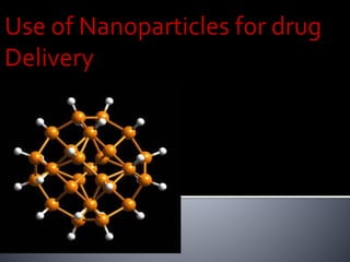
Nanoparticles between 1-100 nanometers in size can be used to deliver drugs in the body. They allow changing the pharmacokinetic properties of drugs without altering the active compound. Biodegradable polymeric nanoparticles have attracted interest as potential drug carriers that can target specific organs and tissues and deliver proteins, peptides, and genes orally. Nanoparticles must be able to travel through blood vessels and cross cell layers to reach their target site. Their small size allows them to potentially penetrate tissues and cells to provide localized drug delivery.


