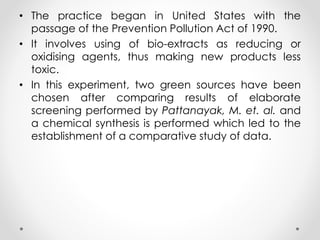
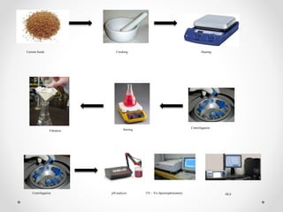
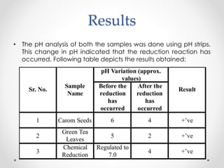
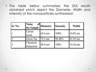
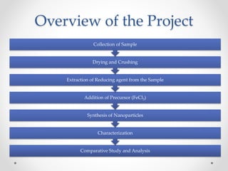
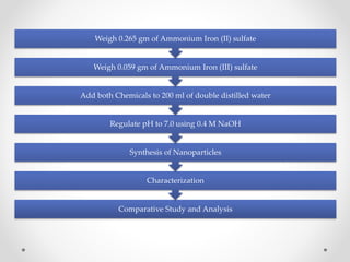
Preparation and
Characterization of Iron
Nanoparticles using
Green Technology and
their comparative study.
Made By -
Prajwal S Bahukhandi
BTB/10/159
A0504110044
Supervisor -
Dr. Kirti Rani Sharma
Amity Institute of
Biotechnology
Co-Supervisor -
Dr. Jagriti
Dr. Nidhi Chauhan
Amity Institute of
Nanotechnology
Iron Nanoparticles!
Overview of the Project
Comparative Study and Analysis
Characterization
Synthesis of Nanoparticles
Addition of Precursor (FeCl3)
Extraction of Reducing agent from the Sample
Drying and Crushing
Collection of Sample
Comparative Study and Analysis
Characterization
Synthesis of Nanoparticles
Regulate pH to 7.0 using 0.4 M NaOH
Add both Chemicals to 200 ml of double distilled water
Weigh 0.059 gm of Ammonium Iron (III) sulfate
Weigh 0.265 gm of Ammonium Iron (II) sulfate
Review of Literature
Application of Iron Nanoparticles:
• Huber Dr. D. L. et. al. described in their paper titled “Synthesis,
Properties, and Applications of Iron Nanoparticles”: Iron as a
nanoparticle has been somewhat neglected in favor of its own
oxides, as well as other metals such as cobalt, nickel, gold, and
platinum. Iron's reactivity is important in macroscopic applications
(particularly rusting), but is a dominant concern at the nanoscale.
Recent work has begun to take advantage of irons potential,
and work in this field appears to be blossoming. [Huber Dr., D. L.
et. al., (2005)]
• Takada Prof. J. et. al. described in their paper titled “Research
and application of iron oxide nanoparticles explored”: Iron oxide
NP of about 100 nm produced yellowish red, and larger particles
sizes led to red and eventually dark purple colors. This study
revealed the application of BIOX as an ecofriendly anode in Li-
ion batteries. [Takada Prof, J. et. al., (2014)] [Laurent, S. et. al.,
(2008)]
Synthesis of Iron Nanoparticles using Green Technology:
• Pattanayak et. al. described in their paper titled “Ecofriendly
synthesis of Iron Nanoparticles from various Plants and Spices
extract”: Biosynthesis from different parts (mostly leaf) of the plant
is found to be the most effective process of synthesis at a very
affordable cost. Appropriate precursors such as Ferric Chloride
can be used for the reduction of plant extracts. Scientists report
the synthesis of nanoparticles, reducing Ferric ions present in the
aqueous solution of Ferric chloride by the help of different plant
extracts. Through elaborate screening process involving about 45
plants, we selected 10 most suitable plants as the potential
candidates for the synthesis of iron nanoparticles. [Pattanayak,
M. et. al., (2013)] [Li, L. et. al., (2006)]
• Iravani, S. et. al. described in their paper titled “Green synthesis of
metal nanoparticles using plants”: This study reveals that in recent
years, the development of efficient green chemistry methods for
synthesis of metal nanoparticles has become a major focus of
researchers. Investigations in order to find out an eco-friendly
technique for production of well-characterized nanoparticles
have been carried out. One of the most considered methods is
production of metal nanoparticles using organisms. Among these
organisms plants seem to be the best candidates and they are
suitable for large-scale biosynthesis of nanoparticles.
Nanoparticles produced by plants are more stable and the rate
of synthesis is faster than in the case of microorganisms. [Iravani,
S., (2011)] [Raveendran, P. et. al., (2013)]
Biomedical Applications of Iron Nanoparticles:
• Gupta, A. K. et. al. described in their paper titled “Synthesis and
surface engineering of iron oxide nanoparticles for biomedical
applications” : Superparamagnetic iron oxide nanoparticles
(SPION) with appropriate surface chemistry have been widely
used experimentally for numerous in vivo applications such as
magnetic resonance imaging contrast enhancement, tissue
repair, immunoassay, detoxification of biological fluids,
hyperthermia, drug delivery and in cell separation, etc. All
these biomedical and bioengineering applications require
that these nanoparticles have high magnetization values and
size smaller than 100 nm with overall narrow particle size
distribution, so that the particles have uniform physical and
chemical properties. To this end, most work in this field has
been done in improving the biocompatibility of the materials,
but only a few scientific investigations and developments
have been carried out in improving the quality of magnetic
particles, their size distribution, their shape and surface in
addition to characterizing them to get a protocol for the
quality control of these particles. [Gupta, A., K. et. al., (2005)]
Characterization:
• Nurmi, J. E. et. al. described in their paper titled “Characterization
and Properties of Metallic Iron Nanoparticles: Spectroscopy,
Electrochemistry, and Kinetics” : Superparamagnetic iron oxide
nanoparticles (SPION) with appropriate surface chemistry
have been widely used experimentally for numerous in vivo.
All these biomedical and bioengineering applications require
that these nanoparticles have high magnetization values and
size smaller than 100 nm with overall narrow particle size
distribution, so that the particles have uniform physical and
chemical properties. [Nurmi, J., E. et. al., (2004)]
Introduction
• What are Nanoparticles? – Particles with sizes in the
range of 10 – 100 nm are called nanoparticles. [Khan, F.
A., et. al., (2012)]
• This range (10 – 100 nm) is known as the Nanoscale.
• Why do they interest us? – Nanoparticle research is
currently an area of intense scientific research due to a
wide variety of potential applications in biomedical,
optical and electronic fields.
• Iron nanoparticles have been found to be an effective
measure to treat several types of ground contamination
and are easily transportable through ground water for in
situ treatment. These factors combined makes this
method cheaper than most methods currently being
used. [Kulkarni, L. et. al., (2009)]
• Iron oxide nanoparticles can easily be reduced to
magnetite and maghemite which preferred in
biomedical in vivo applications because they are
biocompatible and potentially non – toxic to
humans.
• They also show magnetic and paramagnetic
properties which make them a potentially useful
drug delivery system.
• Recent advancements in the field of
nanotechnology have led to the development of
various techniques for the biosynthesis of metal
nanoparticles. [Murphy, C. J. et. al., (2002)]
• Green Technology - Designing chemical products
and processes in a way that reduces or eliminates
hazardous substances from the beginning to end of
a chemical product’s life cycle.
• The practice began in United States with the
passage of the Prevention Pollution Act of 1990.
• It involves using of bio-extracts as reducing or
oxidising agents, thus making new products less
toxic.
• In this experiment, two green sources have been
chosen after comparing results of elaborate
screening performed by Pattanayak, M. et. al. and
a chemical synthesis is performed which led to the
establishment of a comparative study of data.
Objectives
• Green synthesis of Iron Nanoparticles from the
following samples:
1. Carrom Seeds (Trachyspermum ammi)
2. Green Tea (Camellia sinensis)
• Characterization of Iron Nanoparticles produced
from different samples.
• Comparative study and analysis of Iron
Nanoparticles produced from different sources
using Green Technology and Chemical Synthesis
method.
Methodology
Methodologies to be used in the Project
• Sample collection
• Drying: Natural drying in sun and/or Hot air oven.
• Crushing: Using Pestle and Mortar.
• Separation and Purification of plant extract:
Filtration using Whatman No. 1 filter paper and
Centrifugation at 5000 rpm.
• Preparation of 0.001M FeCl3 which is used as a
Precursor
• Mixing of Reducing Agent and Precursor (1:1) under
constant stirring at 50 – 60°C: Using Magnetic Stirrer.
• Characterization:
1. UV – spectroscopy
2. pH analysis
3. Dynamic Light Scattering (DLS): Size of Nanoparticle
• Comparative study and analysis of the result and
data obtained.
Carrom Seeds Crushing Heating
Centrifugation
StirringFiltration
Centrifugation pH analysis UV – Vis Spectrophotometry DLS
Crushing Heating
Centrifugation
StirringFiltration
Centrifugation pH analysis UV – Vis Spectrophotometry DLS
Green Tea Leaves
0.265 gm
Ammonium Iron (II) sulfate
0.059 gm
Ammonium Iron (III) sulfate
pH regulation to 7.0 by dropwise
addition of 0.4 M NaOH
Stirring on magnetic stirrer for 30
minutes
pH analysis UV – Vis
spectrophotometry
DLS
Results
• The pH analysis of both the samples was done using pH strips.
This change in pH indicated that the reduction reaction has
occurred. Following table depicts the results obtained:
• UV – Vis spectroscopy was performed by checking for
absorbance at regular intervals; 200 nm, 300 nm, 400 nm, 500
nm, 600 nm, 700 nm and 800 nm, analyzed the samples. The
following results were obtained:
• The table below summarizes the DLS results
obtained which depict the Diameter, Width and
Intensity of the nanoparticles synthesized:
Interpretation and Discussion
• All the three samples showed a positive pH change,
which confirms the occurrence of the reduction reaction
of Fe3+ ions, which have formed the Iron Nanoparticles
in the samples.
• The absorbance spectra of all three samples show a
common point where the peak was observed, i.e., 500
nm. Which is in relevance with the absorbance spectra
of metallic iron
• The chemically synthesized iron nanoparticles aid the
study of the nanoparticles formed in the plant extract
sample because the absorbance peak observed was at
the same wavelength, confirming the presence of iron
nanoparticles.
• DLS results have made clear that the particles
synthesized are in the nanoscale (1 – 100 nm
range); sizes being 65.6 nm from Carom Seeds
sample, 72.7 nm from Green Tea sample and 88.9
nm from Chemical Synthesis method.
• This proves that synthesis of iron nanoparticles from
green sources, like carom seeds and green tea
leaves is equally efficient as by the chemical
reduction method.
Conclusion and Learning
Outcomes
• The extracts of plants were capable of producing Iron
Nanoparticles efficiently and gave good results in pH analysis
and characterization techniques used (UV – Vis spectroscopy
and DLS).
• Under the UV-Visible wavelength Nanoparticles showed quiet
good surface plasmon resonance behavior. All three samples
showed a peak at the same wavelength, 500 nm.
• The Dynamic Light Spectroscopy results validate the fact that
the particles synthesized were in the nanoscale and thus,
nanoparticles.
• The resultant single sharp peak in the DLS graph states that
there is only one kind of particle present in out sample solution,
i.e., there is no other particle synthesized and there is no
contamination in the sample.
• The conclusion can be reached that Plant Extracts can be an
effective source for synthesis of Iron Nanoparticles and can
produce better nanoparticles to an extent.
• Green Sources prove to be a cheaper source of production of
iron nanoparticles as compared to the chemical process.
• Although, the time taken by the Green Technology method is
a little longer than the Chemical Reduction method, scaling
up the process and synthesizing iron nanoparticles in a larger
quantity by processing larger volumes of the plant extract can
increase its efficiency.
• From the two green sources that were used in the experiment,
Carom Seeds produced nanoparticles of smaller size than the
Green Tea Leaves.
• More stirring and sonication can be done to the samples to
reduce the size of the nanoparticles.
• Also, we can attach a functional group of our interest and
desired properties to the nanoparticle and develop better
applications for the nanoparticle.
References
• Gupta, A., K. and Gupta, M. (2005). Synthesis and
surface engineering of iron oxide nanoparticles for
biomedical applications, Biomaterials, Volume 26,
Issue 18, pp 3995–4021.
• Huber Dr., D. L. (2005). Synthesis, Properties, and
Applications of Iron Nanoparticles., American
Chemical Society.
• Iravani, S. (2011). Green synthesis of metal
nanoparticles using plants, Royal Society of
Chemistry.
• Khan, F. A. (2012). Biotechnology Fundamentals,
CRC Press, 2012, pp 328.
• Kulkarni, L. (2009). Synthesis and Characterization of
Nanoparticles, Industrial and Systems Engineering, pp.
16 – 18.
• Laurent, S., Forge, D., Port, M., Roch, A., Robic, C., Elst,
L. V. and Muller, R. N. (2008). Magnetic Iron Oxide
Nanoparticles: Synthesis, Stabilization, Vectorization,
Physicochemical Characterizations, and Biological
Applications, American Chemical Society, pp 2064–
2110.
• Li, L., Fan, M., Brown, R. C. and Leeuwen, L. V. (2006).
Synthesis, Properties, and Environmental Applications
of Nanoscale Iron Based Materials: A Review, Critical
Reviews in Environmental Science and Technology.
• Murphy, C. J., Sau, T. K. and Gole, A. M. (2005).
Anisotropic Metal Nanoparticles: Synthesis, Assembly,
and Optical Applications, American Chemical
Society, pp 13857–13870.
• Nurmi, J., T., Tratnyek, P., G., Sarathy, V., Baer, D., R.,
Amonette, J., E., Pecher, K. (2004). Characterization
and Properties of Metallic Iron Nanoparticles:
Spectroscopy, Electrochemistry, and Kinetics,
Environmental Science and Technology, 2004, pp
1221–1230
• Pattanayak, M. and Nayak, P. L. (2013). Ecofriendly
synthesis of Iron Nanoparticles from various Plants and
Spices extract, International Journal of Plant, Animal
and Environmental Sciences, Vol. 3, Issue 1.
• Raveendran, P., Fu, J., Wallen, S. L., Kenan and
Venable Laboratories (2003). Completely “Green”
Synthesis and Stabilization of Metal Nanoparticles,
American Chemical Society, pp 13940–13941.
• Takada Prof, J. (2014). Research and application of
iron oxide nanoparticles explored, Science Daily, Vol.
JUL(14), pp. 111 – 129.
Thank You!

Final Presentation on Iron Nanoparticles_Prajwal (1)

  • 1.
    Preparation and Characterization ofIron Nanoparticles using Green Technology and their comparative study. Made By - Prajwal S Bahukhandi BTB/10/159 A0504110044 Supervisor - Dr. Kirti Rani Sharma Amity Institute of Biotechnology Co-Supervisor - Dr. Jagriti Dr. Nidhi Chauhan Amity Institute of Nanotechnology
  • 2.
  • 3.
    Overview of theProject Comparative Study and Analysis Characterization Synthesis of Nanoparticles Addition of Precursor (FeCl3) Extraction of Reducing agent from the Sample Drying and Crushing Collection of Sample
  • 4.
    Comparative Study andAnalysis Characterization Synthesis of Nanoparticles Regulate pH to 7.0 using 0.4 M NaOH Add both Chemicals to 200 ml of double distilled water Weigh 0.059 gm of Ammonium Iron (III) sulfate Weigh 0.265 gm of Ammonium Iron (II) sulfate
  • 5.
    Review of Literature Applicationof Iron Nanoparticles: • Huber Dr. D. L. et. al. described in their paper titled “Synthesis, Properties, and Applications of Iron Nanoparticles”: Iron as a nanoparticle has been somewhat neglected in favor of its own oxides, as well as other metals such as cobalt, nickel, gold, and platinum. Iron's reactivity is important in macroscopic applications (particularly rusting), but is a dominant concern at the nanoscale. Recent work has begun to take advantage of irons potential, and work in this field appears to be blossoming. [Huber Dr., D. L. et. al., (2005)] • Takada Prof. J. et. al. described in their paper titled “Research and application of iron oxide nanoparticles explored”: Iron oxide NP of about 100 nm produced yellowish red, and larger particles sizes led to red and eventually dark purple colors. This study revealed the application of BIOX as an ecofriendly anode in Li- ion batteries. [Takada Prof, J. et. al., (2014)] [Laurent, S. et. al., (2008)]
  • 6.
    Synthesis of IronNanoparticles using Green Technology: • Pattanayak et. al. described in their paper titled “Ecofriendly synthesis of Iron Nanoparticles from various Plants and Spices extract”: Biosynthesis from different parts (mostly leaf) of the plant is found to be the most effective process of synthesis at a very affordable cost. Appropriate precursors such as Ferric Chloride can be used for the reduction of plant extracts. Scientists report the synthesis of nanoparticles, reducing Ferric ions present in the aqueous solution of Ferric chloride by the help of different plant extracts. Through elaborate screening process involving about 45 plants, we selected 10 most suitable plants as the potential candidates for the synthesis of iron nanoparticles. [Pattanayak, M. et. al., (2013)] [Li, L. et. al., (2006)] • Iravani, S. et. al. described in their paper titled “Green synthesis of metal nanoparticles using plants”: This study reveals that in recent years, the development of efficient green chemistry methods for synthesis of metal nanoparticles has become a major focus of researchers. Investigations in order to find out an eco-friendly technique for production of well-characterized nanoparticles have been carried out. One of the most considered methods is production of metal nanoparticles using organisms. Among these organisms plants seem to be the best candidates and they are suitable for large-scale biosynthesis of nanoparticles. Nanoparticles produced by plants are more stable and the rate of synthesis is faster than in the case of microorganisms. [Iravani, S., (2011)] [Raveendran, P. et. al., (2013)]
  • 7.
    Biomedical Applications ofIron Nanoparticles: • Gupta, A. K. et. al. described in their paper titled “Synthesis and surface engineering of iron oxide nanoparticles for biomedical applications” : Superparamagnetic iron oxide nanoparticles (SPION) with appropriate surface chemistry have been widely used experimentally for numerous in vivo applications such as magnetic resonance imaging contrast enhancement, tissue repair, immunoassay, detoxification of biological fluids, hyperthermia, drug delivery and in cell separation, etc. All these biomedical and bioengineering applications require that these nanoparticles have high magnetization values and size smaller than 100 nm with overall narrow particle size distribution, so that the particles have uniform physical and chemical properties. To this end, most work in this field has been done in improving the biocompatibility of the materials, but only a few scientific investigations and developments have been carried out in improving the quality of magnetic particles, their size distribution, their shape and surface in addition to characterizing them to get a protocol for the quality control of these particles. [Gupta, A., K. et. al., (2005)]
  • 8.
    Characterization: • Nurmi, J.E. et. al. described in their paper titled “Characterization and Properties of Metallic Iron Nanoparticles: Spectroscopy, Electrochemistry, and Kinetics” : Superparamagnetic iron oxide nanoparticles (SPION) with appropriate surface chemistry have been widely used experimentally for numerous in vivo. All these biomedical and bioengineering applications require that these nanoparticles have high magnetization values and size smaller than 100 nm with overall narrow particle size distribution, so that the particles have uniform physical and chemical properties. [Nurmi, J., E. et. al., (2004)]
  • 9.
    Introduction • What areNanoparticles? – Particles with sizes in the range of 10 – 100 nm are called nanoparticles. [Khan, F. A., et. al., (2012)] • This range (10 – 100 nm) is known as the Nanoscale. • Why do they interest us? – Nanoparticle research is currently an area of intense scientific research due to a wide variety of potential applications in biomedical, optical and electronic fields. • Iron nanoparticles have been found to be an effective measure to treat several types of ground contamination and are easily transportable through ground water for in situ treatment. These factors combined makes this method cheaper than most methods currently being used. [Kulkarni, L. et. al., (2009)]
  • 10.
    • Iron oxidenanoparticles can easily be reduced to magnetite and maghemite which preferred in biomedical in vivo applications because they are biocompatible and potentially non – toxic to humans. • They also show magnetic and paramagnetic properties which make them a potentially useful drug delivery system. • Recent advancements in the field of nanotechnology have led to the development of various techniques for the biosynthesis of metal nanoparticles. [Murphy, C. J. et. al., (2002)] • Green Technology - Designing chemical products and processes in a way that reduces or eliminates hazardous substances from the beginning to end of a chemical product’s life cycle.
  • 11.
    • The practicebegan in United States with the passage of the Prevention Pollution Act of 1990. • It involves using of bio-extracts as reducing or oxidising agents, thus making new products less toxic. • In this experiment, two green sources have been chosen after comparing results of elaborate screening performed by Pattanayak, M. et. al. and a chemical synthesis is performed which led to the establishment of a comparative study of data.
  • 12.
    Objectives • Green synthesisof Iron Nanoparticles from the following samples: 1. Carrom Seeds (Trachyspermum ammi) 2. Green Tea (Camellia sinensis) • Characterization of Iron Nanoparticles produced from different samples. • Comparative study and analysis of Iron Nanoparticles produced from different sources using Green Technology and Chemical Synthesis method.
  • 13.
    Methodology Methodologies to beused in the Project • Sample collection • Drying: Natural drying in sun and/or Hot air oven. • Crushing: Using Pestle and Mortar. • Separation and Purification of plant extract: Filtration using Whatman No. 1 filter paper and Centrifugation at 5000 rpm. • Preparation of 0.001M FeCl3 which is used as a Precursor • Mixing of Reducing Agent and Precursor (1:1) under constant stirring at 50 – 60°C: Using Magnetic Stirrer.
  • 14.
    • Characterization: 1. UV– spectroscopy 2. pH analysis 3. Dynamic Light Scattering (DLS): Size of Nanoparticle • Comparative study and analysis of the result and data obtained.
  • 15.
    Carrom Seeds CrushingHeating Centrifugation StirringFiltration Centrifugation pH analysis UV – Vis Spectrophotometry DLS
  • 16.
    Crushing Heating Centrifugation StirringFiltration Centrifugation pHanalysis UV – Vis Spectrophotometry DLS Green Tea Leaves
  • 17.
    0.265 gm Ammonium Iron(II) sulfate 0.059 gm Ammonium Iron (III) sulfate pH regulation to 7.0 by dropwise addition of 0.4 M NaOH Stirring on magnetic stirrer for 30 minutes pH analysis UV – Vis spectrophotometry DLS
  • 18.
    Results • The pHanalysis of both the samples was done using pH strips. This change in pH indicated that the reduction reaction has occurred. Following table depicts the results obtained:
  • 19.
    • UV –Vis spectroscopy was performed by checking for absorbance at regular intervals; 200 nm, 300 nm, 400 nm, 500 nm, 600 nm, 700 nm and 800 nm, analyzed the samples. The following results were obtained:
  • 20.
    • The tablebelow summarizes the DLS results obtained which depict the Diameter, Width and Intensity of the nanoparticles synthesized:
  • 24.
    Interpretation and Discussion •All the three samples showed a positive pH change, which confirms the occurrence of the reduction reaction of Fe3+ ions, which have formed the Iron Nanoparticles in the samples. • The absorbance spectra of all three samples show a common point where the peak was observed, i.e., 500 nm. Which is in relevance with the absorbance spectra of metallic iron • The chemically synthesized iron nanoparticles aid the study of the nanoparticles formed in the plant extract sample because the absorbance peak observed was at the same wavelength, confirming the presence of iron nanoparticles.
  • 25.
    • DLS resultshave made clear that the particles synthesized are in the nanoscale (1 – 100 nm range); sizes being 65.6 nm from Carom Seeds sample, 72.7 nm from Green Tea sample and 88.9 nm from Chemical Synthesis method. • This proves that synthesis of iron nanoparticles from green sources, like carom seeds and green tea leaves is equally efficient as by the chemical reduction method.
  • 26.
    Conclusion and Learning Outcomes •The extracts of plants were capable of producing Iron Nanoparticles efficiently and gave good results in pH analysis and characterization techniques used (UV – Vis spectroscopy and DLS). • Under the UV-Visible wavelength Nanoparticles showed quiet good surface plasmon resonance behavior. All three samples showed a peak at the same wavelength, 500 nm. • The Dynamic Light Spectroscopy results validate the fact that the particles synthesized were in the nanoscale and thus, nanoparticles. • The resultant single sharp peak in the DLS graph states that there is only one kind of particle present in out sample solution, i.e., there is no other particle synthesized and there is no contamination in the sample.
  • 27.
    • The conclusioncan be reached that Plant Extracts can be an effective source for synthesis of Iron Nanoparticles and can produce better nanoparticles to an extent. • Green Sources prove to be a cheaper source of production of iron nanoparticles as compared to the chemical process. • Although, the time taken by the Green Technology method is a little longer than the Chemical Reduction method, scaling up the process and synthesizing iron nanoparticles in a larger quantity by processing larger volumes of the plant extract can increase its efficiency. • From the two green sources that were used in the experiment, Carom Seeds produced nanoparticles of smaller size than the Green Tea Leaves. • More stirring and sonication can be done to the samples to reduce the size of the nanoparticles. • Also, we can attach a functional group of our interest and desired properties to the nanoparticle and develop better applications for the nanoparticle.
  • 28.
    References • Gupta, A.,K. and Gupta, M. (2005). Synthesis and surface engineering of iron oxide nanoparticles for biomedical applications, Biomaterials, Volume 26, Issue 18, pp 3995–4021. • Huber Dr., D. L. (2005). Synthesis, Properties, and Applications of Iron Nanoparticles., American Chemical Society. • Iravani, S. (2011). Green synthesis of metal nanoparticles using plants, Royal Society of Chemistry. • Khan, F. A. (2012). Biotechnology Fundamentals, CRC Press, 2012, pp 328.
  • 29.
    • Kulkarni, L.(2009). Synthesis and Characterization of Nanoparticles, Industrial and Systems Engineering, pp. 16 – 18. • Laurent, S., Forge, D., Port, M., Roch, A., Robic, C., Elst, L. V. and Muller, R. N. (2008). Magnetic Iron Oxide Nanoparticles: Synthesis, Stabilization, Vectorization, Physicochemical Characterizations, and Biological Applications, American Chemical Society, pp 2064– 2110. • Li, L., Fan, M., Brown, R. C. and Leeuwen, L. V. (2006). Synthesis, Properties, and Environmental Applications of Nanoscale Iron Based Materials: A Review, Critical Reviews in Environmental Science and Technology. • Murphy, C. J., Sau, T. K. and Gole, A. M. (2005). Anisotropic Metal Nanoparticles: Synthesis, Assembly, and Optical Applications, American Chemical Society, pp 13857–13870.
  • 30.
    • Nurmi, J.,T., Tratnyek, P., G., Sarathy, V., Baer, D., R., Amonette, J., E., Pecher, K. (2004). Characterization and Properties of Metallic Iron Nanoparticles: Spectroscopy, Electrochemistry, and Kinetics, Environmental Science and Technology, 2004, pp 1221–1230 • Pattanayak, M. and Nayak, P. L. (2013). Ecofriendly synthesis of Iron Nanoparticles from various Plants and Spices extract, International Journal of Plant, Animal and Environmental Sciences, Vol. 3, Issue 1. • Raveendran, P., Fu, J., Wallen, S. L., Kenan and Venable Laboratories (2003). Completely “Green” Synthesis and Stabilization of Metal Nanoparticles, American Chemical Society, pp 13940–13941. • Takada Prof, J. (2014). Research and application of iron oxide nanoparticles explored, Science Daily, Vol. JUL(14), pp. 111 – 129.
  • 31.