SlideShare a Scribd company logo
Borrelia burgdorferi
Lyme Disease
Dr. Suprakash Das
Assist. Prof.
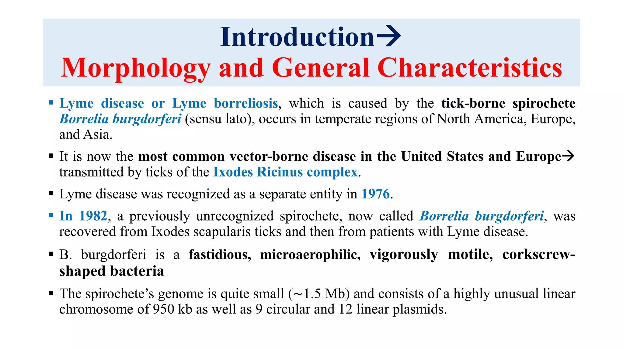
Introduction
Morphology and General Characteristics
 Lyme disease or Lyme borreliosis, which is caused by the tick-borne spirochete
Borrelia burgdorferi (sensu lato), occurs in temperate regions of North America, Europe,
and Asia.
 It is now the most common vector-borne disease in the United States and Europe
transmitted by ticks of the Ixodes Ricinus complex.
 Lyme disease was recognized as a separate entity in 1976.
 In 1982, a previously unrecognized spirochete, now called Borrelia burgdorferi, was
recovered from Ixodes scapularis ticks and then from patients with Lyme disease.
 B. burgdorferi is a fastidious, microaerophilic, vigorously motile, corkscrew-
shaped bacteria
 The spirochete’s genome is quite small (∼1.5 Mb) and consists of a highly unusual linear
chromosome of 950 kb as well as 9 circular and 12 linear plasmids.
Introduction
Morphology and General Characteristics
 The spirochete cell wall consists of a cytoplasmic membrane surrounded by peptidoglycan and
flagella and then by a loosely associated outer membrane.
 Of the Borrelia spp., B. burgdorferi is the longest (20–30 μm) and narrowest (0.2–0.3 μm), and
it has fewer flagella.
 The organism has few proteins with biosynthetic activity and apparently depends on the host for
much of its nutritional requirements.
 The only known virulence factors of B. burgdorferi are surface-exposed lipoproteins that
allow the spirochete to attach to mammalian cells.
 The vectors of Lyme borreliosis are closely related ixodid tick species that are part of the Ixodes
ricinus complex (also called the Ixodes persulcatus complex).
In the northeastern and midwestern United States Ixodes scapularis
West USAIxodes pacificus.
In Europe Ixodes ricinus, and
In Asia Ixodes persulcatus.
Introduction
Morphology and General Characteristics
 Ixodid ticks have larval, nymphal, and adult stages, and they require a blood meal at
each stage.
 A highly efficient, horizontal cycle of B. burgdorferi transmission occurs among
larval and nymphal I. scapularis ticks and certain rodents, particularly white-footed
mice and chipmunks.
 This cycle results in high rates of infection among rodents and nymphal ticks
 Primarily nymphal tick bites cause many new human cases of Lyme disease during
the late spring and summer months.
 White-tailed deer, which are not involved in the life cycle of the spirochete, are the
preferred host of adult I. scapularis, and they seem to be critical for the survival of
ticks.
Pathology
 To maintain its complex enzootic cycle, B. burgdorferi must adapt to two
markedly different environments: the tick and the mammalian or avian host.
 The spirochete survives in a dormant state in the nymphal tick midgut during
the fall, winter, and spring, where it expresses primarily OspA and certain other
proteins.
 When the tick feeds during the late spring or summer, these proteins are
downregulated, and another set of proteins, including OspC, is upregulated.
 OspC binds mammalian plasminogen and its activators present in the blood
meal, which facilitates spreading of the organism in the tick.
 In addition, within the tick salivary gland, OspC binds a tick salivary gland
protein and coating of the spirochete in this tick protein is essential for initial
immune evasion in the mammalian host.
 The spirochete, which has few proteins with biosynthetic activity, appears to meet
its nutritional requirements by infection of a mammalian or avian host.
Pathology
 After injection of B. burgdorferi by the tick (and a clinical incubation period of 3–32
days), the spirochete usually first multiplies locally in the skin at the site of the tick
bite.
 In most patients immune cells first encounter B. burgdorferi at this site.
 During the initial infection B. burgdorferi induces potent proinflammatory and
compensatory anti inflammatory responses in cells in EM lesions
 B. burgdorferi–stimulated peripheral blood mononuclear cells (PBMCs) produce
primarily proinflammatory cytokines, particularly interferon (IFN)-γ.
 Thus both innate and adaptive cellular elements are mobilized to fight the infection.
 Within days to weeks, B. burgdorferi strains often disseminate to many distant anatomic
sites.
 During this period the spirochete has been recovered from blood and cerebrospinal fluid
(CSF) and it has been seen in small numbers in specimens of myocardium, retina,
muscle, bone, spleen, liver, meninges, and brain.
Pathology
 To disseminate, B. burgdorferi adheres to integrins, proteoglycans, or
glycoproteins on host cells or tissue matrices.
 Spreading through the skin and other tissue matrixes may be facilitated by the
binding of plasminogen and its activators to the surface of the spirochete.
 During dissemination, a 66-kilodalton (kDa) spirochetal protein binds the
platelet-specific integrin αIIbβ3 and the vitronectin receptor (αvβ5).
 A 26-kDa glycosaminoglycan binding protein binds heparan sulfate and
dermatan sulfate, which are expressed on endothelial cells.
 A 47-kDa fibronectin-binding protein (BBK32) binds fibronectin
 Finally, decorin-binding proteins A and B (DbpA and DbpB) of the spirochete
bind decorin, a proteoglycan on the surface of collagen
 alignment of spirochetes with collagen fibrils in the extracellular matrix in
Pathology
 All affected tissues show an infiltration of lymphocytes and plasma cells.
 Some degree of vascular damage, including mild vasculitis or hypervascular occlusion, may be
seen in multiple sites, suggesting that spirochetes may have been in or around blood vessels.
 Despite an active immune response, B. burgdorferi may survive during dissemination by
changing or minimizing antigenic expression of surface proteins and by inhibiting certain critical
host immune responses.
 Two linear plasmids (lps) seem to be essential, including lp25, which encodes a nicotinamidase,
and lp28-1, which encodes the VlsE lipoprotein, the protein that undergoes extensive antigenic
variation.
 In addition, the spirochete has a number of highly homologous, differentially expressed
lipoproteins, including OspE/F paralogs, which further contribute to antigenic diversity.
 Finally, B. afzelii and, to a lesser degree, B. burgdorferi have complement regulator-acquiring
surface proteins that bind complement factor H and factor H–like protein 1.
 These complement factors inactivate C3b, which protects the organism from complement-
mediated killing.
Pathology
 Both innate and adaptive immune responses are required for optimal control of spirochetal
dissemination.
 The specific immunoglobulin M (IgM) response is often associated with polyclonal activation
of B cells, including
Elevated total serum IgM levels,
Circulating immune complexes, and
Cryoglobulins.
 Spirochetal killing seems to be accomplished primarily by bactericidal B-cell responses,
which promote spirochetal killing by complement fixation and opsonization.
 Infection of humans is a dead-end event for the spirochete.
 Within several weeks to months, innate and adaptive immune mechanisms control widely
disseminated infection.
 Without antibiotic therapy, spirochetes may survive in localized niches for several more
years.
Clinical Manifestations
 Human Lyme borreliosis generally occurs in stages, with remissions and
exacerbation and different clinical manifestations at each stage.
 Early infection consists of stage 1 (localized EM), followed within days or
weeks by stage 2 (disseminated infection).
 Late infection, or stage 3 (persistent infection), usually begins months to
years after the disease onset, sometimes following long periods of latent
infection.
 In an individual patient, however, the infection is highly variable, ranging
from brief involvement in only one system to chronic, multisystem
involvement of the skin, nerves, or joints.
 In the United States about 10% of individuals have asymptomatic B.
burgdorferi infection and seem to cure the infection without antibiotic
therapy
Clinical Manifestations
Early Infection: Stage 1 (Localized Infection)
 In about 70% to 80% of patients EM(erythema migrans) develops at the site of the tick
bite.
 During the first several days the lesion often has a homogeneous red appearance.
 In addition, the centers of early lesions sometimes become intensely erythematous
and indurated, vesicular, or necrotic.
 As the area of redness around the center expands, most lesions continue to have bright-
red outer borders (usually flat, but occasionally raised) and partial central clearing.
 In some instances migrating lesions remain an even, intense red; several red rings are
found within the outside one; or the central area turns blue before it clears.
 Although the lesion can be located anywhere, the thigh, groin, and axilla are
particularly common sites.
 If EM is on the head, only a linear streak might be seen to emerge from the hairline.
 The lesion is hot to the touch, and patients often describe it as burning or occasionally
itching or painful.
(A) An early erythema migrans (EM) skin lesion is seen 4 days after
detection.
(B) Four days after the onset of the initial skin lesion, secondary
lesions have appeared, and several of their borders have merged.
Clinical Manifestations
Early Infection: Stage 2 (Disseminated Infection)
 Within several days to weeks of the onset of the initial EM lesion, patients may develop
multiple annular secondary skin lesions a sign of hematogenous dissemination.
 Although their appearance is similar to that of the initial lesions, they are generally smaller,
migrate less, and lack indurated centers; they are not associated with previous tick bites,
their borders sometimes merge.
 During this period some patients develop malar rash, conjunctivitis, or, rarely, diffuse
urticaria.
 EM and secondary lesions usually fade within 3 to 4 weeks (range, 1 day–14 months).
 EM is often accompanied by malaise and fatigue, headache, fever and chills, generalized
achiness, and regional lymphadenopathy.
 n about 18% of patients these symptoms are the presenting picture of the infection.
 In addition, patients sometimes have evidence of meningeal irritation with
Clinical Manifestations
Early Infection: Stage 2 (Disseminated Infection)
Episodic attacks of excruciating headache and neck pain,
Mild encephalopathy with difficulty with mentation,
Migratory musculoskeletal pain,
Hepatitis,
Generalized lymphadenopathy or
Splenomegaly,
Sore throat, or rarely,
Non-productive cough
 A few patients have had microscopic hematuria, sometimes with mild
proteinuria (dipstick).
Clinical Manifestations
Early Infection: Stage 2 (Disseminated Infection)
 During the first days of illness headache and neck stiffness are not associated
with a spinal fluid pleocytosis or objective neurologic deficit.
 Except for fatigue and lethargy, which are often constant, the early signs and
symptoms are typically intermittent and changing.
 After several weeks to months, about 15% of untreated patients in the United
States develop frank neurologic abnormalities, including 
Meningitis,
Encephalitis,
Cranial neuritis (including bilateral facial palsy),
Motor and sensory radiculoneuritis,
Mononeuritis multiplex,
Cerebellar ataxia or myelitis—alone or in various combinations.
Clinical Manifestations
Early Infection: Stage 2 (Disseminated Infection)
 The usual pattern consists of fluctuating symptoms of meningitis with superimposed
cranial (particularly facial palsy) or peripheral radiculoneuropathy.
 In Europe the most common neurologic manifestation is
Bannwarth syndrome 
Neuritic pain,
Lymphocytic pleocytosis without headache,
sometimes Cranial neuritis
On examination such patients usually have neck stiffness only on extreme flexion;
Kernig and Brudzinski signs are not present.
Facial palsy may occur alone and in rare instances, it may be the presenting
manifestation of the disease.
Clinical Manifestations
Early Infection: Stage 2 (Disseminated Infection)
 In children the optic nerve may be affected by inflammation or
increased intracranial pressure, which may lead to blindness
 In patients with meningitis, CSF typically has a lymphocytic
pleocytosis of about 100 cells/mm,
 Often with an elevated protein but a normal glucose level.
 Specific IgG, IgM, or IgA antibody to the spirochete is produced
intrathecally.
 Histologically, the lesions show axonal nerve injury with
perivascular infiltration of lymphocytes and plasmocytes around
epineural blood vessels.
Brainstem (IH22 doxycycline
treated); minimal localized
lymphoplasmacytic perivascular
cuffing adjacent to the fourth
ventricle
Clinical Manifestations
Early Infection: Stage 2 (Disseminated Infection)
 Stage 2 neurologic abnormalities usually last for weeks or months, but
they may recur or become chronic.
 Within several weeks after the onset of illness, about 5% of untreated
patients develop cardiac involvement.
 The most common abnormality is fluctuating degrees of atrioventricular
block (first-degree, Wenckebach, or complete heart block).
 However, some patients have evidence of more diffuse cardiac
involvement Acute myopericarditis, Mild left ventricular dysfunction,
or, rarely, Cardiomegaly.
 No patients have had heart murmurs.
 The duration of cardiac involvement is usually brief (3 days–6 weeks).
Clinical Manifestations
Early Infection: Stage 2 (Disseminated Infection)
 During this stage migratory musculoskeletal pain is common in joints,
tendons, bursae, muscle, or bones.
 In addition, a few patients have been described with osteomyelitis, myositis,
panniculitis, or fasciitis.
 Conjunctivitis is the most common eye abnormality in Lyme disease, but deeper
tissues in the eye may be affected as well Iritis, followed by Panophthalmitis,
choroiditis with exudative retinal detachments, or interstitial keratitis.
 In Europe, Borrelia Lymphocytoma, a subacute skin lesion, typically on the ear
or breast, may occur during this period.
 These lesions have dense infiltrates of B cells, which often form follicular
structures, sometimes with the appearance of germinal centers.
 Borrelia lymphocytomas usually last for months but may persist for 1 year or
longer.
Borrelia lymphocytoma
Clinical Manifestations
Late Infection: Stage 3 (Persistent Infection)
 Months after the onset of the illness, within the context of strong cellular
and humoral immune responses to B. burgdorferi many patients begin to
experience intermittent attacks of joint swelling and pain, primarily in
large joints, especially the knee, usually one or two joints at a time.
 However, both large and small joints may be affected.
 Joint fluid white blood cell counts range from 500 to 110,000 cells/mm,
most of which are polymorphonuclear leukocytes.
 A small percentage of patients have persistent joint inflammation in a
knee for months or even several years after 1 to 2 months or longer of oral
antibiotics and 1 month or longer of IV antibiotics.
 This illness is defined as Postinfectious, Antibiotic-refractory Lyme
arthritis.
Clinical Manifestations
Late Infection: Stage 3 (Persistent Infection)
 It is associated with highly inflammatory RST 1 strains.
 The greatest known risk factors for antibiotic-refractory Lyme arthritis are
specific human leukocyte antigen (HLA)-DR alleles, which are risk factors
for many autoimmune diseases.
 In antibiotic refractory arthritis the alleles that are increased in frequency
(such as the DRB1*0101 and 0401 alleles) encode HLA-DR molecules that
 bind and present an epitope of B. burgdorferi outer-surface protein A
(OspA).
 In rare instances, along with or after episodes of Lyme arthritis, patients
may develop chronic neurologic manifestations of the disorder.
 A chronic axonal polyneuropathy may develop, manifested primarily as
spinal radicular pain or distal paresthesias.
Clinical Manifestations
Late Infection: Stage 3 (Persistent Infection)
 Even though sensory symptoms are often localized, electrophysiologic testing
frequently shows a diffuse axonal polyneuropathy affecting both proximal and
distal nerve segments.
 In Europe, B. garinii may cause Chronic Encephalomyelitis, characterized by
Spastic paraparesis,
Ataxia,
Cognitive impairment,
Bladder dysfunction,
Cranial neuropathy, particularly of the seventh or eighth cranial nerve
 In the United States, a mild, late neurologic syndrome has been reported, called
Lyme encephalopathy manifested primarily by subtle cognitive disturbances.
Clinical Manifestations
Late Infection: Stage 3 (Persistent Infection)
 Acrodermatitis chronica atrophicans, which sometimes follows
years after EM, has been observed primarily in Europe and Asia in
association with B. afzelii infection.
 Acrodermatitis chronica atrophicans begins with red violaceous
lesions that become sclerotic or atrophic.
 These lesions, which may be the presenting manifestation of the
disease, may last for many years
 B. burgdorferi has been cultured from such lesions as many as 10
years after their onset.
Clinical Manifestations
POST–LYME DISEASE SYMPTOMS
OR SYNDROME
 The term “post-Lyme symptoms” probably consists of more than one Syndrome.
 At one end of the spectrum, one or a few subjective symptoms, such as
Malaise and
Fatigue or
Minor joint symptoms, may persist for several months after antibiotic
treatment of EM.
 At the far end of the spectrum, patients may have
Disabling joint and muscle pain,
Neurocognitive difficulties,
Incapacitating fatigue, and
Sleep disorder for years after Lyme disease.
Clinical Manifestations
POST–LYME DISEASE SYMPTOMS
OR SYNDROME
 This syndrome is similar to or indistinguishable from chronic fatigue
syndrome or fibromyalgia, which is thought to be a centralized pain
syndrome.
 There is currently no evidence that persistent subjective symptoms after
recommended courses of antibiotic therapy for Lyme disease are caused by
active B. bergdorferi infection.
CONGENITAL INFECTION
 In the mid-1980s the transplacental transmission of B. burgdorferi was
reported in two infants whose mothers had Lyme borreliosis during the first
trimester of pregnancy.
 Both infants died during the first week of life.
 In both, spirochetes were seen in various fetal tissues stained with the
Dieterle silver stain
Laboratory Diagnosis
Direct Methods for Detection of B. burgdorferi
 Laboratory tests for direct detection of B. burgdorferi are hampered by very low
numbers of spirochetes in the majority of clinical samples.
 The lack of sensitive, relatively easy, fast, direct tests for the presence of B. burgdorferi
is one of the main challenges in the laboratory diagnosis of Lyme disease.
 The main direct test modalities used are CULTURE and PCR.
 Histopathology has limited utility, being used mostly to exclude other diseases, and in
the evaluation of suspected cases of borrelial lymphocytoma and acrodermatitis chronica
atrophicans.
 Detection of B. burgdorferi is difficult and time- consuming due to the extreme scarcity
of organisms.
 Warthin-Starry and modified Dieterle silver stains, focus-floating microscopy, as well
as direct and indirect immunofluorescence assays with anti-borrelial antibodies have been
used difficult to interpret and require special expertise and careful use of controls
A- Warthin Starry stain
B- Fluorescent stain
Spirochete detected with Dieterle silver stain in culture
of skin sample from Morgellons disease patient.
Laboratory Diagnosis
Direct Methods CULTURE
 Culture is not a routinely available diagnostic method for the diagnosis of Lyme
disease in clinical practice, due to its relatively low sensitivity, long incubation and the
requirement of special media and expertise.
 However, the ability to isolate and culture B. burgdorferi is essential in Lyme disease
research, and culture remains the gold standard to confirm the diagnosis.
 B. burgdorferi has a limited metabolic capacity and requires a complex growth media for
cultivation.
 Media used for culturing B. burgdorferi include variations of the Barbour- Stoenner-
Kelly (BSK) medium and the modified Kelly–Pettenkofer (MKP) medium.
 Cultures are examined using dark-field microscopy or fluorescent microscopy after
staining aliquots with acridine orange, but sensitivity is improved by testing aliquots with
PCR methods.
 B. burgdorferi replicates slowly and cultures are kept for 8 to 12 weeks before being
considered negative.
Laboratory Diagnosis
Direct Methods CULTURE
 The probability of culturing B. burgdorferi depends on the specimen,
the stage of the disease, and the expertise of the laboratory.
 It may also depend on the genotype.
 Antibiotic therapy with agents effective against B. burgdorferi (even a
single dose) will significantly impact the recovery rate.
 Culture of skin biopsies from EM has a sensitivity of 40 to 60%
 Culture is moderately successful in skin biopsies of acrodermatitis
chronica atrophicans lesions.
 Culture of 9-ml plasma samples from untreated patients with early and
early disseminated infection has a sensitivity of around 40%,
Laboratory Diagnosis
Direct Methods CULTURE/ PCR
 Can be increased to 75% by frequently testing culture aliquots with a sensitive
PCR.
 Blood cultures are more likely to be positive in patients with multiple EM.
 B. burgdorferi is seldom cultured from the blood of Lyme disease patients with
later manifestations of the disease.
 Culture of cerebrospinal fluid is rarely positive.
 B. burgdorferi has not been reliably cultivated from synovial fluid.
The main use of PCR assays is for evaluating synovial fluid samples in patients
with Lyme arthritis, where B. burgdorferi DNA can be detected in up to 70 to 85%
of patients.
A positive PCR may not necessarily mean an infection is active.
Sensitivity of PCR in cerebrospinal fluid samples of patients with early
neuroborreliosis is low (10–30%) and even lower in late disease.
Laboratory Diagnosis
Indirect Methods CDC method
 Indirect methods detect the immune response of the host against the causative organism.
 The majority of laboratory tests performed for Lyme disease are based on detection of the
antibody responses against B. burgdorferi in serum.
 Antibody-based assays are the only type of diagnostic testing for Lyme disease
approved by the US FDA.
 To improve the specificity of serologic testing for Lyme disease, a 2-tier approach
was recommended in 1995 by the CDC
 The first step uses a sensitive enzyme immunoassay (EIA) or rarely, an indirect
immunofluorescence assay (IFA).
 If the test is negative, there is no further testing.
 If the test is borderline or positive, the sample is retested using separate IgM and
IgG Western blots (WB, also referred to as immunoblots in the literature) as the
second step.
Laboratory Diagnosis
Indirect Methods CDC/ Other method
 The WB is interpreted using standardized criteria, requiring at
least two of three signature bands for a positive IgM WB, and 5 of
10 signature bands for a positive IgG WB.
 The IgM WB results are used only for disease of less than 4 weeks
of duration.
 The use of antibody assays in synovial fluid is not recommended
 The current 2-tier algorithm works relatively well when used as
recommended, but there are many areas for improvement.
 Problems include the low sensitivity during early infection, subjective
interpretation of bands
Laboratory Diagnosis
Indirect Methods Other methodS
Other Tests
WCS ELISA
C6 ELISA
 Evidence of intrathecal antibody production is considered a gold standard
for the diagnosis of Lyme neuroborreliosis in Europe.
 Overall, the sensitivity of intrathecal antibody production in acute Lyme
neuroborreliosis is around 50%
CXCL13 is a B lymphocyte chemoattractant chemokine that is increased in
the cerebrospinal fluid of patients with acute Lyme neuroborreliosis
Xenodiagnosis, using the natural tick vector (Ixodes scapularis)
Borrelia lyme disease

More Related Content

What's hot

Leishmaniasis
LeishmaniasisLeishmaniasis
Leishmaniasis
Shaibana Said
 
Bacillus anthracis
Bacillus anthracisBacillus anthracis
Bacillus anthracis
Bacillus anthracisBacillus anthracis
Bacillus anthracis
Arsenic Halcyon
 
Superficial mycoses
Superficial mycosesSuperficial mycoses
Superficial mycoses
Sk. Mizanur Rahman
 
Pseudomonas
PseudomonasPseudomonas
Ricketssia
RicketssiaRicketssia
Ricketssia
Kamran Afzal, PhD.
 
Opportunistic mycoses aspergillosis
Opportunistic mycoses  aspergillosisOpportunistic mycoses  aspergillosis
Opportunistic mycoses aspergillosis
Sk. Mizanur Rahman
 
Dematiaceous fungi.pptx
Dematiaceous fungi.pptxDematiaceous fungi.pptx
Dematiaceous fungi.pptx
FarhanDar5
 
Rickettsial diseases
Rickettsial diseasesRickettsial diseases
Rickettsial diseases
mohammed Qazzaz
 
Neisseria meningitidis
Neisseria meningitidisNeisseria meningitidis
Neisseria meningitidis
Mosese HULKSTAH Tuapati JNR
 
RABIES (RHABDOVIRUS)
RABIES (RHABDOVIRUS)RABIES (RHABDOVIRUS)
RABIES (RHABDOVIRUS)
Suraj Dhara
 
Adenoviruses
AdenovirusesAdenoviruses
Hemophilus influenzae by Dr. Rakesh Prasad Sah
Hemophilus influenzae by Dr. Rakesh Prasad SahHemophilus influenzae by Dr. Rakesh Prasad Sah
Hemophilus influenzae by Dr. Rakesh Prasad Sah
Dr. Rakesh Prasad Sah
 
Opportunistic mycoses
Opportunistic mycosesOpportunistic mycoses
Opportunistic mycoses
raghunathp
 
Salmonella presentation
Salmonella presentationSalmonella presentation
Introduction mycology
Introduction mycologyIntroduction mycology
Introduction mycology
Dr.Dinesh Jain
 
Borrelia
BorreliaBorrelia
Bordetella
BordetellaBordetella
Dimorphic fungi
Dimorphic  fungi Dimorphic  fungi
Dimorphic fungi
Prbn Shah
 
Cryptosporidium
CryptosporidiumCryptosporidium
Cryptosporidium
Snehil Gupta
 

What's hot (20)

Leishmaniasis
LeishmaniasisLeishmaniasis
Leishmaniasis
 
Bacillus anthracis
Bacillus anthracisBacillus anthracis
Bacillus anthracis
 
Bacillus anthracis
Bacillus anthracisBacillus anthracis
Bacillus anthracis
 
Superficial mycoses
Superficial mycosesSuperficial mycoses
Superficial mycoses
 
Pseudomonas
PseudomonasPseudomonas
Pseudomonas
 
Ricketssia
RicketssiaRicketssia
Ricketssia
 
Opportunistic mycoses aspergillosis
Opportunistic mycoses  aspergillosisOpportunistic mycoses  aspergillosis
Opportunistic mycoses aspergillosis
 
Dematiaceous fungi.pptx
Dematiaceous fungi.pptxDematiaceous fungi.pptx
Dematiaceous fungi.pptx
 
Rickettsial diseases
Rickettsial diseasesRickettsial diseases
Rickettsial diseases
 
Neisseria meningitidis
Neisseria meningitidisNeisseria meningitidis
Neisseria meningitidis
 
RABIES (RHABDOVIRUS)
RABIES (RHABDOVIRUS)RABIES (RHABDOVIRUS)
RABIES (RHABDOVIRUS)
 
Adenoviruses
AdenovirusesAdenoviruses
Adenoviruses
 
Hemophilus influenzae by Dr. Rakesh Prasad Sah
Hemophilus influenzae by Dr. Rakesh Prasad SahHemophilus influenzae by Dr. Rakesh Prasad Sah
Hemophilus influenzae by Dr. Rakesh Prasad Sah
 
Opportunistic mycoses
Opportunistic mycosesOpportunistic mycoses
Opportunistic mycoses
 
Salmonella presentation
Salmonella presentationSalmonella presentation
Salmonella presentation
 
Introduction mycology
Introduction mycologyIntroduction mycology
Introduction mycology
 
Borrelia
BorreliaBorrelia
Borrelia
 
Bordetella
BordetellaBordetella
Bordetella
 
Dimorphic fungi
Dimorphic  fungi Dimorphic  fungi
Dimorphic fungi
 
Cryptosporidium
CryptosporidiumCryptosporidium
Cryptosporidium
 

Similar to Borrelia lyme disease

Melioidosis presentation
Melioidosis presentationMelioidosis presentation
Melioidosis presentation
Central Department Of Microbiology, TU
 
Marekes.pdf
Marekes.pdfMarekes.pdf
Marekes.pdf
Mohamed Alashram
 
Picornavirus
PicornavirusPicornavirus
Dr Fares El-khayat -Infectious bursal disease
Dr Fares El-khayat  -Infectious bursal diseaseDr Fares El-khayat  -Infectious bursal disease
Dr Fares El-khayat -Infectious bursal disease
دكتور فارس الخياط
 
Viral forensics
Viral forensicsViral forensics
Viral forensics
PRADIP HIRAPURE
 
Arbo Virus by Dr. Rakesh Prasad Sah
Arbo Virus  by Dr. Rakesh Prasad SahArbo Virus  by Dr. Rakesh Prasad Sah
Arbo Virus by Dr. Rakesh Prasad Sah
Dr. Rakesh Prasad Sah
 
Infectious bursal disease
Infectious bursal diseaseInfectious bursal disease
Infectious bursal disease
SumedhaBobade
 
VIROLOGY
VIROLOGYVIROLOGY
VIROLOGY
Suraj Dhara
 
Corynebacterium Diphtheria. Bordetella Pertusis & Bordetella Parapertusis. Pr...
Corynebacterium Diphtheria. Bordetella Pertusis & Bordetella Parapertusis. Pr...Corynebacterium Diphtheria. Bordetella Pertusis & Bordetella Parapertusis. Pr...
Corynebacterium Diphtheria. Bordetella Pertusis & Bordetella Parapertusis. Pr...
Eneutron
 
Neuroviruses. Rabies virus. Encephalitis.
Neuroviruses. Rabies virus. Encephalitis.Neuroviruses. Rabies virus. Encephalitis.
Neuroviruses. Rabies virus. Encephalitis.
Eneutron
 
Mycobacterium leprae
Mycobacterium lepraeMycobacterium leprae
Mycobacterium leprae
Kimzzy_Tyne
 
Babesia microti
Babesia microtiBabesia microti
Babesia microti
Raze Sirwan
 
mareks disease.ppt
mareks disease.pptmareks disease.ppt
mareks disease.ppt
azlankhan22
 
Mycobacterium tuberculosis
Mycobacterium tuberculosisMycobacterium tuberculosis
Mycobacterium tuberculosis
rubaiya kabir
 
Poxviruses
PoxvirusesPoxviruses
Poxviruses
Latha jithin
 
Infectious diseases of camelids
Infectious diseases of camelidsInfectious diseases of camelids
Infectious diseases of camelids
aminrazavi
 
I039060065
I039060065I039060065
I039060065
inventionjournals
 
MLSM 505 Mycobact.pptx
MLSM 505 Mycobact.pptxMLSM 505 Mycobact.pptx
MLSM 505 Mycobact.pptx
victoriakankutebe
 
MLSM 505 Mycobact.ppt
MLSM 505 Mycobact.pptMLSM 505 Mycobact.ppt
MLSM 505 Mycobact.ppt
victoriakankutebe
 
Mycobacterium spp.ppt
Mycobacterium spp.pptMycobacterium spp.ppt
Mycobacterium spp.ppt
obedcudjoe1
 

Similar to Borrelia lyme disease (20)

Melioidosis presentation
Melioidosis presentationMelioidosis presentation
Melioidosis presentation
 
Marekes.pdf
Marekes.pdfMarekes.pdf
Marekes.pdf
 
Picornavirus
PicornavirusPicornavirus
Picornavirus
 
Dr Fares El-khayat -Infectious bursal disease
Dr Fares El-khayat  -Infectious bursal diseaseDr Fares El-khayat  -Infectious bursal disease
Dr Fares El-khayat -Infectious bursal disease
 
Viral forensics
Viral forensicsViral forensics
Viral forensics
 
Arbo Virus by Dr. Rakesh Prasad Sah
Arbo Virus  by Dr. Rakesh Prasad SahArbo Virus  by Dr. Rakesh Prasad Sah
Arbo Virus by Dr. Rakesh Prasad Sah
 
Infectious bursal disease
Infectious bursal diseaseInfectious bursal disease
Infectious bursal disease
 
VIROLOGY
VIROLOGYVIROLOGY
VIROLOGY
 
Corynebacterium Diphtheria. Bordetella Pertusis & Bordetella Parapertusis. Pr...
Corynebacterium Diphtheria. Bordetella Pertusis & Bordetella Parapertusis. Pr...Corynebacterium Diphtheria. Bordetella Pertusis & Bordetella Parapertusis. Pr...
Corynebacterium Diphtheria. Bordetella Pertusis & Bordetella Parapertusis. Pr...
 
Neuroviruses. Rabies virus. Encephalitis.
Neuroviruses. Rabies virus. Encephalitis.Neuroviruses. Rabies virus. Encephalitis.
Neuroviruses. Rabies virus. Encephalitis.
 
Mycobacterium leprae
Mycobacterium lepraeMycobacterium leprae
Mycobacterium leprae
 
Babesia microti
Babesia microtiBabesia microti
Babesia microti
 
mareks disease.ppt
mareks disease.pptmareks disease.ppt
mareks disease.ppt
 
Mycobacterium tuberculosis
Mycobacterium tuberculosisMycobacterium tuberculosis
Mycobacterium tuberculosis
 
Poxviruses
PoxvirusesPoxviruses
Poxviruses
 
Infectious diseases of camelids
Infectious diseases of camelidsInfectious diseases of camelids
Infectious diseases of camelids
 
I039060065
I039060065I039060065
I039060065
 
MLSM 505 Mycobact.pptx
MLSM 505 Mycobact.pptxMLSM 505 Mycobact.pptx
MLSM 505 Mycobact.pptx
 
MLSM 505 Mycobact.ppt
MLSM 505 Mycobact.pptMLSM 505 Mycobact.ppt
MLSM 505 Mycobact.ppt
 
Mycobacterium spp.ppt
Mycobacterium spp.pptMycobacterium spp.ppt
Mycobacterium spp.ppt
 

More from Suprakash Das

Mycobacterium tuberculosis
Mycobacterium tuberculosisMycobacterium tuberculosis
Mycobacterium tuberculosis
Suprakash Das
 
HIV/AIDS
HIV/AIDSHIV/AIDS
HIV/AIDS
Suprakash Das
 
Cestodes
CestodesCestodes
Cestodes
Suprakash Das
 
Hepatitis viruses a & e
Hepatitis viruses a & eHepatitis viruses a & e
Hepatitis viruses a & e
Suprakash Das
 
Coccidian parasites -Cyclospora cayetanensis
Coccidian parasites -Cyclospora cayetanensisCoccidian parasites -Cyclospora cayetanensis
Coccidian parasites -Cyclospora cayetanensis
Suprakash Das
 
Coccidian parasites- Cryptosporidiosis
Coccidian parasites- CryptosporidiosisCoccidian parasites- Cryptosporidiosis
Coccidian parasites- Cryptosporidiosis
Suprakash Das
 
E. histolytica
E. histolyticaE. histolytica
E. histolytica
Suprakash Das
 
Vibrio practical notes
Vibrio practical notesVibrio practical notes
Vibrio practical notes
Suprakash Das
 
Food poisoning
Food poisoningFood poisoning
Food poisoning
Suprakash Das
 
Leptospira weil's disease
Leptospira weil's diseaseLeptospira weil's disease
Leptospira weil's disease
Suprakash Das
 
Infective endocarditis
Infective endocarditisInfective endocarditis
Infective endocarditis
Suprakash Das
 
Leishmaniasis
LeishmaniasisLeishmaniasis
Leishmaniasis
Suprakash Das
 
Trypanosoma
TrypanosomaTrypanosoma
Trypanosoma
Suprakash Das
 
Malaria
MalariaMalaria
Malaria
Suprakash Das
 
Filarisis
FilarisisFilarisis
Filarisis
Suprakash Das
 
Laboratory diagnosis of Blood Stream Infections (BSIs)
Laboratory diagnosis of Blood Stream Infections (BSIs)Laboratory diagnosis of Blood Stream Infections (BSIs)
Laboratory diagnosis of Blood Stream Infections (BSIs)
Suprakash Das
 
Transplant and Cancer Immunology
Transplant and Cancer ImmunologyTransplant and Cancer Immunology
Transplant and Cancer Immunology
Suprakash Das
 
Gram staining
Gram stainingGram staining
Gram staining
Suprakash Das
 
Gram stain viva 1
Gram stain viva 1Gram stain viva 1
Gram stain viva 1
Suprakash Das
 
Immune response
Immune responseImmune response
Immune response
Suprakash Das
 

More from Suprakash Das (20)

Mycobacterium tuberculosis
Mycobacterium tuberculosisMycobacterium tuberculosis
Mycobacterium tuberculosis
 
HIV/AIDS
HIV/AIDSHIV/AIDS
HIV/AIDS
 
Cestodes
CestodesCestodes
Cestodes
 
Hepatitis viruses a & e
Hepatitis viruses a & eHepatitis viruses a & e
Hepatitis viruses a & e
 
Coccidian parasites -Cyclospora cayetanensis
Coccidian parasites -Cyclospora cayetanensisCoccidian parasites -Cyclospora cayetanensis
Coccidian parasites -Cyclospora cayetanensis
 
Coccidian parasites- Cryptosporidiosis
Coccidian parasites- CryptosporidiosisCoccidian parasites- Cryptosporidiosis
Coccidian parasites- Cryptosporidiosis
 
E. histolytica
E. histolyticaE. histolytica
E. histolytica
 
Vibrio practical notes
Vibrio practical notesVibrio practical notes
Vibrio practical notes
 
Food poisoning
Food poisoningFood poisoning
Food poisoning
 
Leptospira weil's disease
Leptospira weil's diseaseLeptospira weil's disease
Leptospira weil's disease
 
Infective endocarditis
Infective endocarditisInfective endocarditis
Infective endocarditis
 
Leishmaniasis
LeishmaniasisLeishmaniasis
Leishmaniasis
 
Trypanosoma
TrypanosomaTrypanosoma
Trypanosoma
 
Malaria
MalariaMalaria
Malaria
 
Filarisis
FilarisisFilarisis
Filarisis
 
Laboratory diagnosis of Blood Stream Infections (BSIs)
Laboratory diagnosis of Blood Stream Infections (BSIs)Laboratory diagnosis of Blood Stream Infections (BSIs)
Laboratory diagnosis of Blood Stream Infections (BSIs)
 
Transplant and Cancer Immunology
Transplant and Cancer ImmunologyTransplant and Cancer Immunology
Transplant and Cancer Immunology
 
Gram staining
Gram stainingGram staining
Gram staining
 
Gram stain viva 1
Gram stain viva 1Gram stain viva 1
Gram stain viva 1
 
Immune response
Immune responseImmune response
Immune response
 

Recently uploaded

Medical Quiz ( Online Quiz for API Meet 2024 ).pdf
Medical Quiz ( Online Quiz for API Meet 2024 ).pdfMedical Quiz ( Online Quiz for API Meet 2024 ).pdf
Medical Quiz ( Online Quiz for API Meet 2024 ).pdf
Jim Jacob Roy
 
Histopathology of Rheumatoid Arthritis: Visual treat
Histopathology of Rheumatoid Arthritis: Visual treatHistopathology of Rheumatoid Arthritis: Visual treat
Histopathology of Rheumatoid Arthritis: Visual treat
DIVYANSHU740006
 
Post-Menstrual Smell- When to Suspect Vaginitis.pptx
Post-Menstrual Smell- When to Suspect Vaginitis.pptxPost-Menstrual Smell- When to Suspect Vaginitis.pptx
Post-Menstrual Smell- When to Suspect Vaginitis.pptx
FFragrant
 
Cardiac Assessment for B.sc Nursing Student.pdf
Cardiac Assessment for B.sc Nursing Student.pdfCardiac Assessment for B.sc Nursing Student.pdf
Cardiac Assessment for B.sc Nursing Student.pdf
shivalingatalekar1
 
Aortic Association CBL Pilot April 19 – 20 Bern
Aortic Association CBL Pilot April 19 – 20 BernAortic Association CBL Pilot April 19 – 20 Bern
Aortic Association CBL Pilot April 19 – 20 Bern
suvadeepdas911
 
Hemodialysis: Chapter 4, Dialysate Circuit - Dr.Gawad
Hemodialysis: Chapter 4, Dialysate Circuit - Dr.GawadHemodialysis: Chapter 4, Dialysate Circuit - Dr.Gawad
Hemodialysis: Chapter 4, Dialysate Circuit - Dr.Gawad
NephroTube - Dr.Gawad
 
Efficacy of Avartana Sneha in Ayurveda
Efficacy of Avartana Sneha in AyurvedaEfficacy of Avartana Sneha in Ayurveda
Efficacy of Avartana Sneha in Ayurveda
Dr. Jyothirmai Paindla
 
Vestibulocochlear Nerve by Dr. Rabia Inam Gandapore.pptx
Vestibulocochlear Nerve by Dr. Rabia Inam Gandapore.pptxVestibulocochlear Nerve by Dr. Rabia Inam Gandapore.pptx
Vestibulocochlear Nerve by Dr. Rabia Inam Gandapore.pptx
Dr. Rabia Inam Gandapore
 
CHEMOTHERAPY_RDP_CHAPTER 1_ANTI TB DRUGS.pdf
CHEMOTHERAPY_RDP_CHAPTER 1_ANTI TB DRUGS.pdfCHEMOTHERAPY_RDP_CHAPTER 1_ANTI TB DRUGS.pdf
CHEMOTHERAPY_RDP_CHAPTER 1_ANTI TB DRUGS.pdf
rishi2789
 
Complementary feeding in infant IAP PROTOCOLS
Complementary feeding in infant IAP PROTOCOLSComplementary feeding in infant IAP PROTOCOLS
Complementary feeding in infant IAP PROTOCOLS
chiranthgowda16
 
Ear and its clinical correlations By Dr. Rabia Inam Gandapore.pptx
Ear and its clinical correlations By Dr. Rabia Inam Gandapore.pptxEar and its clinical correlations By Dr. Rabia Inam Gandapore.pptx
Ear and its clinical correlations By Dr. Rabia Inam Gandapore.pptx
Dr. Rabia Inam Gandapore
 
Top Travel Vaccinations in Manchester
Top Travel Vaccinations in ManchesterTop Travel Vaccinations in Manchester
Top Travel Vaccinations in Manchester
NX Healthcare
 
Abortion PG Seminar Power point presentation
Abortion PG Seminar Power point presentationAbortion PG Seminar Power point presentation
Abortion PG Seminar Power point presentation
AksshayaRajanbabu
 
Identifying Major Symptoms of Slip Disc.
 Identifying Major Symptoms of Slip Disc. Identifying Major Symptoms of Slip Disc.
Identifying Major Symptoms of Slip Disc.
Gokuldas Hospital
 
Integrating Ayurveda into Parkinson’s Management: A Holistic Approach
Integrating Ayurveda into Parkinson’s Management: A Holistic ApproachIntegrating Ayurveda into Parkinson’s Management: A Holistic Approach
Integrating Ayurveda into Parkinson’s Management: A Holistic Approach
Ayurveda ForAll
 
Promoting Wellbeing - Applied Social Psychology - Psychology SuperNotes
Promoting Wellbeing - Applied Social Psychology - Psychology SuperNotesPromoting Wellbeing - Applied Social Psychology - Psychology SuperNotes
Promoting Wellbeing - Applied Social Psychology - Psychology SuperNotes
PsychoTech Services
 
Role of Mukta Pishti in the Management of Hyperthyroidism
Role of Mukta Pishti in the Management of HyperthyroidismRole of Mukta Pishti in the Management of Hyperthyroidism
Role of Mukta Pishti in the Management of Hyperthyroidism
Dr. Jyothirmai Paindla
 
CHEMOTHERAPY_RDP_CHAPTER 6_Anti Malarial Drugs.pdf
CHEMOTHERAPY_RDP_CHAPTER 6_Anti Malarial Drugs.pdfCHEMOTHERAPY_RDP_CHAPTER 6_Anti Malarial Drugs.pdf
CHEMOTHERAPY_RDP_CHAPTER 6_Anti Malarial Drugs.pdf
rishi2789
 
Histololgy of Female Reproductive System.pptx
Histololgy of Female Reproductive System.pptxHistololgy of Female Reproductive System.pptx
Histololgy of Female Reproductive System.pptx
AyeshaZaid1
 
CHEMOTHERAPY_RDP_CHAPTER 3_ANTIFUNGAL AGENT.pdf
CHEMOTHERAPY_RDP_CHAPTER 3_ANTIFUNGAL AGENT.pdfCHEMOTHERAPY_RDP_CHAPTER 3_ANTIFUNGAL AGENT.pdf
CHEMOTHERAPY_RDP_CHAPTER 3_ANTIFUNGAL AGENT.pdf
rishi2789
 

Recently uploaded (20)

Medical Quiz ( Online Quiz for API Meet 2024 ).pdf
Medical Quiz ( Online Quiz for API Meet 2024 ).pdfMedical Quiz ( Online Quiz for API Meet 2024 ).pdf
Medical Quiz ( Online Quiz for API Meet 2024 ).pdf
 
Histopathology of Rheumatoid Arthritis: Visual treat
Histopathology of Rheumatoid Arthritis: Visual treatHistopathology of Rheumatoid Arthritis: Visual treat
Histopathology of Rheumatoid Arthritis: Visual treat
 
Post-Menstrual Smell- When to Suspect Vaginitis.pptx
Post-Menstrual Smell- When to Suspect Vaginitis.pptxPost-Menstrual Smell- When to Suspect Vaginitis.pptx
Post-Menstrual Smell- When to Suspect Vaginitis.pptx
 
Cardiac Assessment for B.sc Nursing Student.pdf
Cardiac Assessment for B.sc Nursing Student.pdfCardiac Assessment for B.sc Nursing Student.pdf
Cardiac Assessment for B.sc Nursing Student.pdf
 
Aortic Association CBL Pilot April 19 – 20 Bern
Aortic Association CBL Pilot April 19 – 20 BernAortic Association CBL Pilot April 19 – 20 Bern
Aortic Association CBL Pilot April 19 – 20 Bern
 
Hemodialysis: Chapter 4, Dialysate Circuit - Dr.Gawad
Hemodialysis: Chapter 4, Dialysate Circuit - Dr.GawadHemodialysis: Chapter 4, Dialysate Circuit - Dr.Gawad
Hemodialysis: Chapter 4, Dialysate Circuit - Dr.Gawad
 
Efficacy of Avartana Sneha in Ayurveda
Efficacy of Avartana Sneha in AyurvedaEfficacy of Avartana Sneha in Ayurveda
Efficacy of Avartana Sneha in Ayurveda
 
Vestibulocochlear Nerve by Dr. Rabia Inam Gandapore.pptx
Vestibulocochlear Nerve by Dr. Rabia Inam Gandapore.pptxVestibulocochlear Nerve by Dr. Rabia Inam Gandapore.pptx
Vestibulocochlear Nerve by Dr. Rabia Inam Gandapore.pptx
 
CHEMOTHERAPY_RDP_CHAPTER 1_ANTI TB DRUGS.pdf
CHEMOTHERAPY_RDP_CHAPTER 1_ANTI TB DRUGS.pdfCHEMOTHERAPY_RDP_CHAPTER 1_ANTI TB DRUGS.pdf
CHEMOTHERAPY_RDP_CHAPTER 1_ANTI TB DRUGS.pdf
 
Complementary feeding in infant IAP PROTOCOLS
Complementary feeding in infant IAP PROTOCOLSComplementary feeding in infant IAP PROTOCOLS
Complementary feeding in infant IAP PROTOCOLS
 
Ear and its clinical correlations By Dr. Rabia Inam Gandapore.pptx
Ear and its clinical correlations By Dr. Rabia Inam Gandapore.pptxEar and its clinical correlations By Dr. Rabia Inam Gandapore.pptx
Ear and its clinical correlations By Dr. Rabia Inam Gandapore.pptx
 
Top Travel Vaccinations in Manchester
Top Travel Vaccinations in ManchesterTop Travel Vaccinations in Manchester
Top Travel Vaccinations in Manchester
 
Abortion PG Seminar Power point presentation
Abortion PG Seminar Power point presentationAbortion PG Seminar Power point presentation
Abortion PG Seminar Power point presentation
 
Identifying Major Symptoms of Slip Disc.
 Identifying Major Symptoms of Slip Disc. Identifying Major Symptoms of Slip Disc.
Identifying Major Symptoms of Slip Disc.
 
Integrating Ayurveda into Parkinson’s Management: A Holistic Approach
Integrating Ayurveda into Parkinson’s Management: A Holistic ApproachIntegrating Ayurveda into Parkinson’s Management: A Holistic Approach
Integrating Ayurveda into Parkinson’s Management: A Holistic Approach
 
Promoting Wellbeing - Applied Social Psychology - Psychology SuperNotes
Promoting Wellbeing - Applied Social Psychology - Psychology SuperNotesPromoting Wellbeing - Applied Social Psychology - Psychology SuperNotes
Promoting Wellbeing - Applied Social Psychology - Psychology SuperNotes
 
Role of Mukta Pishti in the Management of Hyperthyroidism
Role of Mukta Pishti in the Management of HyperthyroidismRole of Mukta Pishti in the Management of Hyperthyroidism
Role of Mukta Pishti in the Management of Hyperthyroidism
 
CHEMOTHERAPY_RDP_CHAPTER 6_Anti Malarial Drugs.pdf
CHEMOTHERAPY_RDP_CHAPTER 6_Anti Malarial Drugs.pdfCHEMOTHERAPY_RDP_CHAPTER 6_Anti Malarial Drugs.pdf
CHEMOTHERAPY_RDP_CHAPTER 6_Anti Malarial Drugs.pdf
 
Histololgy of Female Reproductive System.pptx
Histololgy of Female Reproductive System.pptxHistololgy of Female Reproductive System.pptx
Histololgy of Female Reproductive System.pptx
 
CHEMOTHERAPY_RDP_CHAPTER 3_ANTIFUNGAL AGENT.pdf
CHEMOTHERAPY_RDP_CHAPTER 3_ANTIFUNGAL AGENT.pdfCHEMOTHERAPY_RDP_CHAPTER 3_ANTIFUNGAL AGENT.pdf
CHEMOTHERAPY_RDP_CHAPTER 3_ANTIFUNGAL AGENT.pdf
 

Borrelia lyme disease

  • 1. Borrelia burgdorferi Lyme Disease Dr. Suprakash Das Assist. Prof.
  • 2. Introduction Morphology and General Characteristics  Lyme disease or Lyme borreliosis, which is caused by the tick-borne spirochete Borrelia burgdorferi (sensu lato), occurs in temperate regions of North America, Europe, and Asia.  It is now the most common vector-borne disease in the United States and Europe transmitted by ticks of the Ixodes Ricinus complex.  Lyme disease was recognized as a separate entity in 1976.  In 1982, a previously unrecognized spirochete, now called Borrelia burgdorferi, was recovered from Ixodes scapularis ticks and then from patients with Lyme disease.  B. burgdorferi is a fastidious, microaerophilic, vigorously motile, corkscrew- shaped bacteria  The spirochete’s genome is quite small (∼1.5 Mb) and consists of a highly unusual linear chromosome of 950 kb as well as 9 circular and 12 linear plasmids.
  • 3. Introduction Morphology and General Characteristics  The spirochete cell wall consists of a cytoplasmic membrane surrounded by peptidoglycan and flagella and then by a loosely associated outer membrane.  Of the Borrelia spp., B. burgdorferi is the longest (20–30 μm) and narrowest (0.2–0.3 μm), and it has fewer flagella.  The organism has few proteins with biosynthetic activity and apparently depends on the host for much of its nutritional requirements.  The only known virulence factors of B. burgdorferi are surface-exposed lipoproteins that allow the spirochete to attach to mammalian cells.  The vectors of Lyme borreliosis are closely related ixodid tick species that are part of the Ixodes ricinus complex (also called the Ixodes persulcatus complex). In the northeastern and midwestern United States Ixodes scapularis West USAIxodes pacificus. In Europe Ixodes ricinus, and In Asia Ixodes persulcatus.
  • 4. Introduction Morphology and General Characteristics  Ixodid ticks have larval, nymphal, and adult stages, and they require a blood meal at each stage.  A highly efficient, horizontal cycle of B. burgdorferi transmission occurs among larval and nymphal I. scapularis ticks and certain rodents, particularly white-footed mice and chipmunks.  This cycle results in high rates of infection among rodents and nymphal ticks  Primarily nymphal tick bites cause many new human cases of Lyme disease during the late spring and summer months.  White-tailed deer, which are not involved in the life cycle of the spirochete, are the preferred host of adult I. scapularis, and they seem to be critical for the survival of ticks.
  • 5.
  • 6.
  • 7.
  • 8.
  • 9. Pathology  To maintain its complex enzootic cycle, B. burgdorferi must adapt to two markedly different environments: the tick and the mammalian or avian host.  The spirochete survives in a dormant state in the nymphal tick midgut during the fall, winter, and spring, where it expresses primarily OspA and certain other proteins.  When the tick feeds during the late spring or summer, these proteins are downregulated, and another set of proteins, including OspC, is upregulated.  OspC binds mammalian plasminogen and its activators present in the blood meal, which facilitates spreading of the organism in the tick.  In addition, within the tick salivary gland, OspC binds a tick salivary gland protein and coating of the spirochete in this tick protein is essential for initial immune evasion in the mammalian host.  The spirochete, which has few proteins with biosynthetic activity, appears to meet its nutritional requirements by infection of a mammalian or avian host.
  • 10. Pathology  After injection of B. burgdorferi by the tick (and a clinical incubation period of 3–32 days), the spirochete usually first multiplies locally in the skin at the site of the tick bite.  In most patients immune cells first encounter B. burgdorferi at this site.  During the initial infection B. burgdorferi induces potent proinflammatory and compensatory anti inflammatory responses in cells in EM lesions  B. burgdorferi–stimulated peripheral blood mononuclear cells (PBMCs) produce primarily proinflammatory cytokines, particularly interferon (IFN)-γ.  Thus both innate and adaptive cellular elements are mobilized to fight the infection.  Within days to weeks, B. burgdorferi strains often disseminate to many distant anatomic sites.  During this period the spirochete has been recovered from blood and cerebrospinal fluid (CSF) and it has been seen in small numbers in specimens of myocardium, retina, muscle, bone, spleen, liver, meninges, and brain.
  • 11. Pathology  To disseminate, B. burgdorferi adheres to integrins, proteoglycans, or glycoproteins on host cells or tissue matrices.  Spreading through the skin and other tissue matrixes may be facilitated by the binding of plasminogen and its activators to the surface of the spirochete.  During dissemination, a 66-kilodalton (kDa) spirochetal protein binds the platelet-specific integrin αIIbβ3 and the vitronectin receptor (αvβ5).  A 26-kDa glycosaminoglycan binding protein binds heparan sulfate and dermatan sulfate, which are expressed on endothelial cells.  A 47-kDa fibronectin-binding protein (BBK32) binds fibronectin  Finally, decorin-binding proteins A and B (DbpA and DbpB) of the spirochete bind decorin, a proteoglycan on the surface of collagen  alignment of spirochetes with collagen fibrils in the extracellular matrix in
  • 12. Pathology  All affected tissues show an infiltration of lymphocytes and plasma cells.  Some degree of vascular damage, including mild vasculitis or hypervascular occlusion, may be seen in multiple sites, suggesting that spirochetes may have been in or around blood vessels.  Despite an active immune response, B. burgdorferi may survive during dissemination by changing or minimizing antigenic expression of surface proteins and by inhibiting certain critical host immune responses.  Two linear plasmids (lps) seem to be essential, including lp25, which encodes a nicotinamidase, and lp28-1, which encodes the VlsE lipoprotein, the protein that undergoes extensive antigenic variation.  In addition, the spirochete has a number of highly homologous, differentially expressed lipoproteins, including OspE/F paralogs, which further contribute to antigenic diversity.  Finally, B. afzelii and, to a lesser degree, B. burgdorferi have complement regulator-acquiring surface proteins that bind complement factor H and factor H–like protein 1.  These complement factors inactivate C3b, which protects the organism from complement- mediated killing.
  • 13. Pathology  Both innate and adaptive immune responses are required for optimal control of spirochetal dissemination.  The specific immunoglobulin M (IgM) response is often associated with polyclonal activation of B cells, including Elevated total serum IgM levels, Circulating immune complexes, and Cryoglobulins.  Spirochetal killing seems to be accomplished primarily by bactericidal B-cell responses, which promote spirochetal killing by complement fixation and opsonization.  Infection of humans is a dead-end event for the spirochete.  Within several weeks to months, innate and adaptive immune mechanisms control widely disseminated infection.  Without antibiotic therapy, spirochetes may survive in localized niches for several more years.
  • 14.
  • 15.
  • 16.
  • 17. Clinical Manifestations  Human Lyme borreliosis generally occurs in stages, with remissions and exacerbation and different clinical manifestations at each stage.  Early infection consists of stage 1 (localized EM), followed within days or weeks by stage 2 (disseminated infection).  Late infection, or stage 3 (persistent infection), usually begins months to years after the disease onset, sometimes following long periods of latent infection.  In an individual patient, however, the infection is highly variable, ranging from brief involvement in only one system to chronic, multisystem involvement of the skin, nerves, or joints.  In the United States about 10% of individuals have asymptomatic B. burgdorferi infection and seem to cure the infection without antibiotic therapy
  • 18. Clinical Manifestations Early Infection: Stage 1 (Localized Infection)  In about 70% to 80% of patients EM(erythema migrans) develops at the site of the tick bite.  During the first several days the lesion often has a homogeneous red appearance.  In addition, the centers of early lesions sometimes become intensely erythematous and indurated, vesicular, or necrotic.  As the area of redness around the center expands, most lesions continue to have bright- red outer borders (usually flat, but occasionally raised) and partial central clearing.  In some instances migrating lesions remain an even, intense red; several red rings are found within the outside one; or the central area turns blue before it clears.  Although the lesion can be located anywhere, the thigh, groin, and axilla are particularly common sites.  If EM is on the head, only a linear streak might be seen to emerge from the hairline.  The lesion is hot to the touch, and patients often describe it as burning or occasionally itching or painful.
  • 19. (A) An early erythema migrans (EM) skin lesion is seen 4 days after detection. (B) Four days after the onset of the initial skin lesion, secondary lesions have appeared, and several of their borders have merged.
  • 20. Clinical Manifestations Early Infection: Stage 2 (Disseminated Infection)  Within several days to weeks of the onset of the initial EM lesion, patients may develop multiple annular secondary skin lesions a sign of hematogenous dissemination.  Although their appearance is similar to that of the initial lesions, they are generally smaller, migrate less, and lack indurated centers; they are not associated with previous tick bites, their borders sometimes merge.  During this period some patients develop malar rash, conjunctivitis, or, rarely, diffuse urticaria.  EM and secondary lesions usually fade within 3 to 4 weeks (range, 1 day–14 months).  EM is often accompanied by malaise and fatigue, headache, fever and chills, generalized achiness, and regional lymphadenopathy.  n about 18% of patients these symptoms are the presenting picture of the infection.  In addition, patients sometimes have evidence of meningeal irritation with
  • 21. Clinical Manifestations Early Infection: Stage 2 (Disseminated Infection) Episodic attacks of excruciating headache and neck pain, Mild encephalopathy with difficulty with mentation, Migratory musculoskeletal pain, Hepatitis, Generalized lymphadenopathy or Splenomegaly, Sore throat, or rarely, Non-productive cough  A few patients have had microscopic hematuria, sometimes with mild proteinuria (dipstick).
  • 22. Clinical Manifestations Early Infection: Stage 2 (Disseminated Infection)  During the first days of illness headache and neck stiffness are not associated with a spinal fluid pleocytosis or objective neurologic deficit.  Except for fatigue and lethargy, which are often constant, the early signs and symptoms are typically intermittent and changing.  After several weeks to months, about 15% of untreated patients in the United States develop frank neurologic abnormalities, including  Meningitis, Encephalitis, Cranial neuritis (including bilateral facial palsy), Motor and sensory radiculoneuritis, Mononeuritis multiplex, Cerebellar ataxia or myelitis—alone or in various combinations.
  • 23. Clinical Manifestations Early Infection: Stage 2 (Disseminated Infection)  The usual pattern consists of fluctuating symptoms of meningitis with superimposed cranial (particularly facial palsy) or peripheral radiculoneuropathy.  In Europe the most common neurologic manifestation is Bannwarth syndrome  Neuritic pain, Lymphocytic pleocytosis without headache, sometimes Cranial neuritis On examination such patients usually have neck stiffness only on extreme flexion; Kernig and Brudzinski signs are not present. Facial palsy may occur alone and in rare instances, it may be the presenting manifestation of the disease.
  • 24. Clinical Manifestations Early Infection: Stage 2 (Disseminated Infection)  In children the optic nerve may be affected by inflammation or increased intracranial pressure, which may lead to blindness  In patients with meningitis, CSF typically has a lymphocytic pleocytosis of about 100 cells/mm,  Often with an elevated protein but a normal glucose level.  Specific IgG, IgM, or IgA antibody to the spirochete is produced intrathecally.  Histologically, the lesions show axonal nerve injury with perivascular infiltration of lymphocytes and plasmocytes around epineural blood vessels.
  • 25. Brainstem (IH22 doxycycline treated); minimal localized lymphoplasmacytic perivascular cuffing adjacent to the fourth ventricle
  • 26. Clinical Manifestations Early Infection: Stage 2 (Disseminated Infection)  Stage 2 neurologic abnormalities usually last for weeks or months, but they may recur or become chronic.  Within several weeks after the onset of illness, about 5% of untreated patients develop cardiac involvement.  The most common abnormality is fluctuating degrees of atrioventricular block (first-degree, Wenckebach, or complete heart block).  However, some patients have evidence of more diffuse cardiac involvement Acute myopericarditis, Mild left ventricular dysfunction, or, rarely, Cardiomegaly.  No patients have had heart murmurs.  The duration of cardiac involvement is usually brief (3 days–6 weeks).
  • 27. Clinical Manifestations Early Infection: Stage 2 (Disseminated Infection)  During this stage migratory musculoskeletal pain is common in joints, tendons, bursae, muscle, or bones.  In addition, a few patients have been described with osteomyelitis, myositis, panniculitis, or fasciitis.  Conjunctivitis is the most common eye abnormality in Lyme disease, but deeper tissues in the eye may be affected as well Iritis, followed by Panophthalmitis, choroiditis with exudative retinal detachments, or interstitial keratitis.  In Europe, Borrelia Lymphocytoma, a subacute skin lesion, typically on the ear or breast, may occur during this period.  These lesions have dense infiltrates of B cells, which often form follicular structures, sometimes with the appearance of germinal centers.  Borrelia lymphocytomas usually last for months but may persist for 1 year or longer.
  • 29. Clinical Manifestations Late Infection: Stage 3 (Persistent Infection)  Months after the onset of the illness, within the context of strong cellular and humoral immune responses to B. burgdorferi many patients begin to experience intermittent attacks of joint swelling and pain, primarily in large joints, especially the knee, usually one or two joints at a time.  However, both large and small joints may be affected.  Joint fluid white blood cell counts range from 500 to 110,000 cells/mm, most of which are polymorphonuclear leukocytes.  A small percentage of patients have persistent joint inflammation in a knee for months or even several years after 1 to 2 months or longer of oral antibiotics and 1 month or longer of IV antibiotics.  This illness is defined as Postinfectious, Antibiotic-refractory Lyme arthritis.
  • 30. Clinical Manifestations Late Infection: Stage 3 (Persistent Infection)  It is associated with highly inflammatory RST 1 strains.  The greatest known risk factors for antibiotic-refractory Lyme arthritis are specific human leukocyte antigen (HLA)-DR alleles, which are risk factors for many autoimmune diseases.  In antibiotic refractory arthritis the alleles that are increased in frequency (such as the DRB1*0101 and 0401 alleles) encode HLA-DR molecules that  bind and present an epitope of B. burgdorferi outer-surface protein A (OspA).  In rare instances, along with or after episodes of Lyme arthritis, patients may develop chronic neurologic manifestations of the disorder.  A chronic axonal polyneuropathy may develop, manifested primarily as spinal radicular pain or distal paresthesias.
  • 31. Clinical Manifestations Late Infection: Stage 3 (Persistent Infection)  Even though sensory symptoms are often localized, electrophysiologic testing frequently shows a diffuse axonal polyneuropathy affecting both proximal and distal nerve segments.  In Europe, B. garinii may cause Chronic Encephalomyelitis, characterized by Spastic paraparesis, Ataxia, Cognitive impairment, Bladder dysfunction, Cranial neuropathy, particularly of the seventh or eighth cranial nerve  In the United States, a mild, late neurologic syndrome has been reported, called Lyme encephalopathy manifested primarily by subtle cognitive disturbances.
  • 32. Clinical Manifestations Late Infection: Stage 3 (Persistent Infection)  Acrodermatitis chronica atrophicans, which sometimes follows years after EM, has been observed primarily in Europe and Asia in association with B. afzelii infection.  Acrodermatitis chronica atrophicans begins with red violaceous lesions that become sclerotic or atrophic.  These lesions, which may be the presenting manifestation of the disease, may last for many years  B. burgdorferi has been cultured from such lesions as many as 10 years after their onset.
  • 33.
  • 34.
  • 35. Clinical Manifestations POST–LYME DISEASE SYMPTOMS OR SYNDROME  The term “post-Lyme symptoms” probably consists of more than one Syndrome.  At one end of the spectrum, one or a few subjective symptoms, such as Malaise and Fatigue or Minor joint symptoms, may persist for several months after antibiotic treatment of EM.  At the far end of the spectrum, patients may have Disabling joint and muscle pain, Neurocognitive difficulties, Incapacitating fatigue, and Sleep disorder for years after Lyme disease.
  • 36. Clinical Manifestations POST–LYME DISEASE SYMPTOMS OR SYNDROME  This syndrome is similar to or indistinguishable from chronic fatigue syndrome or fibromyalgia, which is thought to be a centralized pain syndrome.  There is currently no evidence that persistent subjective symptoms after recommended courses of antibiotic therapy for Lyme disease are caused by active B. bergdorferi infection. CONGENITAL INFECTION  In the mid-1980s the transplacental transmission of B. burgdorferi was reported in two infants whose mothers had Lyme borreliosis during the first trimester of pregnancy.  Both infants died during the first week of life.  In both, spirochetes were seen in various fetal tissues stained with the Dieterle silver stain
  • 37. Laboratory Diagnosis Direct Methods for Detection of B. burgdorferi  Laboratory tests for direct detection of B. burgdorferi are hampered by very low numbers of spirochetes in the majority of clinical samples.  The lack of sensitive, relatively easy, fast, direct tests for the presence of B. burgdorferi is one of the main challenges in the laboratory diagnosis of Lyme disease.  The main direct test modalities used are CULTURE and PCR.  Histopathology has limited utility, being used mostly to exclude other diseases, and in the evaluation of suspected cases of borrelial lymphocytoma and acrodermatitis chronica atrophicans.  Detection of B. burgdorferi is difficult and time- consuming due to the extreme scarcity of organisms.  Warthin-Starry and modified Dieterle silver stains, focus-floating microscopy, as well as direct and indirect immunofluorescence assays with anti-borrelial antibodies have been used difficult to interpret and require special expertise and careful use of controls
  • 38. A- Warthin Starry stain B- Fluorescent stain Spirochete detected with Dieterle silver stain in culture of skin sample from Morgellons disease patient.
  • 39. Laboratory Diagnosis Direct Methods CULTURE  Culture is not a routinely available diagnostic method for the diagnosis of Lyme disease in clinical practice, due to its relatively low sensitivity, long incubation and the requirement of special media and expertise.  However, the ability to isolate and culture B. burgdorferi is essential in Lyme disease research, and culture remains the gold standard to confirm the diagnosis.  B. burgdorferi has a limited metabolic capacity and requires a complex growth media for cultivation.  Media used for culturing B. burgdorferi include variations of the Barbour- Stoenner- Kelly (BSK) medium and the modified Kelly–Pettenkofer (MKP) medium.  Cultures are examined using dark-field microscopy or fluorescent microscopy after staining aliquots with acridine orange, but sensitivity is improved by testing aliquots with PCR methods.  B. burgdorferi replicates slowly and cultures are kept for 8 to 12 weeks before being considered negative.
  • 40.
  • 41. Laboratory Diagnosis Direct Methods CULTURE  The probability of culturing B. burgdorferi depends on the specimen, the stage of the disease, and the expertise of the laboratory.  It may also depend on the genotype.  Antibiotic therapy with agents effective against B. burgdorferi (even a single dose) will significantly impact the recovery rate.  Culture of skin biopsies from EM has a sensitivity of 40 to 60%  Culture is moderately successful in skin biopsies of acrodermatitis chronica atrophicans lesions.  Culture of 9-ml plasma samples from untreated patients with early and early disseminated infection has a sensitivity of around 40%,
  • 42. Laboratory Diagnosis Direct Methods CULTURE/ PCR  Can be increased to 75% by frequently testing culture aliquots with a sensitive PCR.  Blood cultures are more likely to be positive in patients with multiple EM.  B. burgdorferi is seldom cultured from the blood of Lyme disease patients with later manifestations of the disease.  Culture of cerebrospinal fluid is rarely positive.  B. burgdorferi has not been reliably cultivated from synovial fluid. The main use of PCR assays is for evaluating synovial fluid samples in patients with Lyme arthritis, where B. burgdorferi DNA can be detected in up to 70 to 85% of patients. A positive PCR may not necessarily mean an infection is active. Sensitivity of PCR in cerebrospinal fluid samples of patients with early neuroborreliosis is low (10–30%) and even lower in late disease.
  • 43. Laboratory Diagnosis Indirect Methods CDC method  Indirect methods detect the immune response of the host against the causative organism.  The majority of laboratory tests performed for Lyme disease are based on detection of the antibody responses against B. burgdorferi in serum.  Antibody-based assays are the only type of diagnostic testing for Lyme disease approved by the US FDA.  To improve the specificity of serologic testing for Lyme disease, a 2-tier approach was recommended in 1995 by the CDC  The first step uses a sensitive enzyme immunoassay (EIA) or rarely, an indirect immunofluorescence assay (IFA).  If the test is negative, there is no further testing.  If the test is borderline or positive, the sample is retested using separate IgM and IgG Western blots (WB, also referred to as immunoblots in the literature) as the second step.
  • 44. Laboratory Diagnosis Indirect Methods CDC/ Other method  The WB is interpreted using standardized criteria, requiring at least two of three signature bands for a positive IgM WB, and 5 of 10 signature bands for a positive IgG WB.  The IgM WB results are used only for disease of less than 4 weeks of duration.  The use of antibody assays in synovial fluid is not recommended  The current 2-tier algorithm works relatively well when used as recommended, but there are many areas for improvement.  Problems include the low sensitivity during early infection, subjective interpretation of bands
  • 45. Laboratory Diagnosis Indirect Methods Other methodS Other Tests WCS ELISA C6 ELISA  Evidence of intrathecal antibody production is considered a gold standard for the diagnosis of Lyme neuroborreliosis in Europe.  Overall, the sensitivity of intrathecal antibody production in acute Lyme neuroborreliosis is around 50% CXCL13 is a B lymphocyte chemoattractant chemokine that is increased in the cerebrospinal fluid of patients with acute Lyme neuroborreliosis Xenodiagnosis, using the natural tick vector (Ixodes scapularis)