This document provides information on pulmonary tuberculosis (PTB), including:
- Causative agents, epidemiology, pathogenesis, diagnosis, treatment of drug-susceptible and drug-resistant PTB
- Clinical manifestations and spread of primary and post-primary PTB
- Interactions and management of PTB in HIV co-infected individuals
- Potential adverse drug reactions to anti-tuberculosis medications and their management
![Introduction
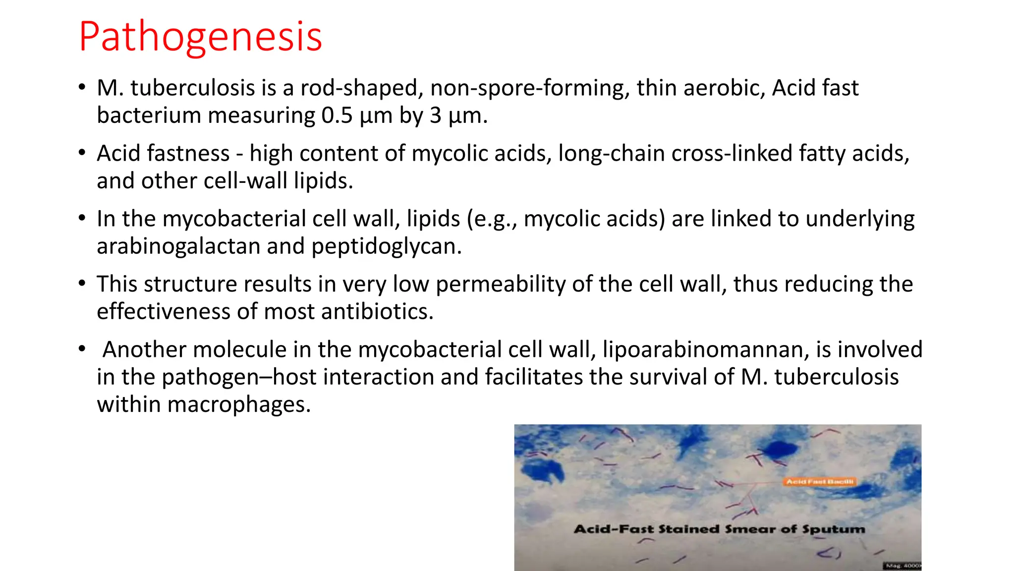
• Tuberculosis (TB), which is caused by bacteria of the Mycobacterium
tuberculosis complex.
• Chronic granulomatous inflammation, induces cell mediated
immunity and delayed hypersensitivity.
• Oldest diseases[70000 Years ago] known to affect humans and the
top cause of infectious death [untreated fatality 70% ] worldwide
excluding COVID-19.
• This disease most often affects the lungs, although other organs are
involved in up to one-third of the cases.
• First discovered in 1882 by Robert Koch.](https://image.slidesharecdn.com/ptb6-240407064917-5e3f4954/75/Pulmonary-tuberculosis-latest-guidelines-and-treatment-2-2048.jpg)