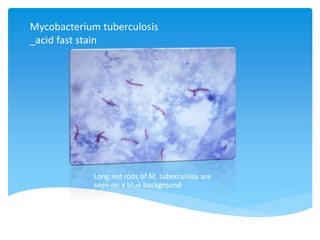
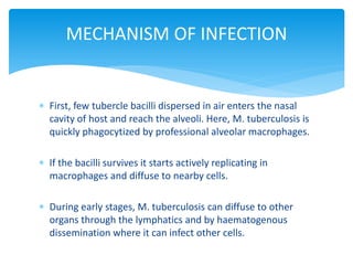
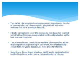
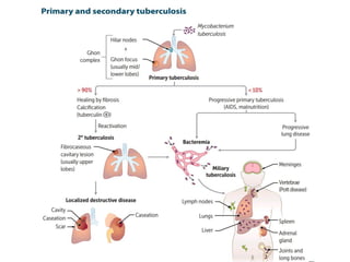
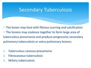
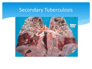
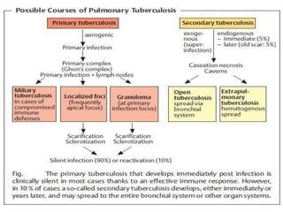
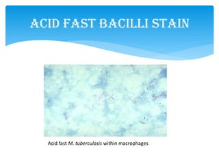
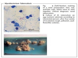
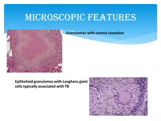
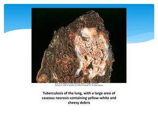
MYCOBACTERIUM
TUBERCULOSIS
by M. MOHSIN JAVED
INTRODUCTION
 Aerobic Bacilli - non spore forming
 Non motile, rod shape
 Cell wall - rich in lipids (prevent dyes used in gram stain)
 Acid fast bacilli (retain carbolfuschin stain despite
subsequent treatment with an ethanol-hydrochloric
acid mixture)
 Very slow growing (doubling time of 18 Hrs.)
Mycobacterium tuberculosis
_acid fast stain
Long red rods of M. tuberculosis are
seen on a blue background
DISEASE
Causes tuberculosis
 Worldwide, M. tuberculosis causes more deaths than
any other single microbial agent
 Approximately one-third of the world's population is
infected with this organism
IMPORTANT PROPERTIES
 Grows slowly (in contrast to most bacteria)
 Media used for it's growth is Lowenstein-Jensen
medium contains complex nutrients ane dyes.
 Obligated aerobe ; causing disease in highly oxygenated
tissue.
 The acid-fast property is attributed to long-chain fatty
acids called mycolic acids in the cell wall.
 Resistant to acids and alkalis.
Continue... IMPORTANT PROPERTIES
 Cord factor is correlated with virulence of the organism.
Virulent strains grow in a characteristic chord like
pattern.
 M. tuberculosis is resistant to dehydration and
therefore survive in dried exported sputum.
TRANSMISSION
Mycobacterium tuberculosis is transmitted from person to person by
respiratory aerosols produced by coughing
Continue... TRANSMISSION
 The portal of entry is respiratory tract and the initial site
of infection is lungs.
 In tissue, it resides chiefly within reticuloendothelial
cells
 For example ; Macrophages
EPIDEMIOLOGY
 Humans are the natural reservoirs of M. tuberculosis.
 Habitat is human lungs
Pathogenesis of
Mycobacterium Tuberculosis
 First, few tubercle bacilli dispersed in air enters the nasal
cavity of host and reach the alveoli. Here, M. tuberculosis is
quickly phagocytized by professional alveolar macrophages.
 If the bacilli survives it starts actively replicating in
macrophages and diffuse to nearby cells.
 During early stages, M. tuberculosis can diffuse to other
organs through the lymphatics and by haematogenous
dissemination where it can infect other cells.
MECHANISM OF INFECTION
 Thereafter, the adaptive immune response , migration to the site
of primary infection of neutrophils, lymphocytes and other
immune cells form a cellular infiltrate.
 Fibrotic components cover the granuloma that becomes calcified
such that bacilli remain encapsulated inside and protected by the
host immune response.
 This primary lesion, classically termed the Ghon complex, within
this complex bacilli persisting in a dormant, non-metabolically
active state, for years, decades, or most often for lifetime.
 Sometimes, during latent infection, bacilli would start replicating
inside this primary lesion, cause the reactivation of disease.
Pathogenesis of Mycobacterium tuberculosis is divided
into two stages;
1. Primary Tuberculosis
2. Secondary Tuberculosis
PATHOGENESIS
 The infection of an individual who has not been previously
infected or immunised is called primary tuberculosis or Ghon’s
complex or childhood tuberculosis.
 The most commonly involved tissues for primary complex are
lungs and lymph nodes.
 Other tissues are tonsils and cervical lymph nodes, lesions may be
found in small intestine and mesenteric lymph nodes.
 Progressive primary tuberculosis is particularly high in
immunocompromised host e.g. in patients of AIDS
Primary Tuberculosis
 Inhaled bacilli implant in the distal
airspaces of lower part of upper
lobe or upper part of lower lobe.
 1-1.5 cm area of grey white
inflammation with consolidation
develops, called as Ghon’s complex which
often caseates.
Ghon’s complex: Subpleural granuloma at right
along with Granuloma in the hilar lymph node
Primary Tuberculosis
Histopathological changes
Granulomatous inflammation
forms both caseating and non
caseating tubercles.
Tuberculous granuloma has the
following criteria:
1. Rounded outlines
2. Central caseous necrosis
3. Transformed macrophages called
epithelioid cells
4. Lymphocytes, plasma cells, and
fibroblasts
 The infection of an individual who has been previously infected or
sensitised is called secondary, or post-primary or reinfection, or
chronic tuberculosis.
 The infection may be
1. Endogenous source such as reactivation of dormant primary
complex.
2. Exogenous source such as fresh dose of reinfection by the tubercle
bacilli.
Secondary tuberculosis occurs most commonly in lungs in the region
of apex. Other sites and tissues which can be involved are tonsils,
pharynx, larynx, small intestine and skin.
Secondary Tuberculosis
 The lesion may heal with fibrous scarring and calcification
 The lesions may coalesce together to form large area of
tuberculous pneumonia and produce progressive secondary
pulmonary tuberculosis or extra pulmonary lesions:
1. Tuberculous caseous pneumonia
2. Fibrocaseous tuberculosis
3. Miliary tuberculosis
Secondary Tuberculosis
EXTRA PULMONARY TUBERCULOSIS
 In tissues or organs seeded hematogenously
 Commonly involved organs includes:
Intestinal tuberculosis
Meninges
Kidneys
Adrenals
Bones
Vertebrae
Fallopian tubes
Secondary Tuberculosis
Hypersensitivity or allergy, and immunity or resistance,
play a major role in the development of lesions in
tuberculosis. Tubercle bacilli as such do not produce any
toxins.
 Mycosides such as ‘cord factor’ which are essential for
growth and virulence of the organism in the animals
 Glycolipids present in the mycobacterial cell wall like
‘Wax-D’ which acts as an adjuvant acting along with
tuberculoprotein.
Immunity And Hypersensitivity
As a part of body’s immune response, T and B cells are activated.
Activated CD4+T cells develop the cell-mediated delayed type
hypersensitivity reaction, while B cells result in formation of
antibodies which play no role in body’s defence against tubercle
bacilli.
In 2-3 days, the macrophages undergo structural changes as a result
of immune mechanisms to epithelioid cells.
The epithelioid cells in time aggregate into tight clusters
or granulomas. Release of cytokines in response to
sensitized CD4+T cells and some constituents of
mycobacterial cell wall play a role in formation of
granuloma.
The development of caseation necrosis is possibly due to
interaction of mycobacteria with activated T cells (CD4+
helper T cells via IFN-γ and CD8+ suppressor T cells
directly)
Disease:- Tuberculosis
 Primary TB
 Progressive Primary TB
 Secondary TB
 Miliary TB
Clinical Manifestations
Primary TB
 Most individuals (~75%) are asymptomatic or have flu-
like symptoms along with:
fever and
chest pain
 Around 3 weeks after infection, they become (skin test,
see “Diagnosis”)
Progressive Primary TB
 Some individuals (5-15%) don’t contain the primary
infection and develop a progressive disease that resembles
a necrotizing bacterial pneumonia
 This presents with:
 fever,
 productive cough, and
 chest pain
Secondary TB
 Pattern of disease that arises in a previously infected
and sensitized patient
 The lesions typically localize to the apex of the upper
lobes
 Symptoms include:
Low grade fever,
Night sweats, and
weight loss
 Without therapy, miliary TB may develop
Miliary TB
 This refers to the uncontrolled hematogenous
dissemination of M. tuberculosis.
 Infection may involve any organ and the course is
usually rapid when it occurs with primary or
secondary progressive disease.
 Multiorgan failure, septic shock, and respiratory
distress, followed by death, may occur.
 X-ray
 Sputum acid fast stain
 Culture
 rRNA or DNA in sputum by nucleic acid amplification
(results in 2-7 hours, but does NOT replace culture)
Diagnosis
Chest Radiograph
Upper lobe infiltrate
with air fluid level
The most common abnormality associated
with primary TB on chest radiography is hilar
adenopathy (Black arrows). Subpleural
granulomas (yellow arrow) are also common.
These two findings constitute the Ghon
complex. This is also shown in the gross
specimen to the right.
Acid Fast Bacilli Stain
Acid fast M. tuberculosis within macrophages
Microscopic Features
Granulomas with central caseation
Epithelioid granulomas with Langhans giant
cells typically associated with TB
Tuberculosis of the lung, with a large area of
caseous necrosis containing yellow-white and
cheesy debris
Treatment of latent TB (asymptomatic, but with
radiographic evidence)
Treatment
Drugs Duration Interval Minimum doses
Isoniazid 9 months Daily 270
Twice weekly* 76
Isoniazid 6 months Daily 180
Twice weekly* 52
Isoniazid and
Rifapentine
3 months Once weekly* 12
Rifampin 4 months Daily 120
 There are currently 10 drugs used for active TB disease
 The first lines drugs are isoniazid, rifampin, ethambutol, pyrazinamide
 Preferred regimen is the aforementioned drugs for 8 weeks
 Afterward, maintenance therapy includes daily isoniazid and rifampin for
18 weeks
IMPORTANT STEP 1 INFORMATION ABOUT TB DRUGS!
 Rifampin turns urine red, be sure to tell patient to expect it!
 Isoniazid can cause peripheral neuropathy, be sure to pretreat with B6
Treatment
Preventions and control:
 Prompt and effective treatment of patients with active
tuberculosis and careful follow up of their contacts with
tuberculin test and CXR.
 Immunization with BCG (Bacillus-Calmette-Guerin)
vaccine.
 Pasteurization of milk and milk products
Reference:
 [Levinson W.] Review of Medical Microbiology and Immunology.
 [Jaypee] Review of Microbiology and Immunology.
 Color Atlas of Microbiology .

Mycobacterium Tuberculosis

  • 1.
  • 2.
    INTRODUCTION  Aerobic Bacilli- non spore forming  Non motile, rod shape  Cell wall - rich in lipids (prevent dyes used in gram stain)  Acid fast bacilli (retain carbolfuschin stain despite subsequent treatment with an ethanol-hydrochloric acid mixture)  Very slow growing (doubling time of 18 Hrs.)
  • 3.
    Mycobacterium tuberculosis _acid faststain Long red rods of M. tuberculosis are seen on a blue background
  • 4.
    DISEASE Causes tuberculosis  Worldwide,M. tuberculosis causes more deaths than any other single microbial agent  Approximately one-third of the world's population is infected with this organism
  • 5.
    IMPORTANT PROPERTIES  Growsslowly (in contrast to most bacteria)  Media used for it's growth is Lowenstein-Jensen medium contains complex nutrients ane dyes.  Obligated aerobe ; causing disease in highly oxygenated tissue.  The acid-fast property is attributed to long-chain fatty acids called mycolic acids in the cell wall.  Resistant to acids and alkalis.
  • 6.
    Continue... IMPORTANT PROPERTIES Cord factor is correlated with virulence of the organism. Virulent strains grow in a characteristic chord like pattern.  M. tuberculosis is resistant to dehydration and therefore survive in dried exported sputum.
  • 7.
    TRANSMISSION Mycobacterium tuberculosis istransmitted from person to person by respiratory aerosols produced by coughing
  • 8.
    Continue... TRANSMISSION  Theportal of entry is respiratory tract and the initial site of infection is lungs.  In tissue, it resides chiefly within reticuloendothelial cells  For example ; Macrophages
  • 9.
    EPIDEMIOLOGY  Humans arethe natural reservoirs of M. tuberculosis.  Habitat is human lungs
  • 10.
  • 11.
     First, fewtubercle bacilli dispersed in air enters the nasal cavity of host and reach the alveoli. Here, M. tuberculosis is quickly phagocytized by professional alveolar macrophages.  If the bacilli survives it starts actively replicating in macrophages and diffuse to nearby cells.  During early stages, M. tuberculosis can diffuse to other organs through the lymphatics and by haematogenous dissemination where it can infect other cells. MECHANISM OF INFECTION
  • 12.
     Thereafter, theadaptive immune response , migration to the site of primary infection of neutrophils, lymphocytes and other immune cells form a cellular infiltrate.  Fibrotic components cover the granuloma that becomes calcified such that bacilli remain encapsulated inside and protected by the host immune response.  This primary lesion, classically termed the Ghon complex, within this complex bacilli persisting in a dormant, non-metabolically active state, for years, decades, or most often for lifetime.  Sometimes, during latent infection, bacilli would start replicating inside this primary lesion, cause the reactivation of disease.
  • 14.
    Pathogenesis of Mycobacteriumtuberculosis is divided into two stages; 1. Primary Tuberculosis 2. Secondary Tuberculosis PATHOGENESIS
  • 15.
     The infectionof an individual who has not been previously infected or immunised is called primary tuberculosis or Ghon’s complex or childhood tuberculosis.  The most commonly involved tissues for primary complex are lungs and lymph nodes.  Other tissues are tonsils and cervical lymph nodes, lesions may be found in small intestine and mesenteric lymph nodes.  Progressive primary tuberculosis is particularly high in immunocompromised host e.g. in patients of AIDS Primary Tuberculosis
  • 16.
     Inhaled bacilliimplant in the distal airspaces of lower part of upper lobe or upper part of lower lobe.  1-1.5 cm area of grey white inflammation with consolidation develops, called as Ghon’s complex which often caseates. Ghon’s complex: Subpleural granuloma at right along with Granuloma in the hilar lymph node Primary Tuberculosis
  • 17.
    Primary Tuberculosis Histopathological changes Granulomatousinflammation forms both caseating and non caseating tubercles. Tuberculous granuloma has the following criteria: 1. Rounded outlines 2. Central caseous necrosis 3. Transformed macrophages called epithelioid cells 4. Lymphocytes, plasma cells, and fibroblasts
  • 18.
     The infectionof an individual who has been previously infected or sensitised is called secondary, or post-primary or reinfection, or chronic tuberculosis.  The infection may be 1. Endogenous source such as reactivation of dormant primary complex. 2. Exogenous source such as fresh dose of reinfection by the tubercle bacilli. Secondary tuberculosis occurs most commonly in lungs in the region of apex. Other sites and tissues which can be involved are tonsils, pharynx, larynx, small intestine and skin. Secondary Tuberculosis
  • 19.
     The lesionmay heal with fibrous scarring and calcification  The lesions may coalesce together to form large area of tuberculous pneumonia and produce progressive secondary pulmonary tuberculosis or extra pulmonary lesions: 1. Tuberculous caseous pneumonia 2. Fibrocaseous tuberculosis 3. Miliary tuberculosis Secondary Tuberculosis
  • 20.
    EXTRA PULMONARY TUBERCULOSIS In tissues or organs seeded hematogenously  Commonly involved organs includes: Intestinal tuberculosis Meninges Kidneys Adrenals Bones Vertebrae Fallopian tubes Secondary Tuberculosis
  • 21.
  • 22.
    Hypersensitivity or allergy,and immunity or resistance, play a major role in the development of lesions in tuberculosis. Tubercle bacilli as such do not produce any toxins.  Mycosides such as ‘cord factor’ which are essential for growth and virulence of the organism in the animals  Glycolipids present in the mycobacterial cell wall like ‘Wax-D’ which acts as an adjuvant acting along with tuberculoprotein. Immunity And Hypersensitivity
  • 23.
    As a partof body’s immune response, T and B cells are activated. Activated CD4+T cells develop the cell-mediated delayed type hypersensitivity reaction, while B cells result in formation of antibodies which play no role in body’s defence against tubercle bacilli. In 2-3 days, the macrophages undergo structural changes as a result of immune mechanisms to epithelioid cells.
  • 25.
    The epithelioid cellsin time aggregate into tight clusters or granulomas. Release of cytokines in response to sensitized CD4+T cells and some constituents of mycobacterial cell wall play a role in formation of granuloma. The development of caseation necrosis is possibly due to interaction of mycobacteria with activated T cells (CD4+ helper T cells via IFN-γ and CD8+ suppressor T cells directly)
  • 26.
    Disease:- Tuberculosis  PrimaryTB  Progressive Primary TB  Secondary TB  Miliary TB Clinical Manifestations
  • 27.
    Clinical Manifestations Primary TB Most individuals (~75%) are asymptomatic or have flu- like symptoms along with: fever and chest pain  Around 3 weeks after infection, they become (skin test, see “Diagnosis”)
  • 28.
    Progressive Primary TB Some individuals (5-15%) don’t contain the primary infection and develop a progressive disease that resembles a necrotizing bacterial pneumonia  This presents with:  fever,  productive cough, and  chest pain
  • 29.
    Secondary TB  Patternof disease that arises in a previously infected and sensitized patient  The lesions typically localize to the apex of the upper lobes  Symptoms include: Low grade fever, Night sweats, and weight loss  Without therapy, miliary TB may develop
  • 30.
    Miliary TB  Thisrefers to the uncontrolled hematogenous dissemination of M. tuberculosis.  Infection may involve any organ and the course is usually rapid when it occurs with primary or secondary progressive disease.  Multiorgan failure, septic shock, and respiratory distress, followed by death, may occur.
  • 32.
     X-ray  Sputumacid fast stain  Culture  rRNA or DNA in sputum by nucleic acid amplification (results in 2-7 hours, but does NOT replace culture) Diagnosis
  • 33.
    Chest Radiograph Upper lobeinfiltrate with air fluid level
  • 34.
    The most commonabnormality associated with primary TB on chest radiography is hilar adenopathy (Black arrows). Subpleural granulomas (yellow arrow) are also common. These two findings constitute the Ghon complex. This is also shown in the gross specimen to the right.
  • 35.
    Acid Fast BacilliStain Acid fast M. tuberculosis within macrophages
  • 37.
    Microscopic Features Granulomas withcentral caseation Epithelioid granulomas with Langhans giant cells typically associated with TB
  • 38.
    Tuberculosis of thelung, with a large area of caseous necrosis containing yellow-white and cheesy debris
  • 39.
    Treatment of latentTB (asymptomatic, but with radiographic evidence) Treatment Drugs Duration Interval Minimum doses Isoniazid 9 months Daily 270 Twice weekly* 76 Isoniazid 6 months Daily 180 Twice weekly* 52 Isoniazid and Rifapentine 3 months Once weekly* 12 Rifampin 4 months Daily 120
  • 40.
     There arecurrently 10 drugs used for active TB disease  The first lines drugs are isoniazid, rifampin, ethambutol, pyrazinamide  Preferred regimen is the aforementioned drugs for 8 weeks  Afterward, maintenance therapy includes daily isoniazid and rifampin for 18 weeks IMPORTANT STEP 1 INFORMATION ABOUT TB DRUGS!  Rifampin turns urine red, be sure to tell patient to expect it!  Isoniazid can cause peripheral neuropathy, be sure to pretreat with B6 Treatment
  • 41.
    Preventions and control: Prompt and effective treatment of patients with active tuberculosis and careful follow up of their contacts with tuberculin test and CXR.  Immunization with BCG (Bacillus-Calmette-Guerin) vaccine.  Pasteurization of milk and milk products
  • 42.
    Reference:  [Levinson W.]Review of Medical Microbiology and Immunology.  [Jaypee] Review of Microbiology and Immunology.  Color Atlas of Microbiology .