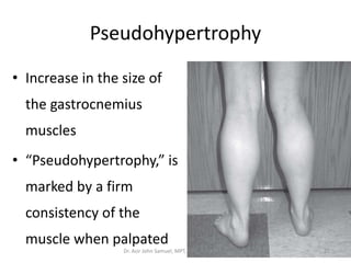
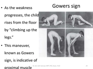
Muscular Dystrophy
Dr. Asir John Samuel, MPT, PhD (Physiotherapy)
Associate Professor,
Maharishi Markandeshwar Institute of Physiotherapy and Rehabilitation
(MMIPR),
Maharishi Markandeshwar (Deemed-to-be University),
Mullana-Ambala, Haryana, India
Muscular dystrophy
• Muscular dystrophy describes a group of
muscle diseases that are genetically
determined and have a steadily progressive
degenerative course
Dr. Asir John Samuel, MPT, PhD, Assoc. Prof. 2
Duchenne muscular dystrophy (DMD)
• Duchenne muscular dystrophy (DMD) in 1987
• Relationship between the different
dystrophies and dystrophin–glycoprotein
complex (DGC)
Dr. Asir John Samuel, MPT, PhD, Assoc. Prof. 3
Dystrophin–glycoprotein complex
(DGC)
• Dystrophin–glycoprotein complex (DGC) is a
group of proteins that links the
subsarcolemmal cytoskeleton and
extracellular matrix with the contractile
apparatus of the muscle and gives stability to
the muscle cell membrane
Dr. Asir John Samuel, MPT, PhD, Assoc. Prof. 4
Muscle cell membrane and associated protein
complexes implicated in muscle disease
Dr. Asir John Samuel, MPT, PhD, Assoc. Prof. 5
Muscular dystrophy - Types
• When dystrophin is deficient, the result is
Duchenne or Becker muscular dystrophy
(BMD)
• When one of the sarcoglycan proteins is
deficient, other limb-girdle muscular
dystrophies (LGMDs) result
• A deficiency of Merosin results in one specific
type of congenital muscular dystrophy (CMD)Dr. Asir John Samuel, MPT, PhD, Assoc. Prof. 6
Muscular dystrophy - Types
• DMD and BMD are also known as
dystrophinopathies because dystrophin is deficient
in these conditions
• LGMDs are also known as sarcoglycanopathies
because one of the sarcoglycan proteins is deficient
in these conditions
• Merosin negative congenital muscular dystrophy is
the result of a deficiency of MerosinDr. Asir John Samuel, MPT, PhD, Assoc. Prof. 7
Spinal muscular atrophy (SMA)
• Spinal muscular atrophy (SMA) refers to
neurogenic disorders whose underlying
pathology affects sensory neurons and spinal
interneurons
• As a result, affects the anterior horn cell
Dr. Asir John Samuel, MPT, PhD, Assoc. Prof. 8
Duchenne muscular dystrophy
• DMD, also known as pseudohypertrophic
muscular dystrophy or progressive muscular
dystrophy
• Most prevalent and severely disabling of the
childhood neuromusclular disorders
• Approximately 1 in 3500 live male births
Dr. Asir John Samuel, MPT, PhD, Assoc. Prof. 9
Duchenne muscular dystrophy
• Dystrophinopathy in which the child becomes
weaker and usually dies of respiratory
insufficiency and/or heart failure due to
myocardial involvement in the second or third
decade of life
Dr. Asir John Samuel, MPT, PhD, Assoc. Prof. 10
Duchenne muscular dystrophy
• X-linked inheritance pattern to DMD whereby
male offspring
• Inherit the disease from their mothers, who
are most often asymptomatic
• Defect to be a mutation at Xp21 in the gene
coding for the protein dystrophin
Dr. Asir John Samuel, MPT, PhD, Assoc. Prof. 11
DMD - Diagnosis
• Clinical presentation gives the first clues to the
diagnosis
• Laboratory findings include an abnormally
high serum creatine kinase (CK) level, which is
50 to 200 times the normal level and usually
ranges from 15,000 to 35,000 IU/L (normal
less than 160 IU/L)Dr. Asir John Samuel, MPT, PhD, Assoc. Prof. 12
DMD - Diagnosis
• Muscle biopsy may be performed to confirm
the diagnosis and shows degenerating and
regenerating fibers, inflammatory infiltrates,
and increased connective tissue and adipose
cells
• Immunohistologic staining of the tissue
reveals the absence of dystrophin along the
muscle cell membranes
Dr. Asir John Samuel, MPT, PhD, Assoc. Prof. 13
After diagnosis - DMD
• With the availability of genetic analysis, all
male family members may be screened for the
disorder
• All female family members may be screened
for their carrier status once a mutation is
identified
Dr. Asir John Samuel, MPT, PhD, Assoc. Prof. 14
DMD - Pathophysiology
• Absence of dystrophin leads to a reduction in
all of the dystrophin-associated proteins in the
muscle cell membrane and
• Causes a disruption in the linkage between
the subsarcolemmal cytoskeleton and the
extracellular matrix
Dr. Asir John Samuel, MPT, PhD, Assoc. Prof. 15
DMD - Pathophysiology
• Lack of dystrophin is thought to cause
sarcolemmal instability and
• An increase susceptibility to membrane
microtears, which may be exacerbated by
muscle contractions.
Dr. Asir John Samuel, MPT, PhD, Assoc. Prof. 16
DMD - Pathophysiology
• Increased calcium channel leaks, and an
increase in reactive oxygen species
• Increase in reactive oxygen species is activated
through a pathway driven by
mechanotransduction by the microtubule
cytoskeleton of the cell
Dr. Asir John Samuel, MPT, PhD, Assoc. Prof. 17
DMD - Pathophysiology
• Activation of this signaling pathway results
from membrane stress and
• Ultimately impacts calcium signaling and
results in an increase in calcium which raises
intracellular calcium levels, leading to muscle
cell necrosis
Dr. Asir John Samuel, MPT, PhD, Assoc. Prof. 18
Clinical Presentation
• Onset of the disorder is insidious
• Symptoms appear between 2 and 5 yrs of age
• If not noticed for months or years, then
• May be misdiagnosed for years
Dr. Asir John Samuel, MPT, PhD, Assoc. Prof. 19
Clinical Presentation
• Reluctance to walk or run at appropriate ages,
• Falling, and difficulty getting up off the floor,
• Toe-walking,
• Clumsiness, and
• An increase in the size of the gastrocnemius
muscles
Dr. Asir John Samuel, MPT, PhD, Assoc. Prof. 20
Pseudohypertrophy
• Increase in the size of
the gastrocnemius
muscles
• “Pseudohypertrophy,” is
marked by a firm
consistency of the
muscle when palpated
Dr. Asir John Samuel, MPT, PhD, Assoc. Prof. 21
Clinical Presentation and Progression
• The weakness is steadily progressive with
proximal muscles tending to be weaker earlier
and to progress faster
• Weakness of the hip and knee extensors
• often results in an exaggerated lumbar
lordosis
Dr. Asir John Samuel, MPT, PhD, Assoc. Prof. 22
Clinical Presentation and Progression
• Child attempts to broaden the base of support
during walking and
• Thus develops a gait that resembles waddling
• Child may develop iliotibial band (ITB)
contractures, which are made worse by this
wide-based stance
Dr. Asir John Samuel, MPT, PhD, Assoc. Prof. 23
Gowers sign• As the weakness
progresses, the child
rises from the floor
by “climbing up the
legs.”
• This maneuver,
known as Gowers
sign, is indicative of
proximal muscle
Dr. Asir John Samuel, MPT, PhD, Assoc. Prof. 24
Contractures
• Plantarflexion at the ankle,
• Inversion of the foot
• Flexion at both the hips and knees
Dr. Asir John Samuel, MPT, PhD, Assoc. Prof. 25
ROM
• Loss of range of motion (ROM),
• Hamstrings,
• Hip flexors,
• ITBs, and
• Heel cords
Dr. Asir John Samuel, MPT, PhD, Assoc. Prof. 26
Contracture
• An increasing degree of contracture is seen at
the hips and knees,
• Upper extremity contractures begin to
develop in the elbows, shoulders and long
finger flexors
• More time sitting
Dr. Asir John Samuel, MPT, PhD, Assoc. Prof. 27
Slow in Functional activities
• Functional activities may be performed more
slowly by children with DMD than by typically
developing children
• Those affected are able to walk, climb stairs
and stand up from the floor without too much
difficulty until 6 or 7 years of age
Dr. Asir John Samuel, MPT, PhD, Assoc. Prof. 28
DMD and BMD
• Loss of unassisted ambulation at 9 to 10 years
of age
• Loss of ambulation should be by the age of 13
after which the patient would be considered
to have either BMD or an intermediate
phenotype
Dr. Asir John Samuel, MPT, PhD, Assoc. Prof. 29
Prognosis
• Child choose to use long-leg braces and
continue to walk as an exercise ambulator for
an additional year
• But will need help getting to and from
standing
• Partial weightbearing walker may also be used
at this stage
Dr. Asir John Samuel, MPT, PhD, Assoc. Prof. 30
Dr. Asir John Samuel, MPT, PhD, Assoc. Prof. 31
Dr. Asir John Samuel, MPT, PhD, Assoc. Prof. 32
Functional Grades: Arms and
Shoulders – Brooke (1983)
1 - Standing with arms at the sides, the patient
can abduct the arms in a full circle until they
touch above the head.
2 - The patient can raise the arms above the
head only by flexing the elbow (i.e., by
shortening the circumference of the movement)
or by using accessory muscles.Dr. Asir John Samuel, MPT, PhD, Assoc. Prof. 33
Functional Grades: Arms and
Shoulders – Brooke (1983)
3 - The patient cannot raise hands above the
head, but can raise an 8-oz glass of water to the
mouth (using both hands if necessary).
4 - The patient can raise hands to the mouth,
but cannot raise an 8-oz glass of water to the
mouth.
Dr. Asir John Samuel, MPT, PhD, Assoc. Prof. 34
Functional Grades: Arms and
Shoulders – Brooke (1983)
5 - The patient cannot raise hands to the mouth,
but can use the hands to hold a pen or to pick
up pennies from a table.
6 - The patient cannot raise hands to the mouth
and has no useful function of the hands.
Dr. Asir John Samuel, MPT, PhD, Assoc. Prof. 35
Problem list
1. Weaknes
2. Decreased active and passive ROM
3. Ambulation dysfunction
4. Decreased functional ability
5. Decreased pulmonary function
6. Emotional trauma—individual and family
7. Progressive scoliosis
8. Pain Dr. Asir John Samuel, MPT, PhD, Assoc. Prof. 36
Physical Therapy Intervention
1. Prevent deformity
2. Prolong functional capacity
3. Improve pulmonary function
4. Facilitate the development and assistance of
family support and support of others
5. Control pain, if necessary
Dr. Asir John Samuel, MPT, PhD, Assoc. Prof. 37
Preventing Deformity
• Tendency for development of plantarflexion
contractures
• Daily stretching of the Achilles tendons should
slow down the development of this deformity
• Night splints
• 10 and 15 reps, holding at least 15 seconds,
performed at least once, and preferably twice,
daily
• Serial casting
Dr. Asir John Samuel, MPT, PhD, Assoc. Prof. 38
Activity Level/Active Exercise
• Care should be taken to avoid overusing muscles
and causing fatigue
• Signs of overuse weakness include feeling weaker
30 minutes postexercise or excessive soreness 24
to 48 hours after exercise
• Other signs include severe muscle cramping,
heaviness in the extremities, and prolonged
shortness of breathDr. Asir John Samuel, MPT, PhD, Assoc. Prof. 39
Activity Level/Active Exercise
• Eccentric muscle activities such as walking or
running downhill and closed chain exercises
such as squats should be avoided
• As they tend to cause more muscle soreness
• Resistive muscle strengthening is not
recommended in boys with DMD because of
the risk of contraction-induced muscle injuryDr. Asir John Samuel, MPT, PhD, Assoc. Prof. 40
Resisted exercise
• Lack of dystrophin increases susceptibility to
muscle cell damage
• Microtears in the muscle cell membrane
increase with muscle contractions and cause
an increase in calcium leak channel activity,
which in turn causes an increase in
intracellular calcium
Dr. Asir John Samuel, MPT, PhD, Assoc. Prof. 41
Resisted exercise
• This increase causes calcium-dependent
proteolysis, which eventually leads to cell
death
• Strength training in boys with DMD has been a
subject of controversy
Dr. Asir John Samuel, MPT, PhD, Assoc. Prof. 42
Minimizing Spinal Deformity
• Child’s sitting time increases, so does
kyphoscoliosis
• Convexity will likely be toward the dominant
extremity
• Spinal orthoses do not delay the development
of the spinal curve and might increase work of
breathing Dr. Asir John Samuel, MPT, PhD, Assoc. Prof. 43
Endurance exercise
• Endurance exercise such as swimming has
been found to be beneficial in mdx mice by
increasing the resistance to fatigue in muscles
by increasing the proportion of type I (slow
oxidative) fibers
• Submaximal endurance training such as
swimming or cycling may be beneficial,
especially in the younger child with DMDDr. Asir John Samuel, MPT, PhD, Assoc. Prof. 44

Muscular dystrophy

  • 1.
    Muscular Dystrophy Dr. AsirJohn Samuel, MPT, PhD (Physiotherapy) Associate Professor, Maharishi Markandeshwar Institute of Physiotherapy and Rehabilitation (MMIPR), Maharishi Markandeshwar (Deemed-to-be University), Mullana-Ambala, Haryana, India
  • 2.
    Muscular dystrophy • Musculardystrophy describes a group of muscle diseases that are genetically determined and have a steadily progressive degenerative course Dr. Asir John Samuel, MPT, PhD, Assoc. Prof. 2
  • 3.
    Duchenne muscular dystrophy(DMD) • Duchenne muscular dystrophy (DMD) in 1987 • Relationship between the different dystrophies and dystrophin–glycoprotein complex (DGC) Dr. Asir John Samuel, MPT, PhD, Assoc. Prof. 3
  • 4.
    Dystrophin–glycoprotein complex (DGC) • Dystrophin–glycoproteincomplex (DGC) is a group of proteins that links the subsarcolemmal cytoskeleton and extracellular matrix with the contractile apparatus of the muscle and gives stability to the muscle cell membrane Dr. Asir John Samuel, MPT, PhD, Assoc. Prof. 4
  • 5.
    Muscle cell membraneand associated protein complexes implicated in muscle disease Dr. Asir John Samuel, MPT, PhD, Assoc. Prof. 5
  • 6.
    Muscular dystrophy -Types • When dystrophin is deficient, the result is Duchenne or Becker muscular dystrophy (BMD) • When one of the sarcoglycan proteins is deficient, other limb-girdle muscular dystrophies (LGMDs) result • A deficiency of Merosin results in one specific type of congenital muscular dystrophy (CMD)Dr. Asir John Samuel, MPT, PhD, Assoc. Prof. 6
  • 7.
    Muscular dystrophy -Types • DMD and BMD are also known as dystrophinopathies because dystrophin is deficient in these conditions • LGMDs are also known as sarcoglycanopathies because one of the sarcoglycan proteins is deficient in these conditions • Merosin negative congenital muscular dystrophy is the result of a deficiency of MerosinDr. Asir John Samuel, MPT, PhD, Assoc. Prof. 7
  • 8.
    Spinal muscular atrophy(SMA) • Spinal muscular atrophy (SMA) refers to neurogenic disorders whose underlying pathology affects sensory neurons and spinal interneurons • As a result, affects the anterior horn cell Dr. Asir John Samuel, MPT, PhD, Assoc. Prof. 8
  • 9.
    Duchenne muscular dystrophy •DMD, also known as pseudohypertrophic muscular dystrophy or progressive muscular dystrophy • Most prevalent and severely disabling of the childhood neuromusclular disorders • Approximately 1 in 3500 live male births Dr. Asir John Samuel, MPT, PhD, Assoc. Prof. 9
  • 10.
    Duchenne muscular dystrophy •Dystrophinopathy in which the child becomes weaker and usually dies of respiratory insufficiency and/or heart failure due to myocardial involvement in the second or third decade of life Dr. Asir John Samuel, MPT, PhD, Assoc. Prof. 10
  • 11.
    Duchenne muscular dystrophy •X-linked inheritance pattern to DMD whereby male offspring • Inherit the disease from their mothers, who are most often asymptomatic • Defect to be a mutation at Xp21 in the gene coding for the protein dystrophin Dr. Asir John Samuel, MPT, PhD, Assoc. Prof. 11
  • 12.
    DMD - Diagnosis •Clinical presentation gives the first clues to the diagnosis • Laboratory findings include an abnormally high serum creatine kinase (CK) level, which is 50 to 200 times the normal level and usually ranges from 15,000 to 35,000 IU/L (normal less than 160 IU/L)Dr. Asir John Samuel, MPT, PhD, Assoc. Prof. 12
  • 13.
    DMD - Diagnosis •Muscle biopsy may be performed to confirm the diagnosis and shows degenerating and regenerating fibers, inflammatory infiltrates, and increased connective tissue and adipose cells • Immunohistologic staining of the tissue reveals the absence of dystrophin along the muscle cell membranes Dr. Asir John Samuel, MPT, PhD, Assoc. Prof. 13
  • 14.
    After diagnosis -DMD • With the availability of genetic analysis, all male family members may be screened for the disorder • All female family members may be screened for their carrier status once a mutation is identified Dr. Asir John Samuel, MPT, PhD, Assoc. Prof. 14
  • 15.
    DMD - Pathophysiology •Absence of dystrophin leads to a reduction in all of the dystrophin-associated proteins in the muscle cell membrane and • Causes a disruption in the linkage between the subsarcolemmal cytoskeleton and the extracellular matrix Dr. Asir John Samuel, MPT, PhD, Assoc. Prof. 15
  • 16.
    DMD - Pathophysiology •Lack of dystrophin is thought to cause sarcolemmal instability and • An increase susceptibility to membrane microtears, which may be exacerbated by muscle contractions. Dr. Asir John Samuel, MPT, PhD, Assoc. Prof. 16
  • 17.
    DMD - Pathophysiology •Increased calcium channel leaks, and an increase in reactive oxygen species • Increase in reactive oxygen species is activated through a pathway driven by mechanotransduction by the microtubule cytoskeleton of the cell Dr. Asir John Samuel, MPT, PhD, Assoc. Prof. 17
  • 18.
    DMD - Pathophysiology •Activation of this signaling pathway results from membrane stress and • Ultimately impacts calcium signaling and results in an increase in calcium which raises intracellular calcium levels, leading to muscle cell necrosis Dr. Asir John Samuel, MPT, PhD, Assoc. Prof. 18
  • 19.
    Clinical Presentation • Onsetof the disorder is insidious • Symptoms appear between 2 and 5 yrs of age • If not noticed for months or years, then • May be misdiagnosed for years Dr. Asir John Samuel, MPT, PhD, Assoc. Prof. 19
  • 20.
    Clinical Presentation • Reluctanceto walk or run at appropriate ages, • Falling, and difficulty getting up off the floor, • Toe-walking, • Clumsiness, and • An increase in the size of the gastrocnemius muscles Dr. Asir John Samuel, MPT, PhD, Assoc. Prof. 20
  • 21.
    Pseudohypertrophy • Increase inthe size of the gastrocnemius muscles • “Pseudohypertrophy,” is marked by a firm consistency of the muscle when palpated Dr. Asir John Samuel, MPT, PhD, Assoc. Prof. 21
  • 22.
    Clinical Presentation andProgression • The weakness is steadily progressive with proximal muscles tending to be weaker earlier and to progress faster • Weakness of the hip and knee extensors • often results in an exaggerated lumbar lordosis Dr. Asir John Samuel, MPT, PhD, Assoc. Prof. 22
  • 23.
    Clinical Presentation andProgression • Child attempts to broaden the base of support during walking and • Thus develops a gait that resembles waddling • Child may develop iliotibial band (ITB) contractures, which are made worse by this wide-based stance Dr. Asir John Samuel, MPT, PhD, Assoc. Prof. 23
  • 24.
    Gowers sign• Asthe weakness progresses, the child rises from the floor by “climbing up the legs.” • This maneuver, known as Gowers sign, is indicative of proximal muscle Dr. Asir John Samuel, MPT, PhD, Assoc. Prof. 24
  • 25.
    Contractures • Plantarflexion atthe ankle, • Inversion of the foot • Flexion at both the hips and knees Dr. Asir John Samuel, MPT, PhD, Assoc. Prof. 25
  • 26.
    ROM • Loss ofrange of motion (ROM), • Hamstrings, • Hip flexors, • ITBs, and • Heel cords Dr. Asir John Samuel, MPT, PhD, Assoc. Prof. 26
  • 27.
    Contracture • An increasingdegree of contracture is seen at the hips and knees, • Upper extremity contractures begin to develop in the elbows, shoulders and long finger flexors • More time sitting Dr. Asir John Samuel, MPT, PhD, Assoc. Prof. 27
  • 28.
    Slow in Functionalactivities • Functional activities may be performed more slowly by children with DMD than by typically developing children • Those affected are able to walk, climb stairs and stand up from the floor without too much difficulty until 6 or 7 years of age Dr. Asir John Samuel, MPT, PhD, Assoc. Prof. 28
  • 29.
    DMD and BMD •Loss of unassisted ambulation at 9 to 10 years of age • Loss of ambulation should be by the age of 13 after which the patient would be considered to have either BMD or an intermediate phenotype Dr. Asir John Samuel, MPT, PhD, Assoc. Prof. 29
  • 30.
    Prognosis • Child chooseto use long-leg braces and continue to walk as an exercise ambulator for an additional year • But will need help getting to and from standing • Partial weightbearing walker may also be used at this stage Dr. Asir John Samuel, MPT, PhD, Assoc. Prof. 30
  • 31.
    Dr. Asir JohnSamuel, MPT, PhD, Assoc. Prof. 31
  • 32.
    Dr. Asir JohnSamuel, MPT, PhD, Assoc. Prof. 32
  • 33.
    Functional Grades: Armsand Shoulders – Brooke (1983) 1 - Standing with arms at the sides, the patient can abduct the arms in a full circle until they touch above the head. 2 - The patient can raise the arms above the head only by flexing the elbow (i.e., by shortening the circumference of the movement) or by using accessory muscles.Dr. Asir John Samuel, MPT, PhD, Assoc. Prof. 33
  • 34.
    Functional Grades: Armsand Shoulders – Brooke (1983) 3 - The patient cannot raise hands above the head, but can raise an 8-oz glass of water to the mouth (using both hands if necessary). 4 - The patient can raise hands to the mouth, but cannot raise an 8-oz glass of water to the mouth. Dr. Asir John Samuel, MPT, PhD, Assoc. Prof. 34
  • 35.
    Functional Grades: Armsand Shoulders – Brooke (1983) 5 - The patient cannot raise hands to the mouth, but can use the hands to hold a pen or to pick up pennies from a table. 6 - The patient cannot raise hands to the mouth and has no useful function of the hands. Dr. Asir John Samuel, MPT, PhD, Assoc. Prof. 35
  • 36.
    Problem list 1. Weaknes 2.Decreased active and passive ROM 3. Ambulation dysfunction 4. Decreased functional ability 5. Decreased pulmonary function 6. Emotional trauma—individual and family 7. Progressive scoliosis 8. Pain Dr. Asir John Samuel, MPT, PhD, Assoc. Prof. 36
  • 37.
    Physical Therapy Intervention 1.Prevent deformity 2. Prolong functional capacity 3. Improve pulmonary function 4. Facilitate the development and assistance of family support and support of others 5. Control pain, if necessary Dr. Asir John Samuel, MPT, PhD, Assoc. Prof. 37
  • 38.
    Preventing Deformity • Tendencyfor development of plantarflexion contractures • Daily stretching of the Achilles tendons should slow down the development of this deformity • Night splints • 10 and 15 reps, holding at least 15 seconds, performed at least once, and preferably twice, daily • Serial casting Dr. Asir John Samuel, MPT, PhD, Assoc. Prof. 38
  • 39.
    Activity Level/Active Exercise •Care should be taken to avoid overusing muscles and causing fatigue • Signs of overuse weakness include feeling weaker 30 minutes postexercise or excessive soreness 24 to 48 hours after exercise • Other signs include severe muscle cramping, heaviness in the extremities, and prolonged shortness of breathDr. Asir John Samuel, MPT, PhD, Assoc. Prof. 39
  • 40.
    Activity Level/Active Exercise •Eccentric muscle activities such as walking or running downhill and closed chain exercises such as squats should be avoided • As they tend to cause more muscle soreness • Resistive muscle strengthening is not recommended in boys with DMD because of the risk of contraction-induced muscle injuryDr. Asir John Samuel, MPT, PhD, Assoc. Prof. 40
  • 41.
    Resisted exercise • Lackof dystrophin increases susceptibility to muscle cell damage • Microtears in the muscle cell membrane increase with muscle contractions and cause an increase in calcium leak channel activity, which in turn causes an increase in intracellular calcium Dr. Asir John Samuel, MPT, PhD, Assoc. Prof. 41
  • 42.
    Resisted exercise • Thisincrease causes calcium-dependent proteolysis, which eventually leads to cell death • Strength training in boys with DMD has been a subject of controversy Dr. Asir John Samuel, MPT, PhD, Assoc. Prof. 42
  • 43.
    Minimizing Spinal Deformity •Child’s sitting time increases, so does kyphoscoliosis • Convexity will likely be toward the dominant extremity • Spinal orthoses do not delay the development of the spinal curve and might increase work of breathing Dr. Asir John Samuel, MPT, PhD, Assoc. Prof. 43
  • 44.
    Endurance exercise • Enduranceexercise such as swimming has been found to be beneficial in mdx mice by increasing the resistance to fatigue in muscles by increasing the proportion of type I (slow oxidative) fibers • Submaximal endurance training such as swimming or cycling may be beneficial, especially in the younger child with DMDDr. Asir John Samuel, MPT, PhD, Assoc. Prof. 44