P G R O R T H O P E D I C D E P A R T M E N T
S H E I K H Z A Y E D H O S P I T A L R A H I M Y A R K H A N
DR HAFIZ USMAN JAMEEL
Topic of Presentation
Muscular dystrophy
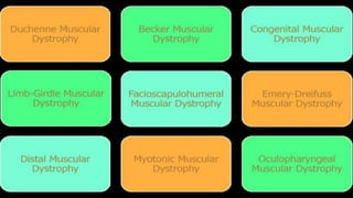
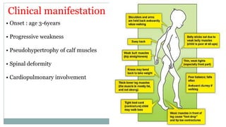
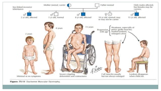
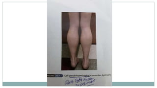
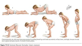
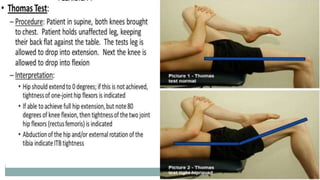
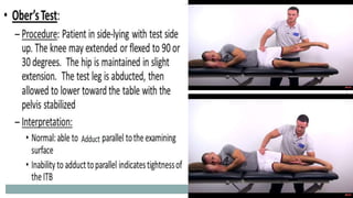
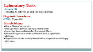
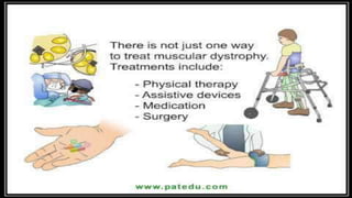
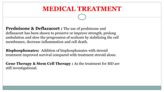
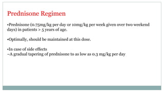
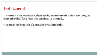
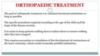
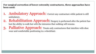
Muscular dystrophies are a group of
hereditary disorders of skeletal muscle
that produce progressive degeneration
of skeletal muscle and associated
weakness
Causes
• Inheritance
• Dominant genes
• Recessive gene
Depends on the age when symptoms appear, and the
types of symptoms that develop.
Risk
• Because these are inherited disorders, risk include a
family history of muscular dystrophy
The disorders differ in terms of the distribution and extent of
muscle weakness depending on the age of onset, rate of
progression, and pattern of inheritance.
Some forms of MD are seen in infancy or childhood while
others might not appear until middle age or later.
Classification
Sex-linked: DMD, BMD, EDMD
Autosomal recessive: LGMD, infantile FSHD
Autosomal dominant: FSHD, Distal MD, Ocular MD,
Oculopharyngeal MD.
DUCHENNE MUSCULAR DYSTROPHY
Also called Pseudo Hypertrophic muscular dystrophy
X LINKED RECESSIVE disorder
INCIDENCE : 1 in 3500 live births
SEX : Males are affected ; Females are Carrier
PATHOGENESIS
Single gene defect
Xp 21 region
Absent dystrophin
•Dystrophin-glycoprotein complex confer stability to the
sarcolemma
•Deficiency of dystrophin (Duchenne's dystrophy) may lead to
secondary loss of the sarcoglycans and dystroglycan
•Disruption of the dystrophin-glycoprotein complexes weakens the
sarcolemma, causing membrane tears and a cascade of events
leading to muscle fiber necrosis
Clinical manifestation
• Onset : age 3-6years
• Progressive weakness
• Pseudohypertrophy of calf muscles
• Spinal deformity
• Cardiopulmonary involvement
Age of onset is between 2-6 years of age
Stage 1 – Pre symptomatic
Creatine kinase usually elevated
Positive family history
CLINICAL FEATURES AND DIAGNOSIS
Stage 2- Early ambulatory
•Clumsy & Waddling gait, manifesting in children aged
•2-6 years; secondary to hip girdle muscle weakness
•Progressive weakness in the proximal musculature, initially in the lower
extremities, but later involving the neck flexors, shoulders, and arms.
•Possible toe-walking , Can climb stairs
•Meryon’s sign:
•Child slips through examiner’s grasp when lifted under arms due to the lack of
adductor muscles tone and severe shoulder girdle muscles weakness.
Stage 3- Late ambulatory
•More difficulty walking
•Around age 8 years, most patients notice difficulty with ascending stairs
and respiratory muscle strength begins a slow but steady decline
•Cannot arise from the floor
•The forced vital capacity begins to gradually wane, leading to symptoms
of nocturnal hypoxemia.
Stage 4 – Non ambulatory
•Possible development of scoliosis
•Scoliosis may progress, especially when more wheelchair dependent
•If wheelchair bound and profoundly weak, patients develop terminal
respiratory or cardiac failure, usually by the early 30s
•Poor nutritional intake can also be a serious complication in
individuals with severe end-stage DMD
•Contractures may develop
•Most are unable to ambulate independently by age 10
•Most are wheelchair dependent by age 15
•Most are died because of cardio respiratory problems by age 25-30
Prognosis
Laboratory Tests
•Serum CK
–Elevated to between 20 and 100 times normal
Diagnostic Procedures
•EMG : Myopathic
Muscle biopsy
•Muscle fibers of varying size
•Small groups of necrotic and regenerating fibers
•Connective tissue and fat replace lost muscle fibers.
•Definitive diagnosis is established on the basis of dystrophin
deficiency.
•Diagnosis can also be made by Western blot analysis of muscle biopsy
•specimens.
MEDICAL TREATMENT
Prednisone & Deflazacort : The use of prednisone and
deflazacort has been shown to preserve or improve strength, prolong
ambulation and slow the pr0gression of scoliosis by stabilizing the cell
membranes, decrease inflammation and cell death.
Bisphosphonates: Addition of bisphosphonates with steroid
treatment improved survival compared with treatment steroid alone.
Gene Therapy & Stem Cell Therapy : As the treatment for MD are
still investigational.
Prednisone Regimen
•Prednisone (0.75mg/kg per day or 10mg/kg per week given over two weekend
days) in patients > 5 years of age.
•Optimally, should be maintained at this dose.
•In case of side effects
–A gradual tapering of prednisone to as low as 0.3 mg/kg per day
Deflazacort
•In contrast with prednisone, alternate day treatment with deflazacort (2mg/kg
every other day) for 2 years was beneficial in one study.
•The mean prolongation of ambulation was 13 months.
ORTHOPAEDIC TREATMENT
The goal of orthopaedic treatment is to maintain functional ambulation as
long as possible.
The specific procedures required according to the age of the child and the
stage of the disease severity.
It is easier to keep patients walking than to induce them to resume walking
after they have stopped.
This requires prevention or retardation of the development of contractures of
the lower extremity, which would eventually prohibit ambulation.
For surgical correction of lower extremity contractures, three approaches have
been used:
1. Ambulatory Approach: Correct any contracture while patient is still
ambulatory.
2. Rehabilitation Approach: Surgery is performed after the patient has
lost the ability to walk but with the intension that walking will resume.
3. Palliative Approach: Treats only contractures that interfere with shoe
wear and comfortable positioning in a wheelchair.
How to prevent contractures formation
1. Stretching Exercises
2. Nightly Bracing
Exercises to stretch the hip muscles and lower extremity braces
worn at night to prevent the child’s sleeping in a frog leg position
are helpful.
Surgical techniques to treat contractures
Percutaneous release of hip flexion and abduction contractures and Ankle
contractures:
GREENE TECHNIQUE
RIDEAU TECHNIQUE: Same as that of Greene but with an open procedure
to release hip flexor contractures and lateral thigh contractures.
Transfer of the Posterior Tibial Tendon to the Dorsum of the Foot
By Greene Technique:
Transfer of the Posterior Tibial Tendon to the Dorsum of the Base of
Second Metatarsal By Mubarak Technique:
To correct the Equinus contracture.
Sartorius, Tensor Fasciae Femoris
and Tensor Fasciae Latae
Tensor Fascia Lata
Intermuscular septum
Achilles Tendon
Postoperative Care
•Patient should be mobilized immediately after surgery.
•Walker assisted ambulation is begun as soon as possible.
•Bilateral polypropylene long leg orthosis are fitted as soon as possible
Transfer of the Posterior Tibial Tendon to the Dorsum of the Foot By
Greene Technique
Transfer of the Posterior Tibial Tendon to the Dorsum of the Base of
Second Metatarsal By Mubarak Technique
Postoperative Care
•Standing and Walking are allowed 24 to 48 hours after surgery.
•Long leg cast is worn for 4 to 6 weeks.
•Knee-ankle-foot orthosis is worn permanently
Becker Muscular Dystrophy
• Milder version of DMD
• Etiology ▫ single gene defect ▫ short arm X chromosome
▫ altered size & decreased amount of dystrophin
• Less common
▫ 2.3 per 100000 live male birth
• Less severe
• Family history: atypical MD
• Similar & less severe than DMD
• Onset: age > 7 years
• Pseudohypertrophy of calf
• Equinous and varus foot
• High rate of scoliosis
• Less frequent cardiac involvement
Clinical features
Diagnosis
• The same as DMD
• Increase CPK (<200x)
• Decrease dystrophin and/or altered size
Natural history
▫ Slower progression
▫ ambulate until adolescence
▫ longer life expectancy
Treatment
▫ the same as in DMD
▫ forefoot equinous: plantar release, midfoot dorsal wedge
osteotomy
Muscular Dystrophy disorder PPT

Muscular Dystrophy disorder PPT

  • 2.
    P G RO R T H O P E D I C D E P A R T M E N T S H E I K H Z A Y E D H O S P I T A L R A H I M Y A R K H A N DR HAFIZ USMAN JAMEEL
  • 3.
  • 4.
    Muscular dystrophies area group of hereditary disorders of skeletal muscle that produce progressive degeneration of skeletal muscle and associated weakness
  • 5.
    Causes • Inheritance • Dominantgenes • Recessive gene Depends on the age when symptoms appear, and the types of symptoms that develop. Risk • Because these are inherited disorders, risk include a family history of muscular dystrophy
  • 7.
    The disorders differin terms of the distribution and extent of muscle weakness depending on the age of onset, rate of progression, and pattern of inheritance. Some forms of MD are seen in infancy or childhood while others might not appear until middle age or later.
  • 9.
    Classification Sex-linked: DMD, BMD,EDMD Autosomal recessive: LGMD, infantile FSHD Autosomal dominant: FSHD, Distal MD, Ocular MD, Oculopharyngeal MD.
  • 11.
    DUCHENNE MUSCULAR DYSTROPHY Alsocalled Pseudo Hypertrophic muscular dystrophy X LINKED RECESSIVE disorder INCIDENCE : 1 in 3500 live births SEX : Males are affected ; Females are Carrier
  • 12.
    PATHOGENESIS Single gene defect Xp21 region Absent dystrophin •Dystrophin-glycoprotein complex confer stability to the sarcolemma •Deficiency of dystrophin (Duchenne's dystrophy) may lead to secondary loss of the sarcoglycans and dystroglycan •Disruption of the dystrophin-glycoprotein complexes weakens the sarcolemma, causing membrane tears and a cascade of events leading to muscle fiber necrosis
  • 14.
    Clinical manifestation • Onset: age 3-6years • Progressive weakness • Pseudohypertrophy of calf muscles • Spinal deformity • Cardiopulmonary involvement
  • 15.
    Age of onsetis between 2-6 years of age Stage 1 – Pre symptomatic Creatine kinase usually elevated Positive family history CLINICAL FEATURES AND DIAGNOSIS
  • 16.
    Stage 2- Earlyambulatory •Clumsy & Waddling gait, manifesting in children aged •2-6 years; secondary to hip girdle muscle weakness •Progressive weakness in the proximal musculature, initially in the lower extremities, but later involving the neck flexors, shoulders, and arms. •Possible toe-walking , Can climb stairs •Meryon’s sign: •Child slips through examiner’s grasp when lifted under arms due to the lack of adductor muscles tone and severe shoulder girdle muscles weakness.
  • 17.
    Stage 3- Lateambulatory •More difficulty walking •Around age 8 years, most patients notice difficulty with ascending stairs and respiratory muscle strength begins a slow but steady decline •Cannot arise from the floor •The forced vital capacity begins to gradually wane, leading to symptoms of nocturnal hypoxemia.
  • 18.
    Stage 4 –Non ambulatory •Possible development of scoliosis •Scoliosis may progress, especially when more wheelchair dependent •If wheelchair bound and profoundly weak, patients develop terminal respiratory or cardiac failure, usually by the early 30s •Poor nutritional intake can also be a serious complication in individuals with severe end-stage DMD •Contractures may develop
  • 19.
    •Most are unableto ambulate independently by age 10 •Most are wheelchair dependent by age 15 •Most are died because of cardio respiratory problems by age 25-30 Prognosis
  • 27.
    Laboratory Tests •Serum CK –Elevatedto between 20 and 100 times normal Diagnostic Procedures •EMG : Myopathic Muscle biopsy •Muscle fibers of varying size •Small groups of necrotic and regenerating fibers •Connective tissue and fat replace lost muscle fibers. •Definitive diagnosis is established on the basis of dystrophin deficiency. •Diagnosis can also be made by Western blot analysis of muscle biopsy •specimens.
  • 29.
    MEDICAL TREATMENT Prednisone &Deflazacort : The use of prednisone and deflazacort has been shown to preserve or improve strength, prolong ambulation and slow the pr0gression of scoliosis by stabilizing the cell membranes, decrease inflammation and cell death. Bisphosphonates: Addition of bisphosphonates with steroid treatment improved survival compared with treatment steroid alone. Gene Therapy & Stem Cell Therapy : As the treatment for MD are still investigational.
  • 30.
    Prednisone Regimen •Prednisone (0.75mg/kgper day or 10mg/kg per week given over two weekend days) in patients > 5 years of age. •Optimally, should be maintained at this dose. •In case of side effects –A gradual tapering of prednisone to as low as 0.3 mg/kg per day
  • 31.
    Deflazacort •In contrast withprednisone, alternate day treatment with deflazacort (2mg/kg every other day) for 2 years was beneficial in one study. •The mean prolongation of ambulation was 13 months.
  • 32.
    ORTHOPAEDIC TREATMENT The goalof orthopaedic treatment is to maintain functional ambulation as long as possible. The specific procedures required according to the age of the child and the stage of the disease severity. It is easier to keep patients walking than to induce them to resume walking after they have stopped. This requires prevention or retardation of the development of contractures of the lower extremity, which would eventually prohibit ambulation.
  • 34.
    For surgical correctionof lower extremity contractures, three approaches have been used: 1. Ambulatory Approach: Correct any contracture while patient is still ambulatory. 2. Rehabilitation Approach: Surgery is performed after the patient has lost the ability to walk but with the intension that walking will resume. 3. Palliative Approach: Treats only contractures that interfere with shoe wear and comfortable positioning in a wheelchair.
  • 35.
    How to preventcontractures formation 1. Stretching Exercises 2. Nightly Bracing Exercises to stretch the hip muscles and lower extremity braces worn at night to prevent the child’s sleeping in a frog leg position are helpful.
  • 36.
    Surgical techniques totreat contractures Percutaneous release of hip flexion and abduction contractures and Ankle contractures: GREENE TECHNIQUE RIDEAU TECHNIQUE: Same as that of Greene but with an open procedure to release hip flexor contractures and lateral thigh contractures. Transfer of the Posterior Tibial Tendon to the Dorsum of the Foot By Greene Technique: Transfer of the Posterior Tibial Tendon to the Dorsum of the Base of Second Metatarsal By Mubarak Technique: To correct the Equinus contracture.
  • 37.
    Sartorius, Tensor FasciaeFemoris and Tensor Fasciae Latae Tensor Fascia Lata Intermuscular septum Achilles Tendon
  • 39.
    Postoperative Care •Patient shouldbe mobilized immediately after surgery. •Walker assisted ambulation is begun as soon as possible. •Bilateral polypropylene long leg orthosis are fitted as soon as possible
  • 40.
    Transfer of thePosterior Tibial Tendon to the Dorsum of the Foot By Greene Technique
  • 41.
    Transfer of thePosterior Tibial Tendon to the Dorsum of the Base of Second Metatarsal By Mubarak Technique
  • 42.
    Postoperative Care •Standing andWalking are allowed 24 to 48 hours after surgery. •Long leg cast is worn for 4 to 6 weeks. •Knee-ankle-foot orthosis is worn permanently
  • 43.
  • 44.
    • Milder versionof DMD • Etiology ▫ single gene defect ▫ short arm X chromosome ▫ altered size & decreased amount of dystrophin
  • 45.
    • Less common ▫2.3 per 100000 live male birth • Less severe • Family history: atypical MD • Similar & less severe than DMD • Onset: age > 7 years • Pseudohypertrophy of calf • Equinous and varus foot • High rate of scoliosis • Less frequent cardiac involvement Clinical features
  • 46.
    Diagnosis • The sameas DMD • Increase CPK (<200x) • Decrease dystrophin and/or altered size Natural history ▫ Slower progression ▫ ambulate until adolescence ▫ longer life expectancy Treatment ▫ the same as in DMD ▫ forefoot equinous: plantar release, midfoot dorsal wedge osteotomy