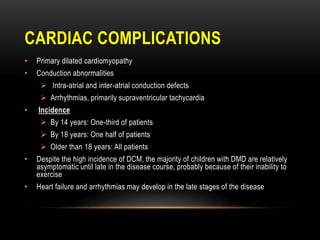
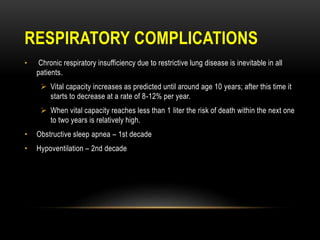
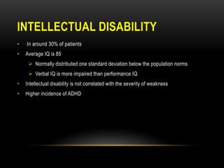
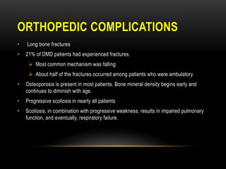
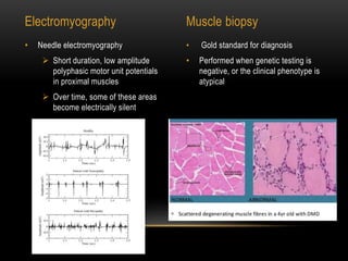
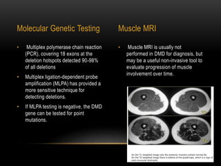
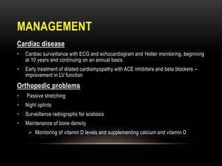
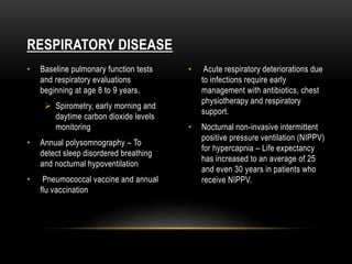
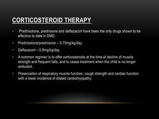
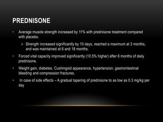
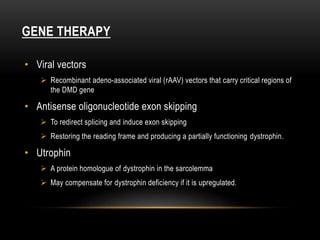
Shyala Chand
DUCHENNE MUSCULAR
DYSTROPHY
INTRODUCTION
• An inherited progressive myopathic disorder
• X-linked recessive form of muscular dystrophy
• Affects 1 in 3600 boys
• Caused by mutations in the dystrophin gene, and hence is termed “dystrophinopathy”
• Duchenne muscular dystrophy (DMD) is associated with the most severe clinical
symptoms
• Becker muscular dystrophy (BMD) has a similar presentation to DMD, but typically
has a later onset and a milder clinical course
• Patients with an intermediate phenotype may be classified clinically as having either
mild DMD or severe BMD
GENETICS AND PATHOGENESIS
• X linked disorder.
• Caused by mutations of the dystrophin gene located on chromosome Xp21.2
 Deletions - Around 72% of patients
 Partial gene duplications - 6 – 10% of patients
 Point mutations - In the coding sequence or the splicing sites.
DYSTROPHIN
• Dystrophin is located on the cytoplasmic face of the plasma membrane of muscle
fibers, functioning as a component of a large, tightly associated glycoprotein complex
• Provides mechanical reinforcement to the sarcolemma and stabilizes the
glycoprotein complex, shielding it from degradation.
 In its absence, the glycoprotein complex is digested by proteases. Loss of these
membrane proteins may initiate the degeneration of muscle fibers, resulting in
muscle weakness.
PATHOGENESIS
• Muscle cell membrane damage related to the loss of dystrophin may permit the
pathologic entry of extracellular calcium into muscle fibers.
• The excess cytosolic calcium can activate calpains, which promote muscle
proteolysis .
• Clinical onset of muscular
weakness usually occurs between 2
and 3 years of age.
• Weakness
• Cardiac Complications
• Respiratory Complications
• Intellectual Disability
• Orthopedic Complications
CLINICAL FEATURES
WEAKNESS
• Proximal before distal limb muscles
• Lower before upper extremities
• Difficulty running, jumping, and walking up steps
• Waddling gait
• Lumbar lordosis
• Pseudohypertrophy of calf muscles, due to fat infiltration
• Patients are usually wheelchair-bound by the age of 12
GOWERS SIGN
• Patient uses his hands and arms to "walk" up his own body from a squatting position
 due to lack of hip and thigh muscle strength.
CARDIAC COMPLICATIONS
• Primary dilated cardiomyopathy
• Conduction abnormalities
 Intra-atrial and inter-atrial conduction defects
 Arrhythmias, primarily supraventricular tachycardia
• Incidence
 By 14 years: One-third of patients
 By 18 years: One half of patients
 Older than 18 years: All patients
• Despite the high incidence of DCM, the majority of children with DMD are relatively
asymptomatic until late in the disease course, probably because of their inability to
exercise
• Heart failure and arrhythmias may develop in the late stages of the disease
RESPIRATORY COMPLICATIONS
• Chronic respiratory insufficiency due to restrictive lung disease is inevitable in all
patients.
 Vital capacity increases as predicted until around age 10 years; after this time it
starts to decrease at a rate of 8-12% per year.
 When vital capacity reaches less than 1 liter the risk of death within the next one
to two years is relatively high.
• Obstructive sleep apnea – 1st decade
• Hypoventilation – 2nd decade
INTELLECTUAL DISABILITY
• In around 30% of patients
• Average IQ is 85
 Normally distributed one standard deviation below the population norms
 Verbal IQ is more impaired than performance IQ
• Intellectual disability is not correlated with the severity of weakness
• Higher incidence of ADHD
ORTHOPEDIC COMPLICATIONS
• Long bone fractures
• 21% of DMD patients had experienced fractures.
 Most common mechanism was falling
 About half of the fractures occurred among patients who were ambulatory
• Osteoporosis is present in most patients. Bone mineral density begins early and
continues to diminish with age.
• Progressive scoliosis in nearly all patients
• Scoliosis, in combination with progressive weakness, results in impaired pulmonary
function, and eventually, respiratory failure.
DIAGNOSIS
• The diagnosis of a dystrophinopathy is suspected based upon:
 Characteristic age and sex
 Presence of symptoms and signs suggestive of a myopathic process
 Markedly increased serum creatine kinase values
 Myopathic changes on electromyography and muscle biopsy
 A positive family history suggesting X-linked recessive inheritance
• Serum muscle enzymes
 Markedly raised serum CK level, 10-20 times the upper limit of normal
 Levels peak at 2-3 years of age and then decline with increasing age, due to
progressive loss of dystrophic muscle fibres
 Elevated serum ALT, AST and LDH
• Gold standard for diagnosis
• Performed when genetic testing is
negative, or the clinical phenotype is
atypical
• Needle electromyography
 Short duration, low amplitude
polyphasic motor unit potentials
in proximal muscles
 Over time, some of these areas
become electrically silent
S
Electromyography Muscle biopsy
• Muscle MRI is usually not
performed in DMD for diagnosis, but
may be a useful non-invasive tool to
evaluate progression of muscle
involvement over time.
• Multiplex polymerase chain reaction
(PCR), covering 18 exons at the
deletion hotspots detected 90-98%
of all deletions
• Multiplex ligation-dependent probe
amplification (MLPA) has provided a
more sensitive technique for
detecting deletions.
• If MLPA testing is negative, the DMD
gene can be tested for point
mutations.
Molecular Genetic Testing Muscle MRI
MANAGEMENT
Cardiac disease
• Cardiac surveillance with ECG and echocardiogram and Holter monitoring, beginning
at 10 years and continuing on an annual basis.
• Early treatment of dilated cardiomyopathy with ACE inhibitors and beta blockers –
improvement in LV function
Orthopedic problems
• Passive stretching
• Night splints
• Surveillance radiographs for scoliosis
• Maintenance of bone density
 Monitoring of vitamin D levels and supplementing calcium and vitamin D
• Baseline pulmonary function tests
and respiratory evaluations
beginning at age 8 to 9 years.
 Spirometry, early morning and
daytime carbon dioxide levels
monitoring
• Annual polysomnography – To
detect sleep disordered breathing
and nocturnal hypoventilation
• Pneumococcal vaccine and annual
flu vaccination
• Acute respiratory deteriorations due
to infections require early
management with antibiotics, chest
physiotherapy and respiratory
support.
• Nocturnal non-invasive intermittent
positive pressure ventilation (NIPPV)
for hypercapnia – Life expectancy
has increased to an average of 25
and even 30 years in patients who
receive NIPPV.
RESPIRATORY DISEASE
CORTICOSTEROID THERAPY
• Prednisolone, prednisone and deflazacort have been the only drugs shown to be
effective to date in DMD.
• Prednisolone/prednisone – 0.75mg/kg/day
• Deflazacort – 0.9mg/kg/day
• A common regimen is to offer corticosteroids at the time of decline of muscle
strength and frequent falls, and to cease treatment when the child is no longer
ambulant.
• Preservation of respiratory muscle function, cough strength and cardiac function,
with a lower incidence of dilated cardiomyopathy.
PREDNISONE
• Average muscle strength increased by 11% with prednisone treatment compared
with placebo.
 Strength increased significantly by 10 days, reached a maximum at 3 months,
and was maintained at 6 and 18 months.
• Forced vital capacity improved significantly (10.5% higher) after 6 months of daily
prednisone.
• Weight gain, diabetes, Cushingoid appearance, hypertension, gastrointestinal
bleeding and compression fractures.
• In case of side effects – A gradual tapering of prednisone to as low as 0.3 mg/kg per
day
DEFLAZACORT
• FDA on Feb. 9, 2017, approved deflazacort (brand name Emflaza) to treat DMD.
• In contrast with prednisone, alternate day treatment with deflazacort (2mg/kg every
other day) for 2 years was beneficial in one study.
• The mean prolongation of ambulation was 13 months.
GENE THERAPY
• Viral vectors
 Recombinant adeno-associated viral (rAAV) vectors that carry critical regions of
the DMD gene
• Antisense oligonucleotide exon skipping
 To redirect splicing and induce exon skipping
 Restoring the reading frame and producing a partially functioning dystrophin.
• Utrophin
 A protein homologue of dystrophin in the sarcolemma
 May compensate for dystrophin deficiency if it is upregulated.
THANK YOU

Duchenne muscular dystrophy

  • 1.
  • 2.
    INTRODUCTION • An inheritedprogressive myopathic disorder • X-linked recessive form of muscular dystrophy • Affects 1 in 3600 boys • Caused by mutations in the dystrophin gene, and hence is termed “dystrophinopathy” • Duchenne muscular dystrophy (DMD) is associated with the most severe clinical symptoms • Becker muscular dystrophy (BMD) has a similar presentation to DMD, but typically has a later onset and a milder clinical course • Patients with an intermediate phenotype may be classified clinically as having either mild DMD or severe BMD
  • 3.
    GENETICS AND PATHOGENESIS •X linked disorder. • Caused by mutations of the dystrophin gene located on chromosome Xp21.2  Deletions - Around 72% of patients  Partial gene duplications - 6 – 10% of patients  Point mutations - In the coding sequence or the splicing sites.
  • 4.
    DYSTROPHIN • Dystrophin islocated on the cytoplasmic face of the plasma membrane of muscle fibers, functioning as a component of a large, tightly associated glycoprotein complex • Provides mechanical reinforcement to the sarcolemma and stabilizes the glycoprotein complex, shielding it from degradation.  In its absence, the glycoprotein complex is digested by proteases. Loss of these membrane proteins may initiate the degeneration of muscle fibers, resulting in muscle weakness.
  • 5.
    PATHOGENESIS • Muscle cellmembrane damage related to the loss of dystrophin may permit the pathologic entry of extracellular calcium into muscle fibers. • The excess cytosolic calcium can activate calpains, which promote muscle proteolysis .
  • 6.
    • Clinical onsetof muscular weakness usually occurs between 2 and 3 years of age. • Weakness • Cardiac Complications • Respiratory Complications • Intellectual Disability • Orthopedic Complications CLINICAL FEATURES
  • 7.
    WEAKNESS • Proximal beforedistal limb muscles • Lower before upper extremities • Difficulty running, jumping, and walking up steps • Waddling gait • Lumbar lordosis • Pseudohypertrophy of calf muscles, due to fat infiltration • Patients are usually wheelchair-bound by the age of 12
  • 8.
    GOWERS SIGN • Patientuses his hands and arms to "walk" up his own body from a squatting position  due to lack of hip and thigh muscle strength.
  • 9.
    CARDIAC COMPLICATIONS • Primarydilated cardiomyopathy • Conduction abnormalities  Intra-atrial and inter-atrial conduction defects  Arrhythmias, primarily supraventricular tachycardia • Incidence  By 14 years: One-third of patients  By 18 years: One half of patients  Older than 18 years: All patients • Despite the high incidence of DCM, the majority of children with DMD are relatively asymptomatic until late in the disease course, probably because of their inability to exercise • Heart failure and arrhythmias may develop in the late stages of the disease
  • 10.
    RESPIRATORY COMPLICATIONS • Chronicrespiratory insufficiency due to restrictive lung disease is inevitable in all patients.  Vital capacity increases as predicted until around age 10 years; after this time it starts to decrease at a rate of 8-12% per year.  When vital capacity reaches less than 1 liter the risk of death within the next one to two years is relatively high. • Obstructive sleep apnea – 1st decade • Hypoventilation – 2nd decade
  • 11.
    INTELLECTUAL DISABILITY • Inaround 30% of patients • Average IQ is 85  Normally distributed one standard deviation below the population norms  Verbal IQ is more impaired than performance IQ • Intellectual disability is not correlated with the severity of weakness • Higher incidence of ADHD
  • 12.
    ORTHOPEDIC COMPLICATIONS • Longbone fractures • 21% of DMD patients had experienced fractures.  Most common mechanism was falling  About half of the fractures occurred among patients who were ambulatory • Osteoporosis is present in most patients. Bone mineral density begins early and continues to diminish with age. • Progressive scoliosis in nearly all patients • Scoliosis, in combination with progressive weakness, results in impaired pulmonary function, and eventually, respiratory failure.
  • 13.
    DIAGNOSIS • The diagnosisof a dystrophinopathy is suspected based upon:  Characteristic age and sex  Presence of symptoms and signs suggestive of a myopathic process  Markedly increased serum creatine kinase values  Myopathic changes on electromyography and muscle biopsy  A positive family history suggesting X-linked recessive inheritance • Serum muscle enzymes  Markedly raised serum CK level, 10-20 times the upper limit of normal  Levels peak at 2-3 years of age and then decline with increasing age, due to progressive loss of dystrophic muscle fibres  Elevated serum ALT, AST and LDH
  • 14.
    • Gold standardfor diagnosis • Performed when genetic testing is negative, or the clinical phenotype is atypical • Needle electromyography  Short duration, low amplitude polyphasic motor unit potentials in proximal muscles  Over time, some of these areas become electrically silent S Electromyography Muscle biopsy
  • 15.
    • Muscle MRIis usually not performed in DMD for diagnosis, but may be a useful non-invasive tool to evaluate progression of muscle involvement over time. • Multiplex polymerase chain reaction (PCR), covering 18 exons at the deletion hotspots detected 90-98% of all deletions • Multiplex ligation-dependent probe amplification (MLPA) has provided a more sensitive technique for detecting deletions. • If MLPA testing is negative, the DMD gene can be tested for point mutations. Molecular Genetic Testing Muscle MRI
  • 16.
    MANAGEMENT Cardiac disease • Cardiacsurveillance with ECG and echocardiogram and Holter monitoring, beginning at 10 years and continuing on an annual basis. • Early treatment of dilated cardiomyopathy with ACE inhibitors and beta blockers – improvement in LV function Orthopedic problems • Passive stretching • Night splints • Surveillance radiographs for scoliosis • Maintenance of bone density  Monitoring of vitamin D levels and supplementing calcium and vitamin D
  • 17.
    • Baseline pulmonaryfunction tests and respiratory evaluations beginning at age 8 to 9 years.  Spirometry, early morning and daytime carbon dioxide levels monitoring • Annual polysomnography – To detect sleep disordered breathing and nocturnal hypoventilation • Pneumococcal vaccine and annual flu vaccination • Acute respiratory deteriorations due to infections require early management with antibiotics, chest physiotherapy and respiratory support. • Nocturnal non-invasive intermittent positive pressure ventilation (NIPPV) for hypercapnia – Life expectancy has increased to an average of 25 and even 30 years in patients who receive NIPPV. RESPIRATORY DISEASE
  • 18.
    CORTICOSTEROID THERAPY • Prednisolone,prednisone and deflazacort have been the only drugs shown to be effective to date in DMD. • Prednisolone/prednisone – 0.75mg/kg/day • Deflazacort – 0.9mg/kg/day • A common regimen is to offer corticosteroids at the time of decline of muscle strength and frequent falls, and to cease treatment when the child is no longer ambulant. • Preservation of respiratory muscle function, cough strength and cardiac function, with a lower incidence of dilated cardiomyopathy.
  • 19.
    PREDNISONE • Average musclestrength increased by 11% with prednisone treatment compared with placebo.  Strength increased significantly by 10 days, reached a maximum at 3 months, and was maintained at 6 and 18 months. • Forced vital capacity improved significantly (10.5% higher) after 6 months of daily prednisone. • Weight gain, diabetes, Cushingoid appearance, hypertension, gastrointestinal bleeding and compression fractures. • In case of side effects – A gradual tapering of prednisone to as low as 0.3 mg/kg per day
  • 20.
    DEFLAZACORT • FDA onFeb. 9, 2017, approved deflazacort (brand name Emflaza) to treat DMD. • In contrast with prednisone, alternate day treatment with deflazacort (2mg/kg every other day) for 2 years was beneficial in one study. • The mean prolongation of ambulation was 13 months.
  • 21.
    GENE THERAPY • Viralvectors  Recombinant adeno-associated viral (rAAV) vectors that carry critical regions of the DMD gene • Antisense oligonucleotide exon skipping  To redirect splicing and induce exon skipping  Restoring the reading frame and producing a partially functioning dystrophin. • Utrophin  A protein homologue of dystrophin in the sarcolemma  May compensate for dystrophin deficiency if it is upregulated.
  • 22.