Downloaded 121 times


![Resting potential
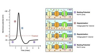
Ion pumps actively maintain concentration
gradients
[Na + ] much higher outside cell
[K + ] much higher inside cell
3 Na + transported out for every 2 K + transported in
Results in net – 70 mV charge inside nerve cell; cell
is polarized](https://image.slidesharecdn.com/muscletissue2-140109092356-phpapp01/85/Muscle-tissue-2-7-320.jpg)


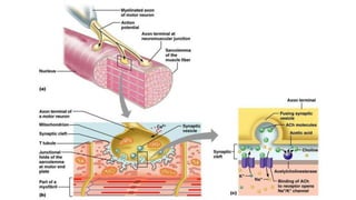

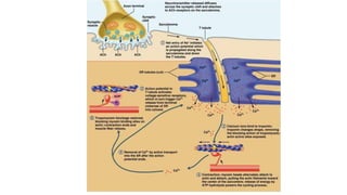


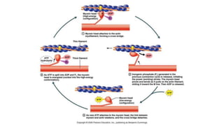

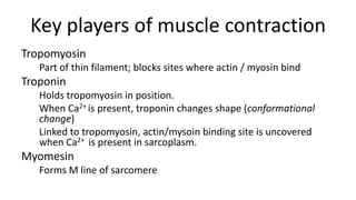


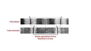
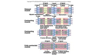
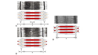

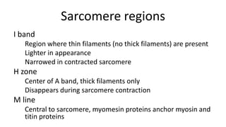


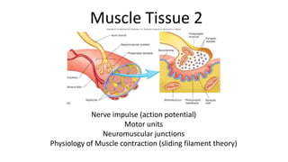
This document summarizes the key components of muscle tissue and muscle contraction. It discusses nerve impulses, motor units, neuromuscular junctions, and the sliding filament theory of muscle contraction. The main components that allow for contraction are described, including myosin, actin, tropomyosin, troponin, and the calcium release initiated by a nerve impulse that allows for crossbridge binding and sarcomere shortening. Regions of the sarcomere such as the Z-line, A-band, I-band, H-zone, and M-line are also outlined.


![Chapt09 Holes Lecture Animation[1]](https://cdn.slidesharecdn.com/ss_thumbnails/chapt09holeslectureanimation1-091122122851-phpapp02-thumbnail.jpg?width=640&height=640&fit=bounds)


![NURS1108_Lecture_7_-_Msuscular_2[1].pptx](https://cdn.slidesharecdn.com/ss_thumbnails/nurs1108lecture7-muscular21-250824235703-73f69db8-thumbnail.jpg?width=640&height=640&fit=bounds)


