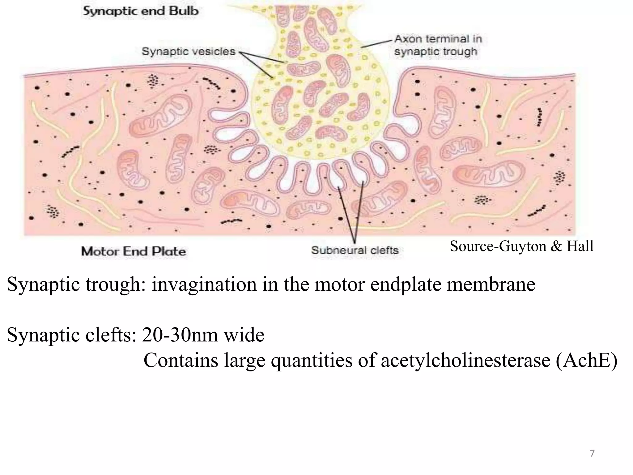
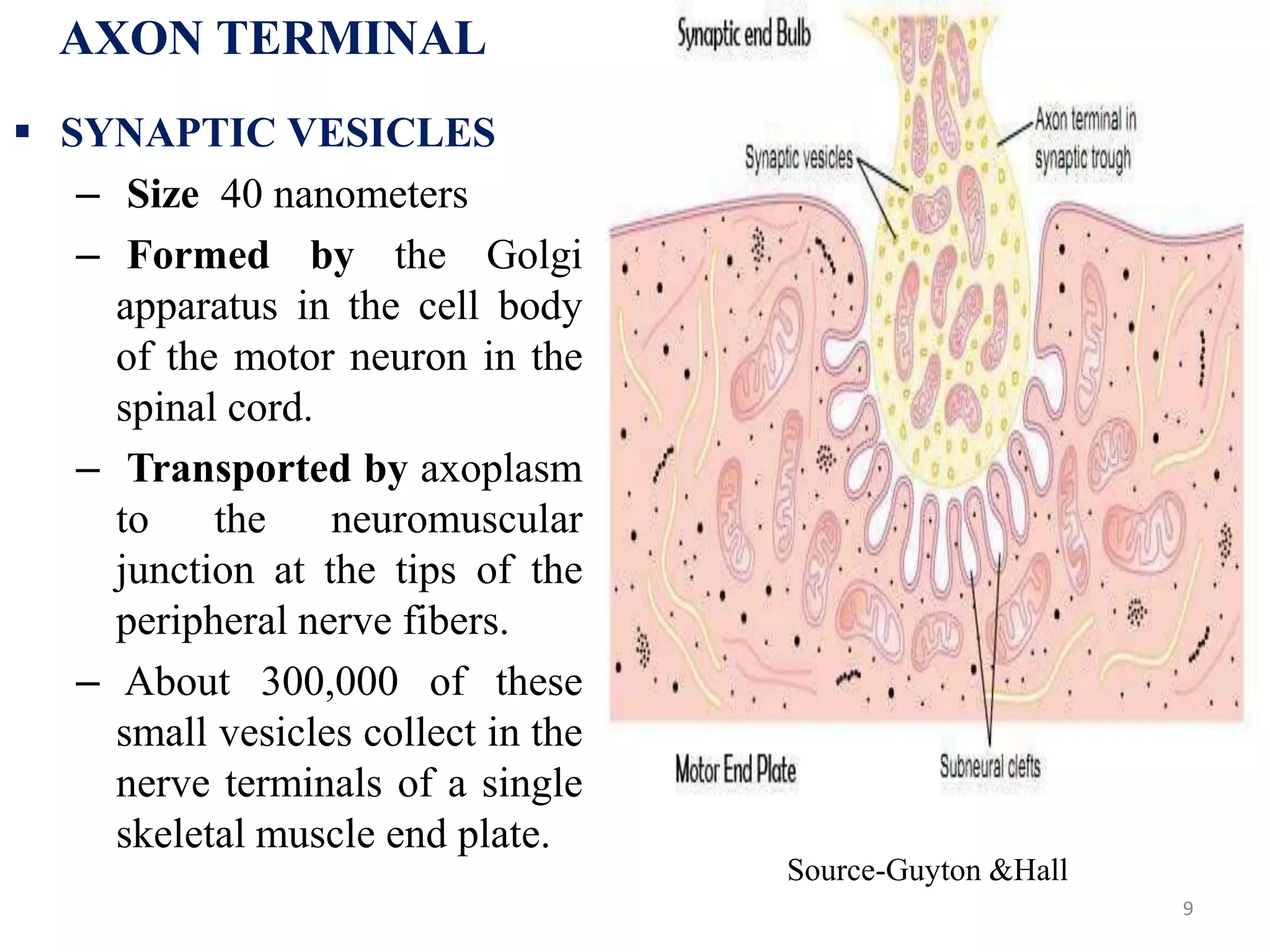
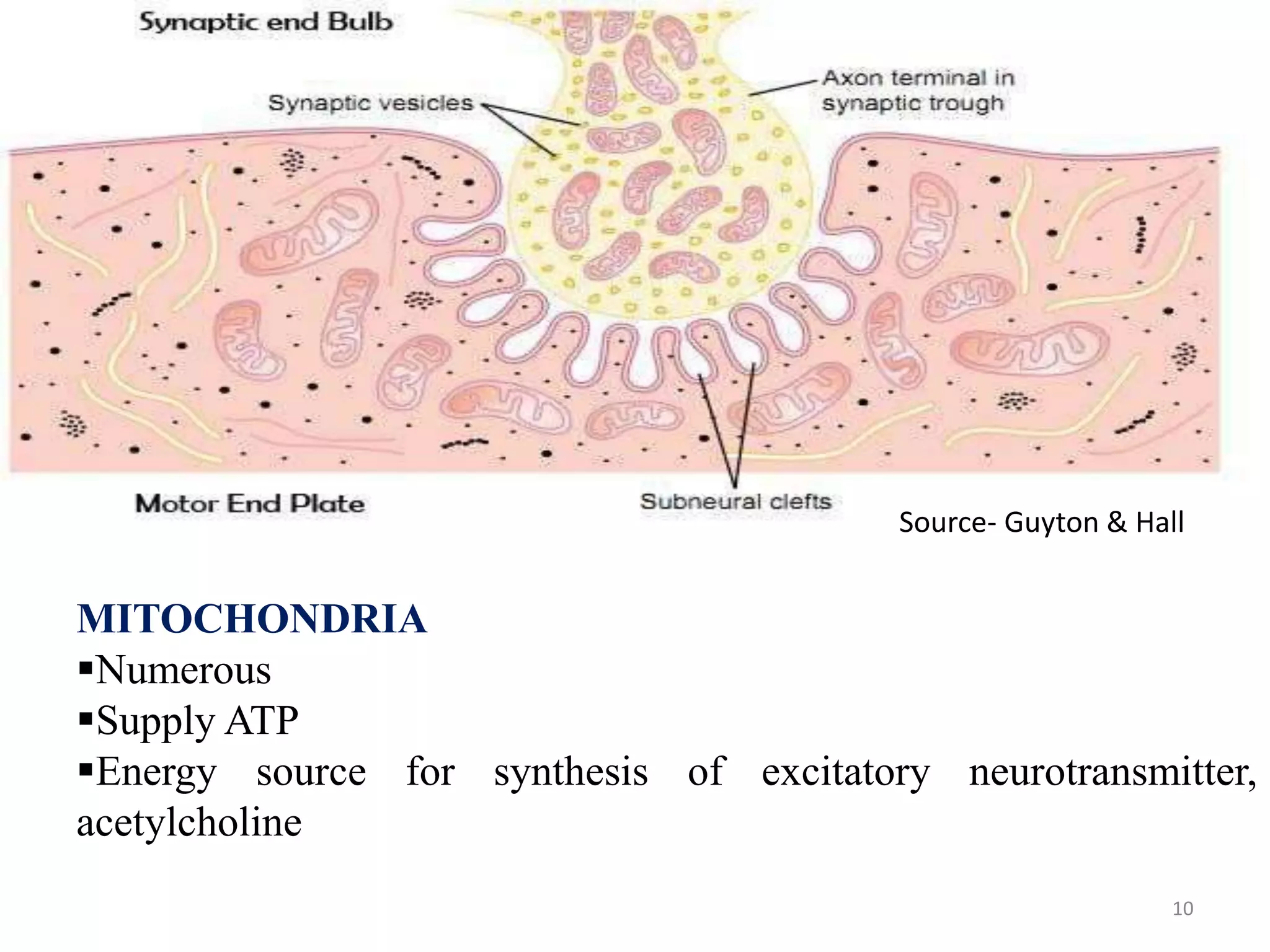
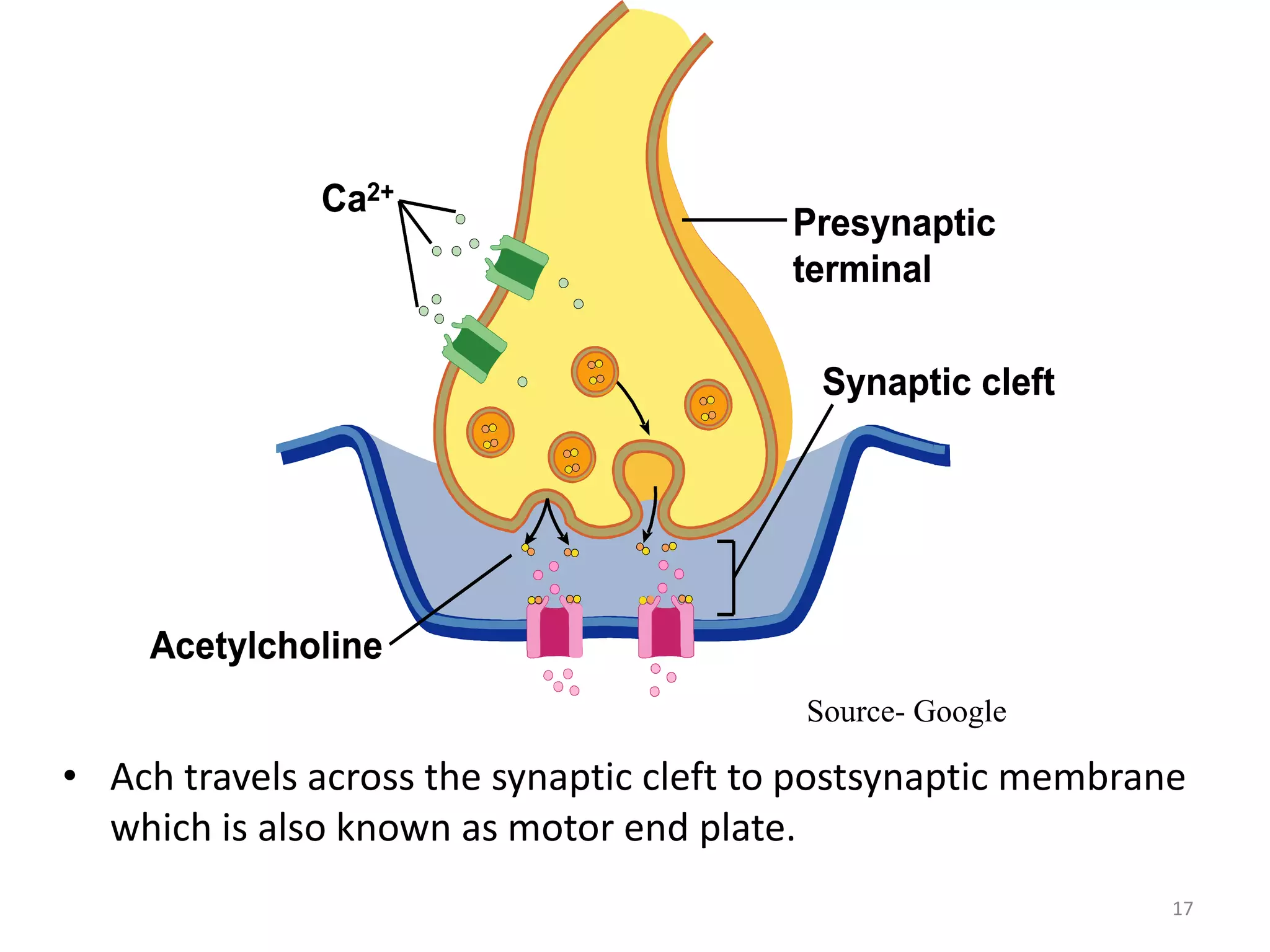
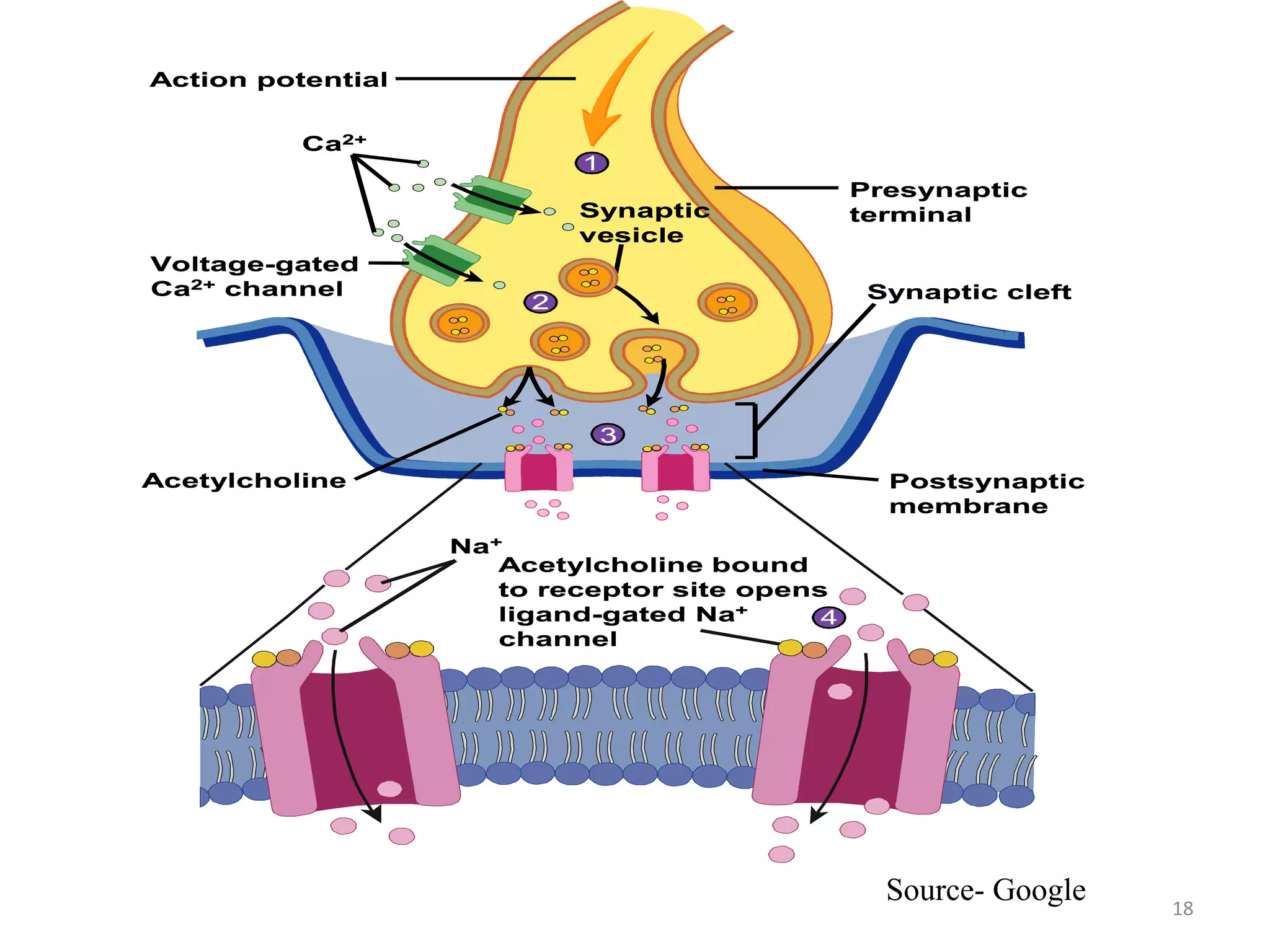
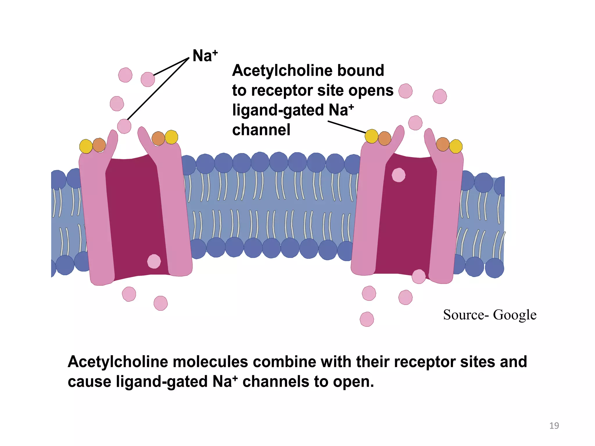
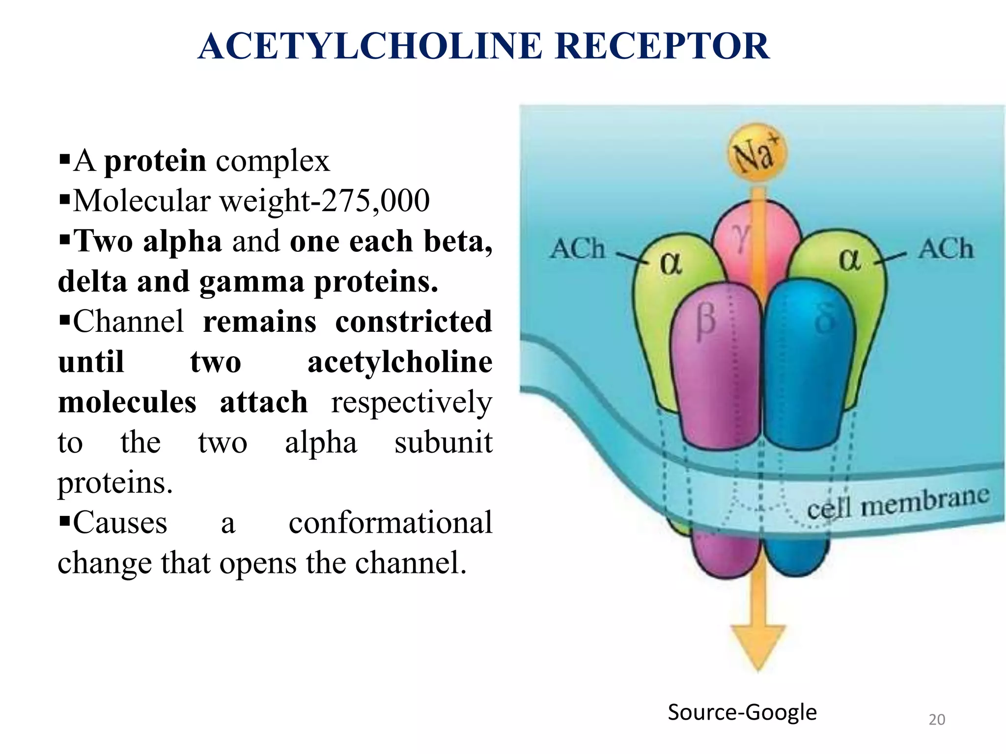
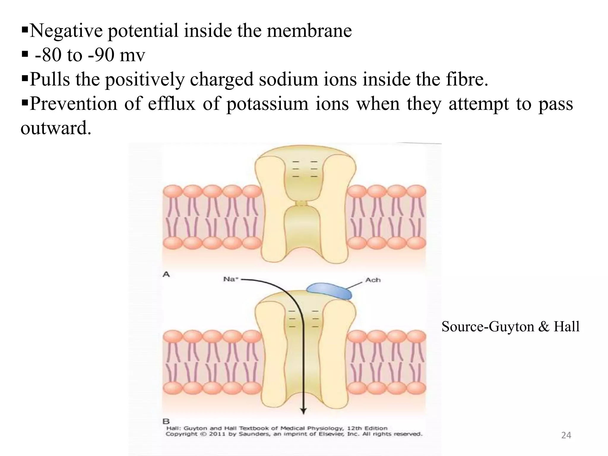
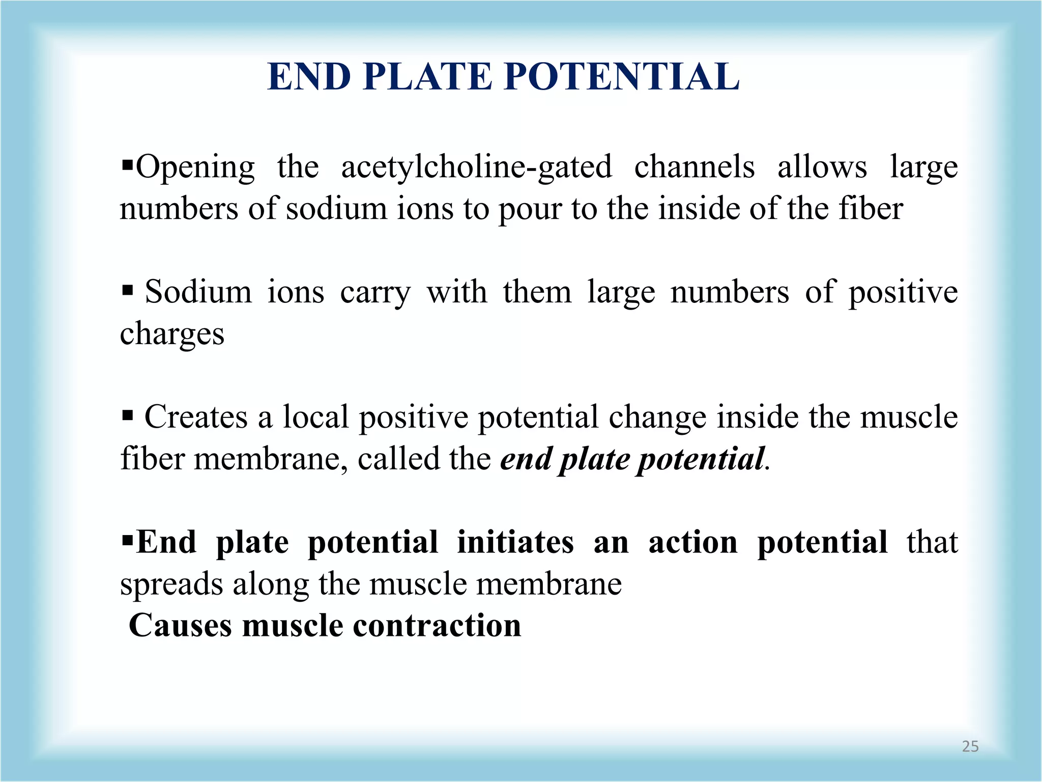
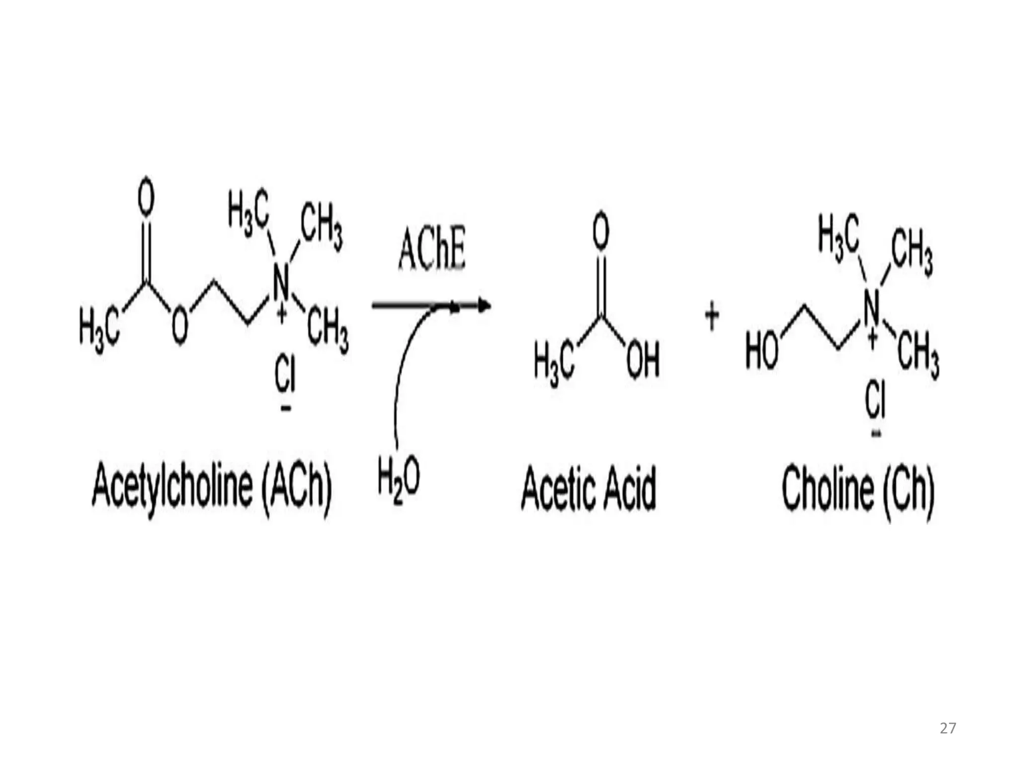
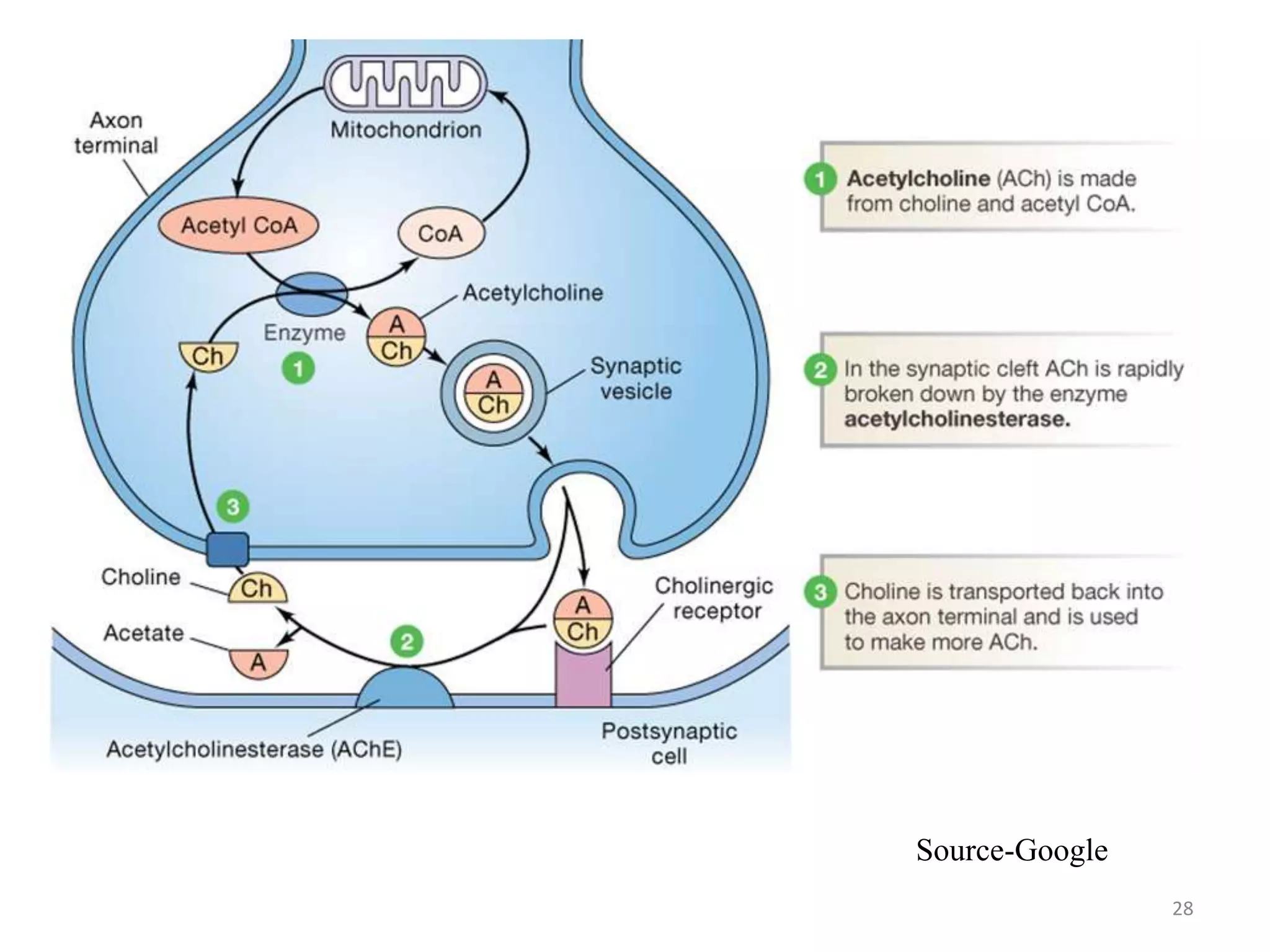
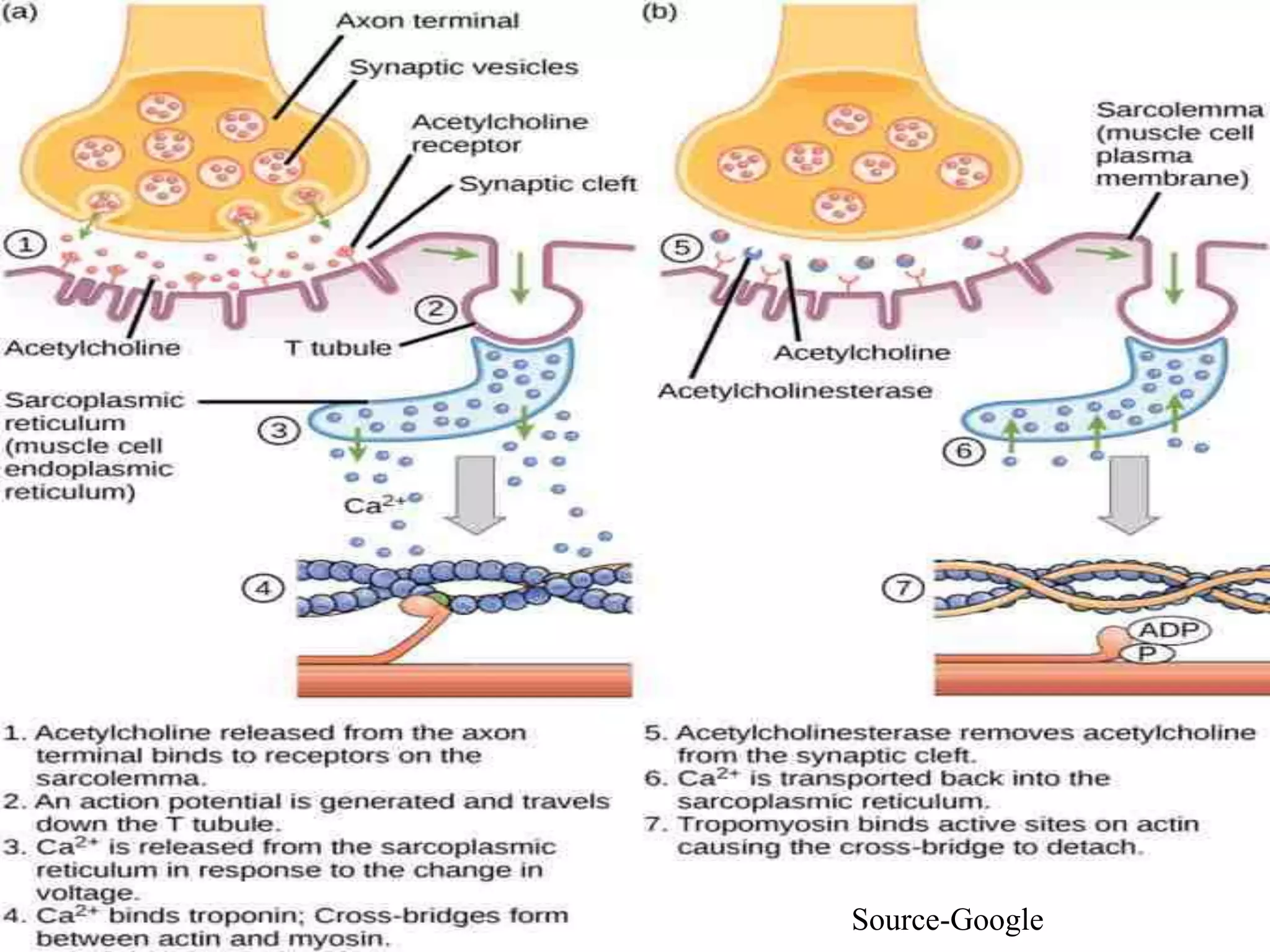
NEUROMUSCULAR JUNCTION
Submitted by:
Preeti Reddy
Komal Pahal
Binal Ramani
Submitted to: Praveen Gupta Sir
1
NEUROMUSCULAR JUNCTION
A neuromuscular junction (or
myoneural junction) is a
chemical synapse formed by the
contact between a motor neuron
and a muscle fibre.
It is at the neuromuscular
junction that a motor neuron is
able to transmit a signal to the
muscle fibre causing muscle
contraction.
Source-Google
2
3
MOTOR NEURON AND FROM WHERE IT
ORIGINATES
Source-Google
A motor neuron is a neuron
whose cell body is located in the
motor cortex, brainstem or the
spinal cord and whose axon
projects to the spinal cord or
outside of the spinal cord to
directly or indirectly control
effector organs mainly muscles
and glands.
4Source-Google
5
Stimulus-response pathway
(Source- Google)
6
ANATOMY OF NMJ
MOTOR END PLATE
 The nerve fiber forms a
complex of branching nerve
terminals that invaginate
into the surface of the
muscle fiber but lie outside
the muscle fiber plasma
membrane
 Entire structure - motor
endplate.
 Covered by one or more
Schwann cells that insulate
it from the surrounding
fluids. Source-Google
7
Synaptic trough: invagination in the motor endplate membrane
Synaptic clefts: 20-30nm wide
Contains large quantities of acetylcholinesterase (AchE)
Source-Guyton & Hall
8
Subneural clefts:
Increase the surface area of the post-synaptic membrane
 Ach gated channels at tops
Voltage gated Na+ channel in bottom half
Source-Guyton &Hall
9
AXON TERMINAL
 SYNAPTIC VESICLES
– Size 40 nanometers
– Formed by the Golgi
apparatus in the cell body
of the motor neuron in the
spinal cord.
– Transported by axoplasm
to the neuromuscular
junction at the tips of the
peripheral nerve fibers.
– About 300,000 of these
small vesicles collect in the
nerve terminals of a single
skeletal muscle end plate.
Source-Guyton &Hall
10
MITOCHONDRIA
Numerous
Supply ATP
Energy source for synthesis of excitatory neurotransmitter,
acetylcholine
Source- Guyton & Hall
11
Secretion Of Acetylcholine By The Nerve Terminals
Nerve impulses reaches at the
neuromuscular junction.
No. of released synaptic vesicles
of acetylcholine are about 125
released from the terminals into
the synaptic space .
NEURAL MEMBRANE
• On inside of neural membrane
linear dense bars
• To each side of dense bars,
protein particles are present
• Voltage gated calcium channels
• Action potential
Source-Guyton & Hall
12
Voltage-gated calcium channels
1.When action potential spreads over the terminal leads to
opening of the channels.
2.Allows calcium ions to diffuse from the synaptic space to the
interior of the nerve terminal.
3.Entry of calcium ions is an effective stimulus for causing the
acetylcholine release from the vesicles is then emptied through
the neural membrane adjacent to the dense bars.
13
Action potentialAction potential
Ca2+Ca2+
Presynaptic
terminal
Presynaptic
terminal
Voltage-gated
Ca2+ channel
Voltage-gated
Ca2+ channel
Action potentials arriving at the presynaptic terminal cause
voltage-gated Ca2+ channels to open.
Source- Google
14
PRESYNAPTIC NERVE TERMINAL OR NERVE
TERMINAL
 Channels opening
 Permission of calcium ions for diffusion
 From synaptic space of interior of terminal
 Calcium ions exert influence on acetylcholine
vesicles
 Carrying and pushing them towards neural membrane
 Fuse with neural membrane
 Exocytosis
15
Ca2+ diffuse into the cell and cause synaptic vesicles to release
acetylcholine, a neurotransmitter molecule.
Ca2+Ca2+
Synaptic
vesicle
Synaptic
vesicle
AcetylcholineAcetylcholine
Ca2+ uptake into the terminal causes release of the neurotransmitter
acetylcholine into synaptic cleft , which has been synthesized and
stored into synaptic vesicles
Source- Google
16
Effect of acetylcholine on postsynaptic muscle fibre
Sub-neural cleft
 Acetylcholine receptors
 Acetylcholine gated ion
channels
 Location is at the mouth of
sub-neural cleft
 Lying immediately below
the dense bars
 Lying side by side in a
circle
 Tubular channels
Source-Guyton &Hall
17
Acetylcholine diffuses from the presynaptic terminal across the
synaptic cleft.
Synaptic cleftSynaptic cleft
AcetylcholineAcetylcholine
Presynaptic
terminal
Presynaptic
terminal
Ca2+Ca2+
• Ach travels across the synaptic cleft to postsynaptic membrane
which is also known as motor end plate.
Source- Google
18
Acetylcholine bound
to receptor site opens
ligand-gated Na+
channel
Acetylcholine bound
to receptor site opens
ligand-gated Na+
channel
Ca2+Ca2+
Voltage-gated
Ca2+ channel
Voltage-gated
Ca2+ channel
Synaptic
vesicle
Synaptic
vesicle
Postsynaptic
membrane
Postsynaptic
membrane
AcetylcholineAcetylcholine
4
Synaptic cleftSynaptic cleft
Action potentialAction potential
Presynaptic
terminal
Presynaptic
terminal
Na+Na+
1
2
3
1
2
3
1
2
3
11
2
3
4
Source- Google
19
Acetylcholine molecules combine with their receptor sites and
cause ligand-gated Na+ channels to open.
Na+Na+
Acetylcholine bound
to receptor site opens
ligand-gated Na+
channel
Acetylcholine bound
to receptor site opens
ligand-gated Na+
channel
Source- Google
20
ACETYLCHOLINE RECEPTOR
A protein complex
Molecular weight-275,000
Two alpha and one each beta,
delta and gamma proteins.
Channel remains constricted
until two acetylcholine
molecules attach respectively
to the two alpha subunit
proteins.
Causes a conformational
change that opens the channel.
Source-Google
21
Source-Guyton & Hall
OPENING OF CHANNEL
 Conformational change
 Opening of channels
 Opened acetylcholine channel
has diameter 0.65nanometer
 Permission to ions
 Na, K and Ca
 Negative ions cannot move such as
Cl
22
Source-Google
23
 Strong negative charges in the mouth of channel
 Repel the negative ions
OPENING OF CHANNEL
WHY Na IONS RUSH INSIDE
 Large amount of Na ions rush inside
 Two positive ions in large concentration
 Sodium and potassium ion
 Sodium ion in extracellular fluid
 Potassium ion in intracellular fluid
24
Negative potential inside the membrane
 -80 to -90 mv
Pulls the positively charged sodium ions inside the fibre.
Prevention of efflux of potassium ions when they attempt to pass
outward.
Source-Guyton & Hall
25
END PLATE POTENTIAL
Opening the acetylcholine-gated channels allows large
numbers of sodium ions to pour to the inside of the fiber
 Sodium ions carry with them large numbers of positive
charges
 Creates a local positive potential change inside the muscle
fiber membrane, called the end plate potential.
End plate potential initiates an action potential that
spreads along the muscle membrane
Causes muscle contraction
26
Acetyl cholinesterase ends Ach activity at N.M junction
• To ensure purposeful movement ,muscle cell electrical response
is turned off by acetylcholinestrase(AchE), which degrade Ach
to choline & acetate
• About 50%of choline is returned to the presynaptic terminal by
Na+choline transport to be reused for Ach synthesis.
• Now muscle fiber can relax ,if sustained contraction is needed
for the desired movement another motor neuron AP leads to
release of more Ach
27
28
Source-Google
29
Synthesis and destruction of acetylcholine (Source-Google)
30
Excitation–Contraction Coupling
A motor neuron connects to a muscle at the neuromuscular
junction, where a synaptic terminal forms a synaptic cleft with
a motor-end plate.
The neurotransmitter acetylcholine diffuses across the
synaptic cleft, causing the depolarization of the sarcolemma.
The depolarization of the sarcolemma stimulates
the sarcoplasmic reticulum to release Ca2+, which causes the
muscle to contract.
31
Source-Google
32
Reference:
Guyton & Hall Textbook Of Medical Physiology
33

Presentation NEUROMUSCULAR JUNCTION

  • 1.
    NEUROMUSCULAR JUNCTION Submitted by: PreetiReddy Komal Pahal Binal Ramani Submitted to: Praveen Gupta Sir 1
  • 2.
    NEUROMUSCULAR JUNCTION A neuromuscularjunction (or myoneural junction) is a chemical synapse formed by the contact between a motor neuron and a muscle fibre. It is at the neuromuscular junction that a motor neuron is able to transmit a signal to the muscle fibre causing muscle contraction. Source-Google 2
  • 3.
    3 MOTOR NEURON ANDFROM WHERE IT ORIGINATES Source-Google A motor neuron is a neuron whose cell body is located in the motor cortex, brainstem or the spinal cord and whose axon projects to the spinal cord or outside of the spinal cord to directly or indirectly control effector organs mainly muscles and glands.
  • 4.
  • 5.
  • 6.
    6 ANATOMY OF NMJ MOTOREND PLATE  The nerve fiber forms a complex of branching nerve terminals that invaginate into the surface of the muscle fiber but lie outside the muscle fiber plasma membrane  Entire structure - motor endplate.  Covered by one or more Schwann cells that insulate it from the surrounding fluids. Source-Google
  • 7.
    7 Synaptic trough: invaginationin the motor endplate membrane Synaptic clefts: 20-30nm wide Contains large quantities of acetylcholinesterase (AchE) Source-Guyton & Hall
  • 8.
    8 Subneural clefts: Increase thesurface area of the post-synaptic membrane  Ach gated channels at tops Voltage gated Na+ channel in bottom half Source-Guyton &Hall
  • 9.
    9 AXON TERMINAL  SYNAPTICVESICLES – Size 40 nanometers – Formed by the Golgi apparatus in the cell body of the motor neuron in the spinal cord. – Transported by axoplasm to the neuromuscular junction at the tips of the peripheral nerve fibers. – About 300,000 of these small vesicles collect in the nerve terminals of a single skeletal muscle end plate. Source-Guyton &Hall
  • 10.
    10 MITOCHONDRIA Numerous Supply ATP Energy sourcefor synthesis of excitatory neurotransmitter, acetylcholine Source- Guyton & Hall
  • 11.
    11 Secretion Of AcetylcholineBy The Nerve Terminals Nerve impulses reaches at the neuromuscular junction. No. of released synaptic vesicles of acetylcholine are about 125 released from the terminals into the synaptic space . NEURAL MEMBRANE • On inside of neural membrane linear dense bars • To each side of dense bars, protein particles are present • Voltage gated calcium channels • Action potential Source-Guyton & Hall
  • 12.
    12 Voltage-gated calcium channels 1.Whenaction potential spreads over the terminal leads to opening of the channels. 2.Allows calcium ions to diffuse from the synaptic space to the interior of the nerve terminal. 3.Entry of calcium ions is an effective stimulus for causing the acetylcholine release from the vesicles is then emptied through the neural membrane adjacent to the dense bars.
  • 13.
    13 Action potentialAction potential Ca2+Ca2+ Presynaptic terminal Presynaptic terminal Voltage-gated Ca2+channel Voltage-gated Ca2+ channel Action potentials arriving at the presynaptic terminal cause voltage-gated Ca2+ channels to open. Source- Google
  • 14.
    14 PRESYNAPTIC NERVE TERMINALOR NERVE TERMINAL  Channels opening  Permission of calcium ions for diffusion  From synaptic space of interior of terminal  Calcium ions exert influence on acetylcholine vesicles  Carrying and pushing them towards neural membrane  Fuse with neural membrane  Exocytosis
  • 15.
    15 Ca2+ diffuse intothe cell and cause synaptic vesicles to release acetylcholine, a neurotransmitter molecule. Ca2+Ca2+ Synaptic vesicle Synaptic vesicle AcetylcholineAcetylcholine Ca2+ uptake into the terminal causes release of the neurotransmitter acetylcholine into synaptic cleft , which has been synthesized and stored into synaptic vesicles Source- Google
  • 16.
    16 Effect of acetylcholineon postsynaptic muscle fibre Sub-neural cleft  Acetylcholine receptors  Acetylcholine gated ion channels  Location is at the mouth of sub-neural cleft  Lying immediately below the dense bars  Lying side by side in a circle  Tubular channels Source-Guyton &Hall
  • 17.
    17 Acetylcholine diffuses fromthe presynaptic terminal across the synaptic cleft. Synaptic cleftSynaptic cleft AcetylcholineAcetylcholine Presynaptic terminal Presynaptic terminal Ca2+Ca2+ • Ach travels across the synaptic cleft to postsynaptic membrane which is also known as motor end plate. Source- Google
  • 18.
    18 Acetylcholine bound to receptorsite opens ligand-gated Na+ channel Acetylcholine bound to receptor site opens ligand-gated Na+ channel Ca2+Ca2+ Voltage-gated Ca2+ channel Voltage-gated Ca2+ channel Synaptic vesicle Synaptic vesicle Postsynaptic membrane Postsynaptic membrane AcetylcholineAcetylcholine 4 Synaptic cleftSynaptic cleft Action potentialAction potential Presynaptic terminal Presynaptic terminal Na+Na+ 1 2 3 1 2 3 1 2 3 11 2 3 4 Source- Google
  • 19.
    19 Acetylcholine molecules combinewith their receptor sites and cause ligand-gated Na+ channels to open. Na+Na+ Acetylcholine bound to receptor site opens ligand-gated Na+ channel Acetylcholine bound to receptor site opens ligand-gated Na+ channel Source- Google
  • 20.
    20 ACETYLCHOLINE RECEPTOR A proteincomplex Molecular weight-275,000 Two alpha and one each beta, delta and gamma proteins. Channel remains constricted until two acetylcholine molecules attach respectively to the two alpha subunit proteins. Causes a conformational change that opens the channel. Source-Google
  • 21.
    21 Source-Guyton & Hall OPENINGOF CHANNEL  Conformational change  Opening of channels  Opened acetylcholine channel has diameter 0.65nanometer  Permission to ions  Na, K and Ca  Negative ions cannot move such as Cl
  • 22.
  • 23.
    23  Strong negativecharges in the mouth of channel  Repel the negative ions OPENING OF CHANNEL WHY Na IONS RUSH INSIDE  Large amount of Na ions rush inside  Two positive ions in large concentration  Sodium and potassium ion  Sodium ion in extracellular fluid  Potassium ion in intracellular fluid
  • 24.
    24 Negative potential insidethe membrane  -80 to -90 mv Pulls the positively charged sodium ions inside the fibre. Prevention of efflux of potassium ions when they attempt to pass outward. Source-Guyton & Hall
  • 25.
    25 END PLATE POTENTIAL Openingthe acetylcholine-gated channels allows large numbers of sodium ions to pour to the inside of the fiber  Sodium ions carry with them large numbers of positive charges  Creates a local positive potential change inside the muscle fiber membrane, called the end plate potential. End plate potential initiates an action potential that spreads along the muscle membrane Causes muscle contraction
  • 26.
    26 Acetyl cholinesterase endsAch activity at N.M junction • To ensure purposeful movement ,muscle cell electrical response is turned off by acetylcholinestrase(AchE), which degrade Ach to choline & acetate • About 50%of choline is returned to the presynaptic terminal by Na+choline transport to be reused for Ach synthesis. • Now muscle fiber can relax ,if sustained contraction is needed for the desired movement another motor neuron AP leads to release of more Ach
  • 27.
  • 28.
  • 29.
    29 Synthesis and destructionof acetylcholine (Source-Google)
  • 30.
    30 Excitation–Contraction Coupling A motorneuron connects to a muscle at the neuromuscular junction, where a synaptic terminal forms a synaptic cleft with a motor-end plate. The neurotransmitter acetylcholine diffuses across the synaptic cleft, causing the depolarization of the sarcolemma. The depolarization of the sarcolemma stimulates the sarcoplasmic reticulum to release Ca2+, which causes the muscle to contract.
  • 31.
  • 32.
    32 Reference: Guyton & HallTextbook Of Medical Physiology
  • 33.