Downloaded 274 times


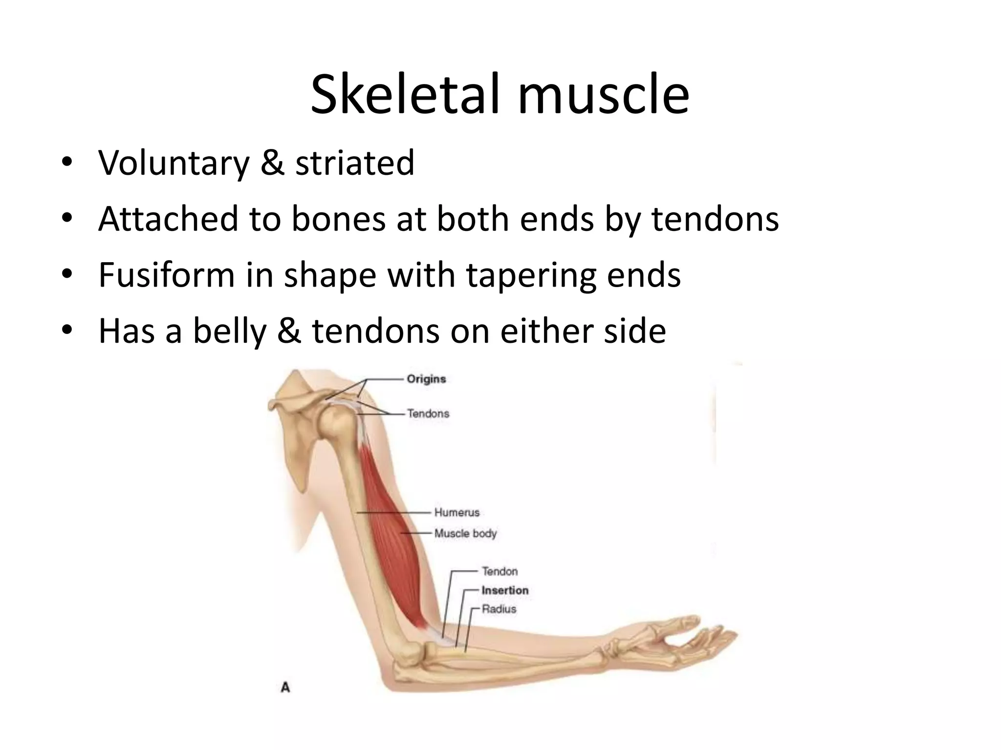


This document provides an introduction to skeletal muscle structure and function. It discusses the following key points in 3 sentences or less: Skeletal muscle forms about 50% of body weight, is voluntary and striated, and its main functions are tension development and shortening under nervous system control. Skeletal muscle is composed of bundles of muscle fibers which contain myofibrils made up of repeating structural units called sarcomeres, the basic contractile units of muscle. Each sarcomere contains thin actin filaments and thick myosin filaments arranged in a repeating pattern that gives skeletal muscle its striated appearance visible under electron microscopy.


