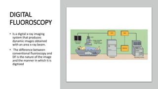
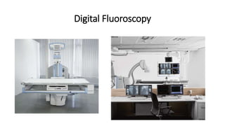
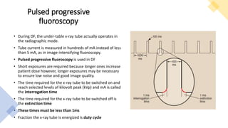
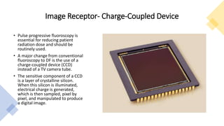
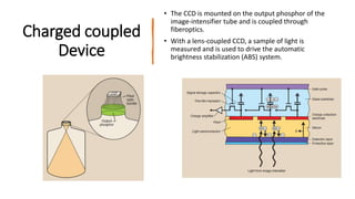
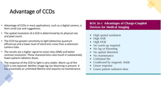
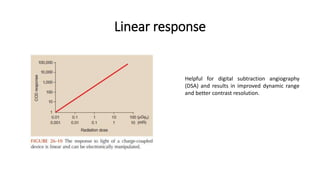
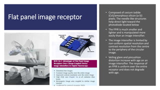
DIGITAL FLUOROSCOPY
By Oliver
Medical Physicist
DIGITAL
FLUOROSCOPY
• Is a digital x-ray imaging
system that produces
dynamic images obtained
with an area x-ray beam.
• The difference between
conventional fluoroscopy and
DF is the nature of the image
and the manner in which it is
digitized
Digital Fluoroscopy
Pulsed progressive
fluoroscopy
• During DF, the under-table x-ray tube actually operates in
the radiographic mode.
• Tube current is measured in hundreds of mA instead of less
than 5 mA, as in image-intensifying fluoroscopy.
• Pulsed progressive fluoroscopy is used in DF
• Short exposures are required because longer ones increase
patient dose however, longer exposures may be necessary
to ensure low noise and good image quality.
• The time required for the x-ray tube to be switched on and
reach selected levels of kilovolt peak (kVp) and mA is called
the interrogation time
• The time required for the x-ray tube to be switched off is
the extinction time
• These times must be less than 1ms
• Fraction the x-ray tube is energized is duty cycle
Image Receptor- Charge-Coupled Device
• Pulse progressive fluoroscopy is
essential for reducing patient
radiation dose and should be
routinely used.
• A major change from conventional
fluoroscopy to DF is the use of a
charge-coupled device (CCD)
instead of a TV camera tube.
• The sensitive component of a CCD
is a layer of crystalline silicon.
When this silicon is illuminated,
electrical charge is generated,
which is then sampled, pixel by
pixel, and manipulated to produce
a digital image.
Charged coupled
Device
• The CCD is mounted on the output phosphor of the
image-intensifier tube and is coupled through
fiberoptics.
• With a lens-coupled CCD, a sample of light is
measured and is used to drive the automatic
brightness stabilization (ABS) system.
Advantage of CCDs
• Advantage of CCDs in most applications, such as a digital camera, is
their small size and ruggedness.
• The spatial resolution of a CCD is determined by its physical size
and pixel count.
• The CCD has greater sensitivity to light (detective quantum
efficiency) and a lower level of electronic noise than a television
camera tube.
• The results are a higher signal-to-noise ratio (SNR) and better
contrast resolution. These characteristics also result in substantially
lower patient radiation doses.
• The response of the CCD to light is very stable. Warm-up of the
CCD is not required. Neither image lag nor blooming is present. It
has essentially an unlimited lifetime and requires no maintenance.
Linear response
Helpful for digital subtraction angiography
(DSA) and results in improved dynamic range
and better contrast resolution.
Flat panel image receptor
• Composed of cesium iodide
(CsI)/amorphous silicon (a-Si)
pixels. The needle-like structures
help direct light toward the
photodiode located below
• The FPIR is much smaller and
lighter and is manipulated more
easily than an image intensifier.
• The image intensifier is limited by
non-uniform spatial resolution and
contrast resolution from the centre
to the periphery of the circular
image.
• Veiling glare and pincushion
distortion increase with age on an
image intensifier. The response of
an FPIR is uniform over the entire
receptor and does not degrade
with age.
Flat panel detector
Flat panel display
• Replaces the cathode ray tube
• They are light in weight, easy to see, and can be readily mounted
suspended in an angiographic room

DIGITAL FLUOROSCOPY DMR.pptx

  • 1.
  • 2.
    DIGITAL FLUOROSCOPY • Is adigital x-ray imaging system that produces dynamic images obtained with an area x-ray beam. • The difference between conventional fluoroscopy and DF is the nature of the image and the manner in which it is digitized
  • 3.
  • 4.
    Pulsed progressive fluoroscopy • DuringDF, the under-table x-ray tube actually operates in the radiographic mode. • Tube current is measured in hundreds of mA instead of less than 5 mA, as in image-intensifying fluoroscopy. • Pulsed progressive fluoroscopy is used in DF • Short exposures are required because longer ones increase patient dose however, longer exposures may be necessary to ensure low noise and good image quality. • The time required for the x-ray tube to be switched on and reach selected levels of kilovolt peak (kVp) and mA is called the interrogation time • The time required for the x-ray tube to be switched off is the extinction time • These times must be less than 1ms • Fraction the x-ray tube is energized is duty cycle
  • 5.
    Image Receptor- Charge-CoupledDevice • Pulse progressive fluoroscopy is essential for reducing patient radiation dose and should be routinely used. • A major change from conventional fluoroscopy to DF is the use of a charge-coupled device (CCD) instead of a TV camera tube. • The sensitive component of a CCD is a layer of crystalline silicon. When this silicon is illuminated, electrical charge is generated, which is then sampled, pixel by pixel, and manipulated to produce a digital image.
  • 6.
    Charged coupled Device • TheCCD is mounted on the output phosphor of the image-intensifier tube and is coupled through fiberoptics. • With a lens-coupled CCD, a sample of light is measured and is used to drive the automatic brightness stabilization (ABS) system.
  • 7.
    Advantage of CCDs •Advantage of CCDs in most applications, such as a digital camera, is their small size and ruggedness. • The spatial resolution of a CCD is determined by its physical size and pixel count. • The CCD has greater sensitivity to light (detective quantum efficiency) and a lower level of electronic noise than a television camera tube. • The results are a higher signal-to-noise ratio (SNR) and better contrast resolution. These characteristics also result in substantially lower patient radiation doses. • The response of the CCD to light is very stable. Warm-up of the CCD is not required. Neither image lag nor blooming is present. It has essentially an unlimited lifetime and requires no maintenance.
  • 8.
    Linear response Helpful fordigital subtraction angiography (DSA) and results in improved dynamic range and better contrast resolution.
  • 9.
    Flat panel imagereceptor • Composed of cesium iodide (CsI)/amorphous silicon (a-Si) pixels. The needle-like structures help direct light toward the photodiode located below • The FPIR is much smaller and lighter and is manipulated more easily than an image intensifier. • The image intensifier is limited by non-uniform spatial resolution and contrast resolution from the centre to the periphery of the circular image. • Veiling glare and pincushion distortion increase with age on an image intensifier. The response of an FPIR is uniform over the entire receptor and does not degrade with age.
  • 10.
  • 11.
    Flat panel display •Replaces the cathode ray tube • They are light in weight, easy to see, and can be readily mounted suspended in an angiographic room