SlideShare a Scribd company logo
1 of 68
MRI SPIN ECHO PULSE SEQUENCES ITS
VARIATIONS AND APPLICATIONS
Presenter :- Yashawant Ku. Yadav
Bsc.MIT 3rd Year
NAMS (Bir Hospital )
Outline
• Introduction P-S
• Terminologies
• Types of Spin echo pulse sequences (Conventional spin echo ,Fast or turbo spin
echo , Inversion recovery ,Fast inversion recovery [STIR (short tau inversion
recovery), FLAIR (fluid attenuated inversion recovery) ] .
• Application of spin echo pulse sequences
• References
Introduction
• Pulse sequence is a series of RF pulses, gradient applications and intervening time
periods.
• Pulse sequences enable us to control the way in which the system applies pulses
and gradients.
• In this way, image weighting and quality is determined.
Intro…
• In MRI, we are imaging properties of hydrogen nuclei (protons) in
tissue.
• With the protons precession around B0, nothing interesting will happen
and we cannot measure any signal.
• In order to measure parameters of the tissue, we need to perturb - excite
- the system
• This is done by applying a transient magnetic field - a radiofrequency
pulse - in a different direction from B0; the direction of this pulse is
often referred to as B1
Contd..
• By applying this RF for a short time, we can rotate the magnetization of
the tissue.
• If the protons are initially aligned along the z (vertical) axis, and we apply
a pulse along the x -axis for the right amount of time, we can rotate the
magnetization from the z-axis onto the y-axis.
Contd..
• This is referred to as a 90-degree pulse, since the magnetization
direction is turned by 90 degrees.
• Once the pulse is turned off, the protons will resume precession along
B0 and will slowly return to their initial equilibrium with the net
magnetization pointing along the z axis.
• The RF pulses are applied for excitation purposes and, in the case of
spin echo, for rephasing purposes too.
Why pulse sequences ??
Image contrast
• All clinical diagnostic images must demonstrate contrast between
normal anatomical features and between anatomy and any pathology.
• And MRI is consider as a high tissue contrast discriminating imaging
modalities among all.
• The contrast characteristic of each image depend on many variables,
Contd..
Basically it is divided into two categories.
• Intrinsic contrast parameters are those that cannot be changed because they are
inherent to the body ’ s tissues.
• Extrinsic contrast parameters are those that can be changed.
Intrinsic contrast PMs Extrinsic contrast PMs
T1 recovery time TR
T2 decay time TE
proton density FALIP ANGLE
flow TI
apparent diffusion coefficient (ADC). TURBO FACTOR / ECHO TRAIN LENGTH
B Value
Terminologies
The repetition time (TR) is the time
from the application of one RF pulse
to the application of the next RF
pulse for each slice and is measured
in milliseconds (ms).
• The TR determines the amount of
longitudinal relaxation that is allowed
to occur between the end of one RF
pulse and the application of the next.
• TR thus determines the amount of T1
relaxation that has occurred when
the signal is read.
Contd..
 The echo time (TE) is the time from
the application of the RF pulse to the
peak of the signal induced in the coil
and is also measured in ms.
• The TE determines how much decay of
transverse magnetization is allowed to
occur.
• TE thus controls the amount of T2
relaxation that has occurred when the
signal is read.
Contd..
• To demonstrate either T1, proton density or T2 contrast, specific values of TR and
TE are selected for a given pulse sequence.
• The selection of appropriate TR and TE weights an image so that one contrast
mechanism predominates over the other two.
Variations
Spin echo pulse sequences (spins are rephased by a 180 ° rephasing
pulse):
• conventional spin echo
• fast or turbo spin echo
• inversion recovery.
Conventional spin echo
• spin echo uses a 90 ° excitation pulse followed by one or more 180 ° rephasing
pulses to generate a spin echo.
• If only one echo is generated, a T1 weighted image can be obtained using a short
TE and a short TR.
Contd…
• The SE represents regeneration of spin phase information apparently
lost during the decay of the FID. The "rebirth" of the FID as a SE is
possible… by Erwin Hahn in 1949
• In fact, most of the FID signal has not been destroyed; it has merely
become "disorganized" because the individual spins comprising it have
lost their phase coherence.
• The system is said to possess a "hidden order" or "atomic memory". By
applying a second RF pulse, certain dephased components of the
original FID can be refocused into a SE.
Contd…
• For proton density and T2
weighting, two RF rephasing
pulses, generating two spin
echoes, are applied.
• The first echo has a short TE and
a long TR to achieve proton
density weighting, and the second
has a long TE and a long TR to
achieve T2 weighting.
Contd..
Parameters
T1 WT
Te short :- 10-30 ms
Tr short :- 300-700ms
Typical scan time :- 4-6 min
T2 WT / PDW
Te short :- 20ms / 80+MS(LONG)
Tr long :- 2000+ms
Sacn time:-
Applications
• Used in any part of the body for any indication.
• To produce T1, T2 (True)and PD weighted images of good quality.
 Disadvantage
Longer scan time
Fast spin echo /turbo
• In this pulse sequence scan time is much shorter than conventional spin
echo .
• In fast spin echo, the scan time is reduced by performing more than one
phase encoding step and subsequently filling more than one line of K
space per TR.
• This is achieved by using several 180 ° rephasing pulses to produce a
train of echoes or echo train.
• At each rephasing, an echo is produced and a different phase encoding
step is performed.
Contd..
• In conventional spin echo, raw image data from each echo are stored in
K space,
• Each echo is used to produce a separate image
• In FSE data from each echo are placed into one image.
• The number of 180 ° rephasing pulses performed per TR corresponds to
the number of echoes produced and the number of lines of K space
filled.
Contd..
• This number is called the turbo factor or the echo train length . The
higher the turbo factor, the shorter the scan time as more phase
encoding steps are performed per TR.
• Number of echoes -- called echo train length (ETL) by GE and Canon; turbo factor by Siemens and
Philips; or shot factor by Hitachi.
• At each 180 ° /phase encoding combination, a different amplitude of
phase encoding gradient slope is applied to fill out a different line of K
space
Contd..
• ETL is the single most important parameter. In general, image acquisition time is
inversely proportional to ETL.
• ETL also has important effects on image quality. Longer ETLs result in more T2-
weighting because more late echoes with longer TE's contribute to the overall
signal.
• Longer ETL's are also decreases overall signal-to-noise ratio (SNR) and
contrast-to-noise ratio (CNR) because the later echoes are weaker.
TR :- 4000MS
OTHER PARAMETERS
UNCHANGED
Contd..
• Time between echoes -- called echo spacing (ESP) by GE, Philips,
Siemens, and Canon; interecho time (ITE) by Hitachi.
• Increasing echo spacing (ESP) permits the use of longer TE's but
adversely impacts SNR and CNR
Weighting in fast spin echo
• The echoes are generated at different TE times and therefore data
collected from them have variable weighting.
• So how is a fast spin echo sequence weighted correctly?
• The TE selected is only an effective TE
• That mean TE at which the operator wishes to weight the resultant
image.
• The system orders the phase encoding steps so that steep or shallow
slopes are applied to the various echoes produced.
Contd…
• fast spin echo has largely replaced spin echo especially for T2
weighting.
• There are, however, two contrast differences between spin echo and fast
spin echo,
• due to the repeated, closely spaced 180 ° pulses of the echo train
• First, fat remains bright on T2 weighted images due to the multiple RF
pulses, which reduce the effects of spin – spin interactions in fat ( J
coupling )
• Second, the repeated 180 ° pulses can increase magnetization transfer
effects so that muscle, appears darker on fast spin echo images than in
conventional spin echo.
Contd..
• In addition, the multiple 180 ° pulses reduce magnetic susceptibility effects,
which can be detrimental when looking for small hemorrhages.
Contd…
• Image blurring may occur in fast spin echo images at the edges of
tissues with different T2 decay values.
• This is because each line of K space filled during an echo train contains
data from echoes with a different TE.
•
• When using long echo trains, late echoes that have a low signal
amplitude contribute to the resolution of K space.
• If these echoes are negligible, then resolution is lost from the image and
blurring occurs.
TYPICAL PARAMETERS
Dual echo
• TR 2500–4500 ms (for weighting and slice number)
• effective TE1 17 ms
• effective TE2 102 ms
• ETL 8 – This may be split so that the PD image is acquired with the first four
echoes and the T2 with the second four.
Single echo
• T2 weighting
• TR 4000–8000 ms
• TE 102 ms
• ETL 16
Single echo
T1 weighting
TR 600 ms
TE 17 ms
ETL 4
USES
• brains, spines , joints, extremities and pelvis
SS-FSE / HASTE
Other vendors have similar sequences under slightly different names: GE
(Single-shot fast spin echo, SS-FSE), Philips (Single-shot turbo spin
echo, SSH-TSE; ultra-fast spin echo, UFSE), HItachi (Single-shot fast
SE), and Canon (Fast Advanced Spin Echo, FASE, SuperFASE).
• This sequence even faster than FSE ,
• In this technique all the lines of K space are acquired in one TR.
• SS - FSE combines a parti al Fourier technique with fast spin echo.
• Half of the lines of K space are acquired in one TR and the other half are transposed.
Contd..
• Currently the highest turbo factor used in single shot imaging is 728
• Another consideration when using long echo trains is that the specific
absorption rate ( SAR ) is significantly increased by applying so many
successive 180 ° pulses.
• slice number can be reduce to manifest SAR but that cause difficult to
get required coverage area … so,
• Refocusing angle is reduced to 120°
• This reduces the SAR significantly (which is proportional to the square
of the flip angle) but also reduces the SNR.
• the benefi t of being able to obtain more slices per acquisition
Contd…
Representative HASTE/SS-FSE pulse sequence.
Note asymmetric sampling in phase-encode direction. The peripheral lines on one side
of k-space are not sampled but estimated by conjugate symmetry.
Application
• routine scout images
• images of the head or body in children or uncooperative patients
• fetal imaging
• non-breath-hold abdominal imaging
• MR cholangiopancreatography
• MR myelography, and
• non-contrast MR angiography.
Examples of conventional SSFSE
compared to half-Fourier vrfSSFSE.
(A) Conventional SSFSE and
(B) half-Fourier vrfSSFSE images
from a patient with renal cell
carcinoma of the left kidney (arrow).
(C) Conventional SSFSE and
(D) half-Fourier vrfSSFSE images
from a patient with a mildly dilated
pancreatic and common bile duct.
Images are mildly cropped from the
full field of view. Note the equivalent
image quality.
IR (Inversion recovery )
• Inversion recovery sequences were
initially designed to produce very heavy
T1 weighting.
• However at present they are mainly
used in conjunction with a FSE
sequence to produce T2 weighted
images.
• It is usually used to suppress the signal
from certain tissues in conjunction with
long TEs and T2 weighting, although at
low field it is still used for T1 contrast.
Contd..
• Inversion recovery is a pulse sequence
that begins with a 180 ° inverting pulse.
This inverts the NMV through 180 ° into
full saturation.
• When the inverting pulse is removed, the
NMV begins to relax back to B 0 .
• A 90 ° excitation pulse is then applied at
a time from the 180 ° inverting pulse
known as the TI ( time from inversion )
Contd…
• A further 180° RF pulse is applied which rephases spins in the
transverse plane and produces an echo at time TE after the excitation
pulse.
• The contrast of the resultant image depends primarily on the length of
the TI.
• If the TI is sufficiently long to allow the NMV to pass through the
transverse plane, the contrast depends on the degree of saturation that is
produced by the 90° pulse (as in spin echo), i.e.
• if the 90° pulse is applied shortly after the NMV has passed through the
transverse plane, heavy saturation and T1 weighting results.
Contd…
TIs of 300 ms to 700 ms result in this type of heavy T1 weighting
FIR (FAST INVERSION RECOVERY )
• It is a combination of inversion recovery and fast spin echo.
• It is usually used to suppress the signal from certain tissues in conjunct on with
long TEs and T2 weighting, so that water and pathology return a high signal.
• Although at low field it is still used for T1 contrast.
• The two main sequences in this category are STIR and FLAIR.
STIR (Short tau inversion recovery )
• This sequences uses short TIs such as 100– 180 ms, depending on field
strength .
• TIs of this magnitude place the 90° excitation pulse at the time that
NMV of fat is passing exactly through the transverse plane.
• At this point (called the null point) there is no longitudinal component
in fat.
• Therefore the 90° excitation pulse produces no transverse component
in fat and therefore no signal.
• In this way a fat suppressed image result.
Uses
• STIR is an extremely
important sequence in
musculoskeletal imaging
because normal bone,
which contains fatty
marrow, is suppressed and
lesions within bone such as
bone bruising and tumors
are seen more clearly.
FLAIR (fluid attenuated inversion recovery )
• It uses long TIs such as 1700–2200 ms,
depending on field strength, to null the
signal from CSF in exactly same way as
STIR.
• Because CSF has a long T1 recovery
time, the TI must be longer to
correspond with its null point.
T1-FLAIR brain image at 3T with TR=2100,
TE=9, TI=880.
T2 FLAIR :TI 2000-2500 very long TRs
(8000) and TEs (140)
For T1-FLAIR both the TR and TE values are kept relatively short to
minimize T2-weighting. Considering these restrictions, the TInull for a T1-
FLAIR sequence is typically in the range of 800-1000 ms.
white matter, and gray matter are
approximately400, and 650 ms
respectively.
TI= 0.69 x T1.
Typical parameters The required TI depends on the field
strength (higher fields).
T1 weighting TI 300–700 ms
• TE 10–20 ms
• TR long
• ETL 4
STIR
• TI 100–180 ms
• TE 70 ms+ (for T2 weighting)
• TR long
• ETL 16
 FLAIR
• TI 1500–2200 ms
• TE 70 ms+ (for T2 weighting)
• TR long
• ETL 12–16
ADVANTGES DISADVANTAGES
Selective Tissue Suppression Longer scan times
Up to Twice the Dynamic Range Increase in flow-related artifacts
Additive T1 and T2 contrast ?? Signal-to-noise can decrease as
tissues are suppressed
Higher specific absorption rate (SAR)
due to additional 180° pulses
Summary if FIR
DOUBLE IR
• Double Inversion Recovery (DIR) is an inversion recovery variant that
uses not one, but two nonselective 180°-inverting pulses.
• Here the first 180°-pulse suppresses CSF and the second suppresses
white matter.
Double IR sequence suppressing CSF and white
matter. TR=11000, TE=30, TI-1=3400, TI-2=325.
Black blood by DIR
• In this method, two 180°-pulses are applied very close together in
time.
• The first 180°-pulse is nonselective, meaning that it inverts the
magnetization for all slices within the imaging volume.
• The second 180°-pulse, following immediately on the heels of the
first 180°-pulse, is slice selective,
• Thus the myocardium and other relatively stationary tissues have their
signals preserved,
• but the blood flowing from adjacent slices has an inverted
magnetization.
Contd…
The TI is adjusted to null the signal from inflowing blood, which
appears "black" on magnitude reconstructed IR images.
Phase sensitive IR ?????
The sequence with the gray background is
called phase-corrected or phase-sensitive
inversion recovery (PSIR).
Phase-corrected IR preserves the information about Mz polarity, with more
negative values rendered increasingly darker.
• If TI is not perfectly chosen, detection of contrast enhancement may be
impaired using magnitude reconstruction.
• Phase-sensitive IR is less sensitive to choice of TI as illustrated in this
example of septal ischemia (arrow).
References
1. https://www.mriquestions.com
2. MRI at a Glance CATHERINE WESTBROOK
3. MRI in Practice Fourth Edition
T2 FLAIR
T1
T1 flair
•Long T1 materials are dark on T1-weighted images, but
long T2 materials are bright on T2-weighted images.
• T2 Relaxation Accompanying T1 Relaxation how ???
• Does T2 Relaxation Occurring Without T1 Relaxation??
1. where it experiences a local static field disturbance (B1oo) in addition to
the main magnetic field (Bo).
2. dipolar interaction also known as a spin-spin "flip-flop".
Mri spin echo pulse sequences its variations and

More Related Content

What's hot

MRI Parameters for Imaging
MRI Parameters for ImagingMRI Parameters for Imaging
MRI Parameters for ImagingAnjan Dangal
 
K Space in MRI
K Space in MRIK Space in MRI
K Space in MRIKajal Jha
 
SPIN ECHO SEQUENCES MRI PPT
SPIN ECHO SEQUENCES MRI PPTSPIN ECHO SEQUENCES MRI PPT
SPIN ECHO SEQUENCES MRI PPTSEHRISH97
 
Gradient echo pulse sequences hjn.pptx
Gradient echo pulse sequences hjn.pptxGradient echo pulse sequences hjn.pptx
Gradient echo pulse sequences hjn.pptxVANI PUSHPA MUDAVATH
 
Mri basic principle and sequences
Mri basic principle and sequencesMri basic principle and sequences
Mri basic principle and sequencesPawan Maurya
 
Fat suppression techniques in MRI
Fat suppression techniques in MRIFat suppression techniques in MRI
Fat suppression techniques in MRISakshamKumar30
 
Perfusion MRI (DSC and DCE perfusion techniques) for radiology residents
Perfusion MRI (DSC and DCE perfusion techniques) for radiology residentsPerfusion MRI (DSC and DCE perfusion techniques) for radiology residents
Perfusion MRI (DSC and DCE perfusion techniques) for radiology residentsRiham Dessouky
 
Ultrasound instrumentation
Ultrasound instrumentationUltrasound instrumentation
Ultrasound instrumentationVnAy Kris
 
Presentation1, mr pulse sequences.
Presentation1, mr pulse sequences.Presentation1, mr pulse sequences.
Presentation1, mr pulse sequences.Abdellah Nazeer
 
K space and parallel imaging
K space and parallel imagingK space and parallel imaging
K space and parallel imagingASHIK E H
 
Basics of mri physics Dr. Muhammad Bin Zulfiqar
Basics of mri physics Dr. Muhammad Bin ZulfiqarBasics of mri physics Dr. Muhammad Bin Zulfiqar
Basics of mri physics Dr. Muhammad Bin ZulfiqarDr. Muhammad Bin Zulfiqar
 
Fat suppression imaging
Fat suppression imagingFat suppression imaging
Fat suppression imagingRoshan Shah
 
Magnetic Resonance Perfusion
Magnetic Resonance PerfusionMagnetic Resonance Perfusion
Magnetic Resonance PerfusionRahman Ud Din
 
Mri image quality gamal mahdaly
Mri image quality gamal mahdalyMri image quality gamal mahdaly
Mri image quality gamal mahdalyGamal Mahdaly
 

What's hot (20)

MRI Parameters for Imaging
MRI Parameters for ImagingMRI Parameters for Imaging
MRI Parameters for Imaging
 
Mri basic sequences
Mri basic sequencesMri basic sequences
Mri basic sequences
 
K Space in MRI
K Space in MRIK Space in MRI
K Space in MRI
 
SPIN ECHO SEQUENCES MRI PPT
SPIN ECHO SEQUENCES MRI PPTSPIN ECHO SEQUENCES MRI PPT
SPIN ECHO SEQUENCES MRI PPT
 
Gradient echo pulse sequences hjn.pptx
Gradient echo pulse sequences hjn.pptxGradient echo pulse sequences hjn.pptx
Gradient echo pulse sequences hjn.pptx
 
Mri basic principle and sequences
Mri basic principle and sequencesMri basic principle and sequences
Mri basic principle and sequences
 
Mri physics
Mri physicsMri physics
Mri physics
 
Mri Pulse Seqs
Mri Pulse SeqsMri Pulse Seqs
Mri Pulse Seqs
 
Mri artifacts
Mri artifactsMri artifacts
Mri artifacts
 
Fat suppression techniques in MRI
Fat suppression techniques in MRIFat suppression techniques in MRI
Fat suppression techniques in MRI
 
Perfusion MRI (DSC and DCE perfusion techniques) for radiology residents
Perfusion MRI (DSC and DCE perfusion techniques) for radiology residentsPerfusion MRI (DSC and DCE perfusion techniques) for radiology residents
Perfusion MRI (DSC and DCE perfusion techniques) for radiology residents
 
Ultrasound instrumentation
Ultrasound instrumentationUltrasound instrumentation
Ultrasound instrumentation
 
Ct
CtCt
Ct
 
Presentation1, mr pulse sequences.
Presentation1, mr pulse sequences.Presentation1, mr pulse sequences.
Presentation1, mr pulse sequences.
 
K space and parallel imaging
K space and parallel imagingK space and parallel imaging
K space and parallel imaging
 
BASIC MRI SEQUENCES
BASIC MRI SEQUENCESBASIC MRI SEQUENCES
BASIC MRI SEQUENCES
 
Basics of mri physics Dr. Muhammad Bin Zulfiqar
Basics of mri physics Dr. Muhammad Bin ZulfiqarBasics of mri physics Dr. Muhammad Bin Zulfiqar
Basics of mri physics Dr. Muhammad Bin Zulfiqar
 
Fat suppression imaging
Fat suppression imagingFat suppression imaging
Fat suppression imaging
 
Magnetic Resonance Perfusion
Magnetic Resonance PerfusionMagnetic Resonance Perfusion
Magnetic Resonance Perfusion
 
Mri image quality gamal mahdaly
Mri image quality gamal mahdalyMri image quality gamal mahdaly
Mri image quality gamal mahdaly
 

Similar to Mri spin echo pulse sequences its variations and

Week 4 to 5 extra
Week 4 to 5 extraWeek 4 to 5 extra
Week 4 to 5 extraChaun Dee
 
1basicpulsesequencesinmri-181215050852.pdf
1basicpulsesequencesinmri-181215050852.pdf1basicpulsesequencesinmri-181215050852.pdf
1basicpulsesequencesinmri-181215050852.pdfsudheendrapv
 
MRI PHYSICS PART 2 BY GKM.pptx
MRI PHYSICS PART 2 BY GKM.pptxMRI PHYSICS PART 2 BY GKM.pptx
MRI PHYSICS PART 2 BY GKM.pptxGulshan Verma
 
Advances in neuroimaging
Advances in neuroimagingAdvances in neuroimaging
Advances in neuroimagingfahad shafi
 
Physics of 3 Tesla MRI & Silent MRI
Physics of 3 Tesla MRI & Silent MRIPhysics of 3 Tesla MRI & Silent MRI
Physics of 3 Tesla MRI & Silent MRIDr Varun Bansal
 
Knobology optimising echo images - dr hafeesh fazulu - pushpagiri
Knobology   optimising echo images - dr hafeesh fazulu - pushpagiriKnobology   optimising echo images - dr hafeesh fazulu - pushpagiri
Knobology optimising echo images - dr hafeesh fazulu - pushpagiriHafeesh Fazulu
 
Magnetic resonance imaging
Magnetic resonance imagingMagnetic resonance imaging
Magnetic resonance imagingAbhinaya Luitel
 
A seminar on INTRODUCTION TO MULTI-RESOLUTION AND WAVELET TRANSFORM
A seminar on INTRODUCTION TO MULTI-RESOLUTION AND WAVELET TRANSFORMA seminar on INTRODUCTION TO MULTI-RESOLUTION AND WAVELET TRANSFORM
A seminar on INTRODUCTION TO MULTI-RESOLUTION AND WAVELET TRANSFORMमनीष राठौर
 

Similar to Mri spin echo pulse sequences its variations and (20)

Week 4 to 5 extra
Week 4 to 5 extraWeek 4 to 5 extra
Week 4 to 5 extra
 
1basicpulsesequencesinmri-181215050852.pdf
1basicpulsesequencesinmri-181215050852.pdf1basicpulsesequencesinmri-181215050852.pdf
1basicpulsesequencesinmri-181215050852.pdf
 
PULSE SCQUENCE-1.pptx
PULSE SCQUENCE-1.pptxPULSE SCQUENCE-1.pptx
PULSE SCQUENCE-1.pptx
 
Mri physics ii
Mri physics iiMri physics ii
Mri physics ii
 
MRI.pptx
MRI.pptxMRI.pptx
MRI.pptx
 
BASIC MRI SEQUENCES
BASIC MRI SEQUENCESBASIC MRI SEQUENCES
BASIC MRI SEQUENCES
 
MRI basic sequences
MRI basic sequencesMRI basic sequences
MRI basic sequences
 
MRI PHYSICS PART 2 BY GKM.pptx
MRI PHYSICS PART 2 BY GKM.pptxMRI PHYSICS PART 2 BY GKM.pptx
MRI PHYSICS PART 2 BY GKM.pptx
 
MRI Physics RV
MRI Physics RVMRI Physics RV
MRI Physics RV
 
9 mri
9 mri9 mri
9 mri
 
Advances in neuroimaging
Advances in neuroimagingAdvances in neuroimaging
Advances in neuroimaging
 
Physics of 3 Tesla MRI & Silent MRI
Physics of 3 Tesla MRI & Silent MRIPhysics of 3 Tesla MRI & Silent MRI
Physics of 3 Tesla MRI & Silent MRI
 
Knobology optimising echo images - dr hafeesh fazulu - pushpagiri
Knobology   optimising echo images - dr hafeesh fazulu - pushpagiriKnobology   optimising echo images - dr hafeesh fazulu - pushpagiri
Knobology optimising echo images - dr hafeesh fazulu - pushpagiri
 
Lect5-FourierSeries.pdf
Lect5-FourierSeries.pdfLect5-FourierSeries.pdf
Lect5-FourierSeries.pdf
 
Mri inversion
Mri inversionMri inversion
Mri inversion
 
MR Spectroscopy
MR SpectroscopyMR Spectroscopy
MR Spectroscopy
 
Magnetic resonance imaging
Magnetic resonance imagingMagnetic resonance imaging
Magnetic resonance imaging
 
A seminar on INTRODUCTION TO MULTI-RESOLUTION AND WAVELET TRANSFORM
A seminar on INTRODUCTION TO MULTI-RESOLUTION AND WAVELET TRANSFORMA seminar on INTRODUCTION TO MULTI-RESOLUTION AND WAVELET TRANSFORM
A seminar on INTRODUCTION TO MULTI-RESOLUTION AND WAVELET TRANSFORM
 
Mri basics
Mri basicsMri basics
Mri basics
 
signal.ppt
signal.pptsignal.ppt
signal.ppt
 

More from Yashawant Yadav

Mr procedure of pelvis and hip joint
Mr procedure of pelvis and hip jointMr procedure of pelvis and hip joint
Mr procedure of pelvis and hip jointYashawant Yadav
 
Positron emission tomography pet scan and its applications
Positron emission tomography pet scan and its applicationsPositron emission tomography pet scan and its applications
Positron emission tomography pet scan and its applicationsYashawant Yadav
 
Application of ultrasound
Application of ultrasoundApplication of ultrasound
Application of ultrasoundYashawant Yadav
 
Ptc )percutaneous transhepatic cholangiography
Ptc )percutaneous transhepatic cholangiographyPtc )percutaneous transhepatic cholangiography
Ptc )percutaneous transhepatic cholangiographyYashawant Yadav
 
Ct coronary angiography edited 1st
Ct   coronary  angiography edited 1stCt   coronary  angiography edited 1st
Ct coronary angiography edited 1stYashawant Yadav
 
Technique of thoracic and lumber spine radiography
Technique  of thoracic and lumber spine radiographyTechnique  of thoracic and lumber spine radiography
Technique of thoracic and lumber spine radiographyYashawant Yadav
 
Digital breast tomosynthesis
Digital breast tomosynthesisDigital breast tomosynthesis
Digital breast tomosynthesisYashawant Yadav
 
Ptc )percutaneous transhepatic cholangiography
Ptc )percutaneous transhepatic cholangiographyPtc )percutaneous transhepatic cholangiography
Ptc )percutaneous transhepatic cholangiographyYashawant Yadav
 
High voltage transformer and high frequency generator
High voltage transformer and high frequency generatorHigh voltage transformer and high frequency generator
High voltage transformer and high frequency generatorYashawant Yadav
 
Radiological contrast media
Radiological contrast mediaRadiological contrast media
Radiological contrast mediaYashawant Yadav
 
Procedure of ercp and t tube cholangiography
Procedure of ercp and t tube cholangiographyProcedure of ercp and t tube cholangiography
Procedure of ercp and t tube cholangiographyYashawant Yadav
 
Presentation1.pptx thoraccic and lumber spine
Presentation1.pptx thoraccic and lumber spinePresentation1.pptx thoraccic and lumber spine
Presentation1.pptx thoraccic and lumber spineYashawant Yadav
 
Technique of dental radiographic
Technique of dental radiographicTechnique of dental radiographic
Technique of dental radiographicYashawant Yadav
 
Presentation2.pptx technique chest 2
Presentation2.pptx technique chest 2Presentation2.pptx technique chest 2
Presentation2.pptx technique chest 2Yashawant Yadav
 
Image characteristics of x ray film
Image characteristics of x ray filmImage characteristics of x ray film
Image characteristics of x ray filmYashawant Yadav
 
Mammographic equipment and its advancement
Mammographic equipment and its advancementMammographic equipment and its advancement
Mammographic equipment and its advancementYashawant Yadav
 
Dacrocystography and sialography
Dacrocystography and sialographyDacrocystography and sialography
Dacrocystography and sialographyYashawant Yadav
 
Ct image quality artifacts and it remedy
Ct image quality artifacts and it remedyCt image quality artifacts and it remedy
Ct image quality artifacts and it remedyYashawant Yadav
 

More from Yashawant Yadav (20)

Mr procedure of pelvis and hip joint
Mr procedure of pelvis and hip jointMr procedure of pelvis and hip joint
Mr procedure of pelvis and hip joint
 
Positron emission tomography pet scan and its applications
Positron emission tomography pet scan and its applicationsPositron emission tomography pet scan and its applications
Positron emission tomography pet scan and its applications
 
Application of ultrasound
Application of ultrasoundApplication of ultrasound
Application of ultrasound
 
Ptc )percutaneous transhepatic cholangiography
Ptc )percutaneous transhepatic cholangiographyPtc )percutaneous transhepatic cholangiography
Ptc )percutaneous transhepatic cholangiography
 
Ct coronary angiography edited 1st
Ct   coronary  angiography edited 1stCt   coronary  angiography edited 1st
Ct coronary angiography edited 1st
 
Ct protocol for ivu
Ct protocol for ivuCt protocol for ivu
Ct protocol for ivu
 
Technique of thoracic and lumber spine radiography
Technique  of thoracic and lumber spine radiographyTechnique  of thoracic and lumber spine radiography
Technique of thoracic and lumber spine radiography
 
Digital breast tomosynthesis
Digital breast tomosynthesisDigital breast tomosynthesis
Digital breast tomosynthesis
 
Ptc )percutaneous transhepatic cholangiography
Ptc )percutaneous transhepatic cholangiographyPtc )percutaneous transhepatic cholangiography
Ptc )percutaneous transhepatic cholangiography
 
High voltage transformer and high frequency generator
High voltage transformer and high frequency generatorHigh voltage transformer and high frequency generator
High voltage transformer and high frequency generator
 
Radiological contrast media
Radiological contrast mediaRadiological contrast media
Radiological contrast media
 
Procedure of ercp and t tube cholangiography
Procedure of ercp and t tube cholangiographyProcedure of ercp and t tube cholangiography
Procedure of ercp and t tube cholangiography
 
Presentation1.pptx thoraccic and lumber spine
Presentation1.pptx thoraccic and lumber spinePresentation1.pptx thoraccic and lumber spine
Presentation1.pptx thoraccic and lumber spine
 
Technique of dental radiographic
Technique of dental radiographicTechnique of dental radiographic
Technique of dental radiographic
 
Presentation2.pptx technique chest 2
Presentation2.pptx technique chest 2Presentation2.pptx technique chest 2
Presentation2.pptx technique chest 2
 
Image characteristics of x ray film
Image characteristics of x ray filmImage characteristics of x ray film
Image characteristics of x ray film
 
Procedure of upper gi
Procedure of upper giProcedure of upper gi
Procedure of upper gi
 
Mammographic equipment and its advancement
Mammographic equipment and its advancementMammographic equipment and its advancement
Mammographic equipment and its advancement
 
Dacrocystography and sialography
Dacrocystography and sialographyDacrocystography and sialography
Dacrocystography and sialography
 
Ct image quality artifacts and it remedy
Ct image quality artifacts and it remedyCt image quality artifacts and it remedy
Ct image quality artifacts and it remedy
 

Recently uploaded

ANG SEKTOR NG agrikultura.pptx QUARTER 4
ANG SEKTOR NG agrikultura.pptx QUARTER 4ANG SEKTOR NG agrikultura.pptx QUARTER 4
ANG SEKTOR NG agrikultura.pptx QUARTER 4MiaBumagat1
 
Choosing the Right CBSE School A Comprehensive Guide for Parents
Choosing the Right CBSE School A Comprehensive Guide for ParentsChoosing the Right CBSE School A Comprehensive Guide for Parents
Choosing the Right CBSE School A Comprehensive Guide for Parentsnavabharathschool99
 
Procuring digital preservation CAN be quick and painless with our new dynamic...
Procuring digital preservation CAN be quick and painless with our new dynamic...Procuring digital preservation CAN be quick and painless with our new dynamic...
Procuring digital preservation CAN be quick and painless with our new dynamic...Jisc
 
Difference Between Search & Browse Methods in Odoo 17
Difference Between Search & Browse Methods in Odoo 17Difference Between Search & Browse Methods in Odoo 17
Difference Between Search & Browse Methods in Odoo 17Celine George
 
Grade 9 Q4-MELC1-Active and Passive Voice.pptx
Grade 9 Q4-MELC1-Active and Passive Voice.pptxGrade 9 Q4-MELC1-Active and Passive Voice.pptx
Grade 9 Q4-MELC1-Active and Passive Voice.pptxChelloAnnAsuncion2
 
Influencing policy (training slides from Fast Track Impact)
Influencing policy (training slides from Fast Track Impact)Influencing policy (training slides from Fast Track Impact)
Influencing policy (training slides from Fast Track Impact)Mark Reed
 
ENGLISH 7_Q4_LESSON 2_ Employing a Variety of Strategies for Effective Interp...
ENGLISH 7_Q4_LESSON 2_ Employing a Variety of Strategies for Effective Interp...ENGLISH 7_Q4_LESSON 2_ Employing a Variety of Strategies for Effective Interp...
ENGLISH 7_Q4_LESSON 2_ Employing a Variety of Strategies for Effective Interp...JhezDiaz1
 
Computed Fields and api Depends in the Odoo 17
Computed Fields and api Depends in the Odoo 17Computed Fields and api Depends in the Odoo 17
Computed Fields and api Depends in the Odoo 17Celine George
 
call girls in Kamla Market (DELHI) 🔝 >༒9953330565🔝 genuine Escort Service 🔝✔️✔️
call girls in Kamla Market (DELHI) 🔝 >༒9953330565🔝 genuine Escort Service 🔝✔️✔️call girls in Kamla Market (DELHI) 🔝 >༒9953330565🔝 genuine Escort Service 🔝✔️✔️
call girls in Kamla Market (DELHI) 🔝 >༒9953330565🔝 genuine Escort Service 🔝✔️✔️9953056974 Low Rate Call Girls In Saket, Delhi NCR
 
AMERICAN LANGUAGE HUB_Level2_Student'sBook_Answerkey.pdf
AMERICAN LANGUAGE HUB_Level2_Student'sBook_Answerkey.pdfAMERICAN LANGUAGE HUB_Level2_Student'sBook_Answerkey.pdf
AMERICAN LANGUAGE HUB_Level2_Student'sBook_Answerkey.pdfphamnguyenenglishnb
 
What is Model Inheritance in Odoo 17 ERP
What is Model Inheritance in Odoo 17 ERPWhat is Model Inheritance in Odoo 17 ERP
What is Model Inheritance in Odoo 17 ERPCeline George
 
Gas measurement O2,Co2,& ph) 04/2024.pptx
Gas measurement O2,Co2,& ph) 04/2024.pptxGas measurement O2,Co2,& ph) 04/2024.pptx
Gas measurement O2,Co2,& ph) 04/2024.pptxDr.Ibrahim Hassaan
 
ENGLISH6-Q4-W3.pptxqurter our high choom
ENGLISH6-Q4-W3.pptxqurter our high choomENGLISH6-Q4-W3.pptxqurter our high choom
ENGLISH6-Q4-W3.pptxqurter our high choomnelietumpap1
 
Visit to a blind student's school🧑‍🦯🧑‍🦯(community medicine)
Visit to a blind student's school🧑‍🦯🧑‍🦯(community medicine)Visit to a blind student's school🧑‍🦯🧑‍🦯(community medicine)
Visit to a blind student's school🧑‍🦯🧑‍🦯(community medicine)lakshayb543
 
ECONOMIC CONTEXT - PAPER 1 Q3: NEWSPAPERS.pptx
ECONOMIC CONTEXT - PAPER 1 Q3: NEWSPAPERS.pptxECONOMIC CONTEXT - PAPER 1 Q3: NEWSPAPERS.pptx
ECONOMIC CONTEXT - PAPER 1 Q3: NEWSPAPERS.pptxiammrhaywood
 
INTRODUCTION TO CATHOLIC CHRISTOLOGY.pptx
INTRODUCTION TO CATHOLIC CHRISTOLOGY.pptxINTRODUCTION TO CATHOLIC CHRISTOLOGY.pptx
INTRODUCTION TO CATHOLIC CHRISTOLOGY.pptxHumphrey A Beña
 

Recently uploaded (20)

OS-operating systems- ch04 (Threads) ...
OS-operating systems- ch04 (Threads) ...OS-operating systems- ch04 (Threads) ...
OS-operating systems- ch04 (Threads) ...
 
Raw materials used in Herbal Cosmetics.pptx
Raw materials used in Herbal Cosmetics.pptxRaw materials used in Herbal Cosmetics.pptx
Raw materials used in Herbal Cosmetics.pptx
 
FINALS_OF_LEFT_ON_C'N_EL_DORADO_2024.pptx
FINALS_OF_LEFT_ON_C'N_EL_DORADO_2024.pptxFINALS_OF_LEFT_ON_C'N_EL_DORADO_2024.pptx
FINALS_OF_LEFT_ON_C'N_EL_DORADO_2024.pptx
 
ANG SEKTOR NG agrikultura.pptx QUARTER 4
ANG SEKTOR NG agrikultura.pptx QUARTER 4ANG SEKTOR NG agrikultura.pptx QUARTER 4
ANG SEKTOR NG agrikultura.pptx QUARTER 4
 
Choosing the Right CBSE School A Comprehensive Guide for Parents
Choosing the Right CBSE School A Comprehensive Guide for ParentsChoosing the Right CBSE School A Comprehensive Guide for Parents
Choosing the Right CBSE School A Comprehensive Guide for Parents
 
Procuring digital preservation CAN be quick and painless with our new dynamic...
Procuring digital preservation CAN be quick and painless with our new dynamic...Procuring digital preservation CAN be quick and painless with our new dynamic...
Procuring digital preservation CAN be quick and painless with our new dynamic...
 
Difference Between Search & Browse Methods in Odoo 17
Difference Between Search & Browse Methods in Odoo 17Difference Between Search & Browse Methods in Odoo 17
Difference Between Search & Browse Methods in Odoo 17
 
Grade 9 Q4-MELC1-Active and Passive Voice.pptx
Grade 9 Q4-MELC1-Active and Passive Voice.pptxGrade 9 Q4-MELC1-Active and Passive Voice.pptx
Grade 9 Q4-MELC1-Active and Passive Voice.pptx
 
Influencing policy (training slides from Fast Track Impact)
Influencing policy (training slides from Fast Track Impact)Influencing policy (training slides from Fast Track Impact)
Influencing policy (training slides from Fast Track Impact)
 
ENGLISH 7_Q4_LESSON 2_ Employing a Variety of Strategies for Effective Interp...
ENGLISH 7_Q4_LESSON 2_ Employing a Variety of Strategies for Effective Interp...ENGLISH 7_Q4_LESSON 2_ Employing a Variety of Strategies for Effective Interp...
ENGLISH 7_Q4_LESSON 2_ Employing a Variety of Strategies for Effective Interp...
 
Computed Fields and api Depends in the Odoo 17
Computed Fields and api Depends in the Odoo 17Computed Fields and api Depends in the Odoo 17
Computed Fields and api Depends in the Odoo 17
 
call girls in Kamla Market (DELHI) 🔝 >༒9953330565🔝 genuine Escort Service 🔝✔️✔️
call girls in Kamla Market (DELHI) 🔝 >༒9953330565🔝 genuine Escort Service 🔝✔️✔️call girls in Kamla Market (DELHI) 🔝 >༒9953330565🔝 genuine Escort Service 🔝✔️✔️
call girls in Kamla Market (DELHI) 🔝 >༒9953330565🔝 genuine Escort Service 🔝✔️✔️
 
AMERICAN LANGUAGE HUB_Level2_Student'sBook_Answerkey.pdf
AMERICAN LANGUAGE HUB_Level2_Student'sBook_Answerkey.pdfAMERICAN LANGUAGE HUB_Level2_Student'sBook_Answerkey.pdf
AMERICAN LANGUAGE HUB_Level2_Student'sBook_Answerkey.pdf
 
What is Model Inheritance in Odoo 17 ERP
What is Model Inheritance in Odoo 17 ERPWhat is Model Inheritance in Odoo 17 ERP
What is Model Inheritance in Odoo 17 ERP
 
LEFT_ON_C'N_ PRELIMS_EL_DORADO_2024.pptx
LEFT_ON_C'N_ PRELIMS_EL_DORADO_2024.pptxLEFT_ON_C'N_ PRELIMS_EL_DORADO_2024.pptx
LEFT_ON_C'N_ PRELIMS_EL_DORADO_2024.pptx
 
Gas measurement O2,Co2,& ph) 04/2024.pptx
Gas measurement O2,Co2,& ph) 04/2024.pptxGas measurement O2,Co2,& ph) 04/2024.pptx
Gas measurement O2,Co2,& ph) 04/2024.pptx
 
ENGLISH6-Q4-W3.pptxqurter our high choom
ENGLISH6-Q4-W3.pptxqurter our high choomENGLISH6-Q4-W3.pptxqurter our high choom
ENGLISH6-Q4-W3.pptxqurter our high choom
 
Visit to a blind student's school🧑‍🦯🧑‍🦯(community medicine)
Visit to a blind student's school🧑‍🦯🧑‍🦯(community medicine)Visit to a blind student's school🧑‍🦯🧑‍🦯(community medicine)
Visit to a blind student's school🧑‍🦯🧑‍🦯(community medicine)
 
ECONOMIC CONTEXT - PAPER 1 Q3: NEWSPAPERS.pptx
ECONOMIC CONTEXT - PAPER 1 Q3: NEWSPAPERS.pptxECONOMIC CONTEXT - PAPER 1 Q3: NEWSPAPERS.pptx
ECONOMIC CONTEXT - PAPER 1 Q3: NEWSPAPERS.pptx
 
INTRODUCTION TO CATHOLIC CHRISTOLOGY.pptx
INTRODUCTION TO CATHOLIC CHRISTOLOGY.pptxINTRODUCTION TO CATHOLIC CHRISTOLOGY.pptx
INTRODUCTION TO CATHOLIC CHRISTOLOGY.pptx
 

Mri spin echo pulse sequences its variations and

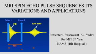
  • 1. MRI SPIN ECHO PULSE SEQUENCES ITS VARIATIONS AND APPLICATIONS Presenter :- Yashawant Ku. Yadav Bsc.MIT 3rd Year NAMS (Bir Hospital )
  • 2. Outline • Introduction P-S • Terminologies • Types of Spin echo pulse sequences (Conventional spin echo ,Fast or turbo spin echo , Inversion recovery ,Fast inversion recovery [STIR (short tau inversion recovery), FLAIR (fluid attenuated inversion recovery) ] . • Application of spin echo pulse sequences • References
  • 3. Introduction • Pulse sequence is a series of RF pulses, gradient applications and intervening time periods. • Pulse sequences enable us to control the way in which the system applies pulses and gradients. • In this way, image weighting and quality is determined.
  • 4. Intro… • In MRI, we are imaging properties of hydrogen nuclei (protons) in tissue. • With the protons precession around B0, nothing interesting will happen and we cannot measure any signal. • In order to measure parameters of the tissue, we need to perturb - excite - the system • This is done by applying a transient magnetic field - a radiofrequency pulse - in a different direction from B0; the direction of this pulse is often referred to as B1
  • 5. Contd.. • By applying this RF for a short time, we can rotate the magnetization of the tissue. • If the protons are initially aligned along the z (vertical) axis, and we apply a pulse along the x -axis for the right amount of time, we can rotate the magnetization from the z-axis onto the y-axis.
  • 6. Contd.. • This is referred to as a 90-degree pulse, since the magnetization direction is turned by 90 degrees. • Once the pulse is turned off, the protons will resume precession along B0 and will slowly return to their initial equilibrium with the net magnetization pointing along the z axis. • The RF pulses are applied for excitation purposes and, in the case of spin echo, for rephasing purposes too.
  • 7. Why pulse sequences ?? Image contrast • All clinical diagnostic images must demonstrate contrast between normal anatomical features and between anatomy and any pathology. • And MRI is consider as a high tissue contrast discriminating imaging modalities among all. • The contrast characteristic of each image depend on many variables,
  • 8. Contd.. Basically it is divided into two categories. • Intrinsic contrast parameters are those that cannot be changed because they are inherent to the body ’ s tissues. • Extrinsic contrast parameters are those that can be changed. Intrinsic contrast PMs Extrinsic contrast PMs T1 recovery time TR T2 decay time TE proton density FALIP ANGLE flow TI apparent diffusion coefficient (ADC). TURBO FACTOR / ECHO TRAIN LENGTH B Value
  • 9. Terminologies The repetition time (TR) is the time from the application of one RF pulse to the application of the next RF pulse for each slice and is measured in milliseconds (ms). • The TR determines the amount of longitudinal relaxation that is allowed to occur between the end of one RF pulse and the application of the next. • TR thus determines the amount of T1 relaxation that has occurred when the signal is read.
  • 10. Contd..  The echo time (TE) is the time from the application of the RF pulse to the peak of the signal induced in the coil and is also measured in ms. • The TE determines how much decay of transverse magnetization is allowed to occur. • TE thus controls the amount of T2 relaxation that has occurred when the signal is read.
  • 11. Contd.. • To demonstrate either T1, proton density or T2 contrast, specific values of TR and TE are selected for a given pulse sequence. • The selection of appropriate TR and TE weights an image so that one contrast mechanism predominates over the other two.
  • 12. Variations Spin echo pulse sequences (spins are rephased by a 180 ° rephasing pulse): • conventional spin echo • fast or turbo spin echo • inversion recovery.
  • 13.
  • 14. Conventional spin echo • spin echo uses a 90 ° excitation pulse followed by one or more 180 ° rephasing pulses to generate a spin echo. • If only one echo is generated, a T1 weighted image can be obtained using a short TE and a short TR.
  • 15. Contd… • The SE represents regeneration of spin phase information apparently lost during the decay of the FID. The "rebirth" of the FID as a SE is possible… by Erwin Hahn in 1949 • In fact, most of the FID signal has not been destroyed; it has merely become "disorganized" because the individual spins comprising it have lost their phase coherence. • The system is said to possess a "hidden order" or "atomic memory". By applying a second RF pulse, certain dephased components of the original FID can be refocused into a SE.
  • 16. Contd… • For proton density and T2 weighting, two RF rephasing pulses, generating two spin echoes, are applied. • The first echo has a short TE and a long TR to achieve proton density weighting, and the second has a long TE and a long TR to achieve T2 weighting.
  • 18.
  • 19.
  • 20. Parameters T1 WT Te short :- 10-30 ms Tr short :- 300-700ms Typical scan time :- 4-6 min T2 WT / PDW Te short :- 20ms / 80+MS(LONG) Tr long :- 2000+ms Sacn time:-
  • 21. Applications • Used in any part of the body for any indication. • To produce T1, T2 (True)and PD weighted images of good quality.  Disadvantage Longer scan time
  • 22. Fast spin echo /turbo • In this pulse sequence scan time is much shorter than conventional spin echo . • In fast spin echo, the scan time is reduced by performing more than one phase encoding step and subsequently filling more than one line of K space per TR. • This is achieved by using several 180 ° rephasing pulses to produce a train of echoes or echo train. • At each rephasing, an echo is produced and a different phase encoding step is performed.
  • 23. Contd.. • In conventional spin echo, raw image data from each echo are stored in K space, • Each echo is used to produce a separate image • In FSE data from each echo are placed into one image. • The number of 180 ° rephasing pulses performed per TR corresponds to the number of echoes produced and the number of lines of K space filled.
  • 24. Contd.. • This number is called the turbo factor or the echo train length . The higher the turbo factor, the shorter the scan time as more phase encoding steps are performed per TR. • Number of echoes -- called echo train length (ETL) by GE and Canon; turbo factor by Siemens and Philips; or shot factor by Hitachi. • At each 180 ° /phase encoding combination, a different amplitude of phase encoding gradient slope is applied to fill out a different line of K space
  • 25. Contd.. • ETL is the single most important parameter. In general, image acquisition time is inversely proportional to ETL. • ETL also has important effects on image quality. Longer ETLs result in more T2- weighting because more late echoes with longer TE's contribute to the overall signal. • Longer ETL's are also decreases overall signal-to-noise ratio (SNR) and contrast-to-noise ratio (CNR) because the later echoes are weaker.
  • 26. TR :- 4000MS OTHER PARAMETERS UNCHANGED
  • 27. Contd.. • Time between echoes -- called echo spacing (ESP) by GE, Philips, Siemens, and Canon; interecho time (ITE) by Hitachi. • Increasing echo spacing (ESP) permits the use of longer TE's but adversely impacts SNR and CNR
  • 28. Weighting in fast spin echo • The echoes are generated at different TE times and therefore data collected from them have variable weighting. • So how is a fast spin echo sequence weighted correctly? • The TE selected is only an effective TE • That mean TE at which the operator wishes to weight the resultant image. • The system orders the phase encoding steps so that steep or shallow slopes are applied to the various echoes produced.
  • 29.
  • 30.
  • 31. Contd… • fast spin echo has largely replaced spin echo especially for T2 weighting. • There are, however, two contrast differences between spin echo and fast spin echo, • due to the repeated, closely spaced 180 ° pulses of the echo train • First, fat remains bright on T2 weighted images due to the multiple RF pulses, which reduce the effects of spin – spin interactions in fat ( J coupling ) • Second, the repeated 180 ° pulses can increase magnetization transfer effects so that muscle, appears darker on fast spin echo images than in conventional spin echo.
  • 32. Contd.. • In addition, the multiple 180 ° pulses reduce magnetic susceptibility effects, which can be detrimental when looking for small hemorrhages.
  • 33.
  • 34. Contd… • Image blurring may occur in fast spin echo images at the edges of tissues with different T2 decay values. • This is because each line of K space filled during an echo train contains data from echoes with a different TE. • • When using long echo trains, late echoes that have a low signal amplitude contribute to the resolution of K space. • If these echoes are negligible, then resolution is lost from the image and blurring occurs.
  • 35. TYPICAL PARAMETERS Dual echo • TR 2500–4500 ms (for weighting and slice number) • effective TE1 17 ms • effective TE2 102 ms • ETL 8 – This may be split so that the PD image is acquired with the first four echoes and the T2 with the second four. Single echo • T2 weighting • TR 4000–8000 ms • TE 102 ms • ETL 16 Single echo T1 weighting TR 600 ms TE 17 ms ETL 4
  • 36. USES • brains, spines , joints, extremities and pelvis
  • 37. SS-FSE / HASTE Other vendors have similar sequences under slightly different names: GE (Single-shot fast spin echo, SS-FSE), Philips (Single-shot turbo spin echo, SSH-TSE; ultra-fast spin echo, UFSE), HItachi (Single-shot fast SE), and Canon (Fast Advanced Spin Echo, FASE, SuperFASE). • This sequence even faster than FSE , • In this technique all the lines of K space are acquired in one TR. • SS - FSE combines a parti al Fourier technique with fast spin echo. • Half of the lines of K space are acquired in one TR and the other half are transposed.
  • 38. Contd.. • Currently the highest turbo factor used in single shot imaging is 728 • Another consideration when using long echo trains is that the specific absorption rate ( SAR ) is significantly increased by applying so many successive 180 ° pulses. • slice number can be reduce to manifest SAR but that cause difficult to get required coverage area … so, • Refocusing angle is reduced to 120° • This reduces the SAR significantly (which is proportional to the square of the flip angle) but also reduces the SNR. • the benefi t of being able to obtain more slices per acquisition
  • 39. Contd… Representative HASTE/SS-FSE pulse sequence. Note asymmetric sampling in phase-encode direction. The peripheral lines on one side of k-space are not sampled but estimated by conjugate symmetry.
  • 40.
  • 41. Application • routine scout images • images of the head or body in children or uncooperative patients • fetal imaging • non-breath-hold abdominal imaging • MR cholangiopancreatography • MR myelography, and • non-contrast MR angiography.
  • 42.
  • 43. Examples of conventional SSFSE compared to half-Fourier vrfSSFSE. (A) Conventional SSFSE and (B) half-Fourier vrfSSFSE images from a patient with renal cell carcinoma of the left kidney (arrow). (C) Conventional SSFSE and (D) half-Fourier vrfSSFSE images from a patient with a mildly dilated pancreatic and common bile duct. Images are mildly cropped from the full field of view. Note the equivalent image quality.
  • 44. IR (Inversion recovery ) • Inversion recovery sequences were initially designed to produce very heavy T1 weighting. • However at present they are mainly used in conjunction with a FSE sequence to produce T2 weighted images. • It is usually used to suppress the signal from certain tissues in conjunction with long TEs and T2 weighting, although at low field it is still used for T1 contrast.
  • 45. Contd.. • Inversion recovery is a pulse sequence that begins with a 180 ° inverting pulse. This inverts the NMV through 180 ° into full saturation. • When the inverting pulse is removed, the NMV begins to relax back to B 0 . • A 90 ° excitation pulse is then applied at a time from the 180 ° inverting pulse known as the TI ( time from inversion )
  • 46. Contd… • A further 180° RF pulse is applied which rephases spins in the transverse plane and produces an echo at time TE after the excitation pulse. • The contrast of the resultant image depends primarily on the length of the TI. • If the TI is sufficiently long to allow the NMV to pass through the transverse plane, the contrast depends on the degree of saturation that is produced by the 90° pulse (as in spin echo), i.e. • if the 90° pulse is applied shortly after the NMV has passed through the transverse plane, heavy saturation and T1 weighting results.
  • 47. Contd… TIs of 300 ms to 700 ms result in this type of heavy T1 weighting
  • 48.
  • 49. FIR (FAST INVERSION RECOVERY ) • It is a combination of inversion recovery and fast spin echo. • It is usually used to suppress the signal from certain tissues in conjunct on with long TEs and T2 weighting, so that water and pathology return a high signal. • Although at low field it is still used for T1 contrast. • The two main sequences in this category are STIR and FLAIR.
  • 50. STIR (Short tau inversion recovery ) • This sequences uses short TIs such as 100– 180 ms, depending on field strength . • TIs of this magnitude place the 90° excitation pulse at the time that NMV of fat is passing exactly through the transverse plane. • At this point (called the null point) there is no longitudinal component in fat. • Therefore the 90° excitation pulse produces no transverse component in fat and therefore no signal. • In this way a fat suppressed image result.
  • 51.
  • 52. Uses • STIR is an extremely important sequence in musculoskeletal imaging because normal bone, which contains fatty marrow, is suppressed and lesions within bone such as bone bruising and tumors are seen more clearly.
  • 53. FLAIR (fluid attenuated inversion recovery ) • It uses long TIs such as 1700–2200 ms, depending on field strength, to null the signal from CSF in exactly same way as STIR. • Because CSF has a long T1 recovery time, the TI must be longer to correspond with its null point.
  • 54. T1-FLAIR brain image at 3T with TR=2100, TE=9, TI=880. T2 FLAIR :TI 2000-2500 very long TRs (8000) and TEs (140) For T1-FLAIR both the TR and TE values are kept relatively short to minimize T2-weighting. Considering these restrictions, the TInull for a T1- FLAIR sequence is typically in the range of 800-1000 ms.
  • 55. white matter, and gray matter are approximately400, and 650 ms respectively. TI= 0.69 x T1.
  • 56. Typical parameters The required TI depends on the field strength (higher fields). T1 weighting TI 300–700 ms • TE 10–20 ms • TR long • ETL 4 STIR • TI 100–180 ms • TE 70 ms+ (for T2 weighting) • TR long • ETL 16  FLAIR • TI 1500–2200 ms • TE 70 ms+ (for T2 weighting) • TR long • ETL 12–16
  • 57. ADVANTGES DISADVANTAGES Selective Tissue Suppression Longer scan times Up to Twice the Dynamic Range Increase in flow-related artifacts Additive T1 and T2 contrast ?? Signal-to-noise can decrease as tissues are suppressed Higher specific absorption rate (SAR) due to additional 180° pulses
  • 59. DOUBLE IR • Double Inversion Recovery (DIR) is an inversion recovery variant that uses not one, but two nonselective 180°-inverting pulses. • Here the first 180°-pulse suppresses CSF and the second suppresses white matter. Double IR sequence suppressing CSF and white matter. TR=11000, TE=30, TI-1=3400, TI-2=325.
  • 60. Black blood by DIR • In this method, two 180°-pulses are applied very close together in time. • The first 180°-pulse is nonselective, meaning that it inverts the magnetization for all slices within the imaging volume. • The second 180°-pulse, following immediately on the heels of the first 180°-pulse, is slice selective, • Thus the myocardium and other relatively stationary tissues have their signals preserved, • but the blood flowing from adjacent slices has an inverted magnetization.
  • 61. Contd… The TI is adjusted to null the signal from inflowing blood, which appears "black" on magnitude reconstructed IR images.
  • 62. Phase sensitive IR ????? The sequence with the gray background is called phase-corrected or phase-sensitive inversion recovery (PSIR). Phase-corrected IR preserves the information about Mz polarity, with more negative values rendered increasingly darker.
  • 63. • If TI is not perfectly chosen, detection of contrast enhancement may be impaired using magnitude reconstruction. • Phase-sensitive IR is less sensitive to choice of TI as illustrated in this example of septal ischemia (arrow).
  • 64. References 1. https://www.mriquestions.com 2. MRI at a Glance CATHERINE WESTBROOK 3. MRI in Practice Fourth Edition
  • 66. •Long T1 materials are dark on T1-weighted images, but long T2 materials are bright on T2-weighted images.
  • 67. • T2 Relaxation Accompanying T1 Relaxation how ??? • Does T2 Relaxation Occurring Without T1 Relaxation?? 1. where it experiences a local static field disturbance (B1oo) in addition to the main magnetic field (Bo). 2. dipolar interaction also known as a spin-spin "flip-flop".

Editor's Notes

  1. Radio waves have frequencies as high as 300 gigahertz (GHz) to as low as 30 hertz (Hz). "spin-spin" interactions are just one of several mechanisms by which T2 relaxation can occur.)
  2. Manipulation of external contrast parameters to achieve particular pathology based contrast image is pulse sequence The b-value is a factor that reflects the strength and timing of the gradients used to generate diffusion-weighted images.
  3. TR determines amount of T1 and proton density weighting and Flip angle controls the amount of T1 and proton density weighting
  4. As the scan time is a function of the TR, NEX and number of phase encodings, to reduce the scan time one or more of these factors should be reduced. ‘ Decreasing the TR and the NEX affects image weighting and SNR, which is undesirable. Reducing the number of phase encodings reduces the spatial resolution, which is also a disadvantage
  5. Motion, susceptibility, and edge-related artifacts increase.  the shortest permitted ESP should therefore be chosen in most applications.   A typical ESP is on the order of 16 to 20 msec  
  6. The shallowest phase encodings are performed on echoes occurring around 100 ms. Data acquired from these phase encodings have a TE at or close to 100 ms. Phase encodings performed at the very beginning and end of the echo train are steep, and the signal amplitude of these echoes is small. They contain either proton density or very heavily T2 weighted data, which are present in the image but whose impact is less predominant.
  7. Contrast difference SE AND FSE IS similar so it is used in every clinical examinations , MSKS, PELVIC ,CNS However, fat saturation techniques can be used to compensate for this
  8. This, however, may be reduced by decreasing the spacing between echoes and/or the turbo factor…. In additi on, artefact from metal implants is significantly reduced when using fast spin echo because the repeated 180 ° RF pulses compensate for fi eld inhomogeneity
  9. As FSE is incompatible with respiratory compensation techniques, it can only be used in the chest and abdomen with respiratory triggering or multiple NEX.
  10. there is a SNR penalty.
  11. Because CSF has a long intrinsic T1 value (3000-4000 ms), its TInull is strongly dependent on the TR selected. T2 flair proved itself by revealing a wide range of lesions, including cortical, periventricular, and meningeal diseases  T2-FLAIR became a standard protocol for routine imaging. (True flair )
  12. DIR technique has been for brain imaging, especially for detection of multiple sclerosis plaques and lesions of the cerebral cortex.
  13. commonly used in cardiovascular MRI.  is slice selective, meaning it returns the magnetization of all tissues in that slice only back to the +z-direction.
  14. It is possible to use a third (or even fourth) 180°-pulse in conjunction with a black blood DIR technique. This third pulse is commonly used to suppress pericardial fat,
  15.  Several major MR vendors offer this sequence and some call it "Real IR" or "True IR". Magnitude reconstruction does not require such sophisticated post-processing.  Additionally, magnitude reconstruction allows setting of TI for selective nulling of tissues such as fat (STIR) or CSF (FLAIR). Selective nulling techniques are not possible with phase-sensitive IR. 
  16. Longer t1 tissue recover slowly so MZ is suppress and signal is low , but longer t2 tissue diphase slowly so Mxy is maintained and signal is high
  17. Although T2 relaxation always accompanies T1 relaxation, T2 relaxation may also occur without T1 relaxation. This is sometimes referred to as the "secular contribution to T2."