Download to read offline

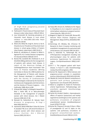
![•Pregnancy
•Sicklecellanaemia
These recommendations for preoperative
testing are set out in ‘Look-up tables’ cross
referenced by the type of surgery, common
6
chronicillnessesandage.
7.Testresultobtainedfromthemedicalrecord,
within 6 months of surgery are generally
acceptable if the patient medical history has not
changed substantially. [1] Ausset et al had
formulated modified Global Quality index which
include 15 criteria to be incorporated in
preoperative assessment as shown in (Table1.)
below.
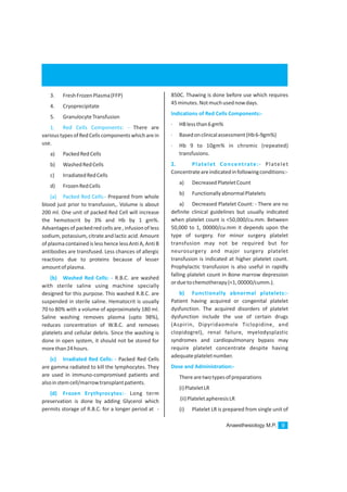
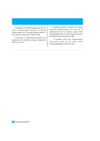
2
Table 1: Modified GQI * Criteria
Name
Age
Cardiopulmonarystatus
Surgicalprocedure
Preoperativediagnosis
Preoperativevitalsigns
Peroralstatus
Medications
Allergies
Weight
ASA
Anesthetichistoryandcomplications
Assessment
Plan
*GQI=100X (a-b)/a5 = the percentage of
proportion of criteria present on the PEF to the 15
GQI criteria, where a is the total number of criteria
selected (a=15) and b is the number of criteria
lacking.
The “set mind” of surgeons, anesthesiologist,
and other clinical personnel need to be changed
regarding PAE through communication and
educationandasystemofprinciplesandmethods
should be employed to perform pre anesthetic
evaluation of patients of all ages scheduled to
receive general anesthesia, regional anesthesia,
moderate or deep sedation for elective surgical
andnonsurgicalprocedures.
One should not expertise ourselves in
dictating the series of investigation that do not
make any important contribution to preoperative
assessment of patient and for safe Anesthesia.
Specific tests should be individualized based upon
information retrieved from the patients past
medical record, physical examination, and the
type and severity of planned, major or minor
surgical procedure.PAE not only aids in adequate
documentation and record keeping but as well
establishes a good familiarity with the concerned
anesthesiologist which allays anxiety and
apprehension. At a minimum, assessment of
airway, lungs and heart should not be out of sight,
outofmindofanesthesiologist
1. American society of anesthesiologist Task
force on pre-anesthesia evaluation. Practice
advisory for pre -anesthesia evaluation: An
updated report by the American society of
anesthesiologist task force on pre-anesthesia
evaluation.Anesthesiology2012;116:522-38
2. Ausset S, Bouaziz H, Brossea M, Kinirons B,
Benhamou D. Improvement of information
gained from the pre-anesthetic visit through a
quality of assurance programme. Br J Anaesth
2002;88:280-283.
3. Kluger MT,Tham EJ, ColemanNA, Runciman
WB, Bullock MF . Inadequate pre-operative
evaluation and preparation: A review of 197
reports from the Australian incident monitoring
study.Anesthesia2000;55:1173-1178
CONCLUSION
References
Anaesthesiology M.P. 17](https://image.slidesharecdn.com/mpanaesthesiologyoctober2016-161107142226/85/Mp-anaesthesiology-october-2016-18-320.jpg)

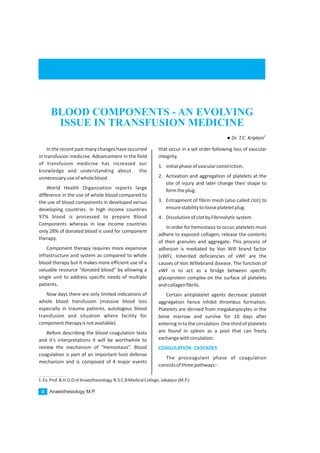
This document discusses recent changes in transfusion medicine regarding the use of blood components instead of whole blood. It notes that high income countries now process 97% of donated blood into components, while low income countries only process 28% into components. Component therapy allows one blood donation to help multiple patients. The document then provides details on the coagulation cascade and factors, describing the intrinsic, extrinsic, and common pathways. It also briefly discusses the fibrinolytic system and the historical naming of coagulation factors.


