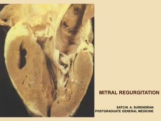
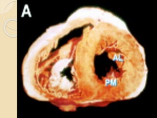
MITRAL REGURGITATION
SATCHI. A. SURENDRAN
POSTGRADUATE GENERAL MEDICINE
ANATOMY
The mitral valve apparatus consists of
• the anterior and the posterior leaflets ,
• the mitral annulus (fixed and dynamic)
• The chordae tendinae
• Papillary muscles
• Underlying myocardium
Flail MV
Common Causes
 Mitral Valve Prolapse (MVPS)
 Rheumatic Heart Disease
 Infective Endocarditis
 Annular Calcification
 Cardiomyopathy
 Ischemic Heart Disease
Rheumatic Heart Disease
 Usually associated with stenosis and
fusion of the commissures
 Retractile fibrosis of leaflets and
chordae, causing loss of coaptation.
 Secondary dilatation of the mitral
annulus tends to further decrease the
coaptation.
Degenerative
 Mitral annular calcification is common
in the elderly population
 Accelerated by hypertension, Old
Age and diabetes.
 Connective tissue diseases such as
Marfan and Hurler syndromes.
Mechanisms
 Abnormal movement of the leaflets
into the left atrium (LA) during systole
 Caused by inadequate chordal
support (elongation or rupture) and
excessive valvular tissue.
 Myxomatous degeneration -
characterized by redundant, floppy
leaflets and associated with
progressive MR.
MVP MR Myxomatous
Infective Endocarditis
 approx. 5 %of cases of severe MR.
 Vegetations may produce mild MR by
interposition between leaflets.
 Severe endocarditic MR is usually
related to ruptured chordae and less
frequently to destruction of mitral tissue
(edges or perforation)
Ischemic and Functional MR
 Consequence of LV wall dysfunction (ischemia,
scarring, aneurysm, cardiomyopathy, or
myocarditis,)
 The coaptation of intrinsically normal leaflets is
incomplete.
 The pattern of mitral valve (MV) deformation from
the medial to the lateral side is asymmetrical in
ischemic MR, whereas it was symmetrical in
nonischemic MR of cardiomyopathy.
 Responds to vasodilators or improvement
of ischemia-is a result of a reversal of
adverse LV geometry.
 Rupture of papillary muscle produces MR
because of the flail leaflet.
 80 percent of cases involves the
posteromedial papillary muscle.
 Complete rupture is rapidly fatal without
surgery, and partial- or single-head rupture
of the papillary allows emergency surgery.
Other causes
 (1) connective tissue disorders Marfan
syndrome, Ehlers-Danlos syndrome,
pseudoxanthoma elasticum, osteogenesis
imperfecta, Hurler disease, SLE, and
anticardiolipin syndrome;
 (2) cardiac trauma penetrating or
nonpenetrating
 (3) myocardial disease—hypertrophic
cardiomyopathy, amyloidosis, or sarcoidosis;
 (4) endocardial lesions caused by
hypereosinophilic syndrome, endocardial
fibroelastosis, carcinoid tumors, ergot toxicity,
radiation toxicity, diet or drug toxicity
 (5) congenital cleft mitral valve, corrected
transposition
 (6) cardiac tumors.
Hemodynamics – Severity
Parameters
 The abnormal coaptation creates a
regurgitant orifice during systole.
 The systolic pressure gradient between the
LV and LA is the driving force of the
regurgitant flow, which results in a regurgitant
volume.
 May be expressed as the regurgitant
fraction.
 Torricelli principle, states that flow through
an orifice varies by the square root of the
pressure gradient across that orifice:
MRV= MROA* constant* Ts*√ (LVP-LAP)
 The pressure gradient between the LV and
LA begins with mitral closure (simultaneous
to S1) and persists after closure of the aortic
valve (S2) until the mitral valve opens.
 MR induces a low-pressure form of
volume overload as a result of ejection
into the LA.
 Moderate MR when the regurgitant
fraction is in the range of 30 to 50 %
 severe MR - regurgitant fraction >50%
Orifice Area
 In Functional MR (dilated cardiomyopathy) -
constant decrease in OA throughout systole.
 In MVP, OA is small in early systole,
increasing substantially in midsystole, and
decreasing mildly during LV relaxation.
 In Rheumatic MR, constant regurgitant OA
during most of systole.
Determinants of Volume
Overload
 Area of the regurgitant orifice
 Regurgitant gradient
 Regurgitant duration
MR & AR Comparison
 The volume overload is usually less severe in MR than
in AR, despite a larger regurgitant gradient and orifice
 Due to shorter duration of regurgitation during the
cardiac cycle in MR than in AR
 The LV is unloaded in both early and late systole by
ejection into the low-pressure LA; whereas in AR, the
excess volume is ejected into the high-pressure aorta
only in early systole.
 The lower LV mass and mass-to-volume ratio in
patients with MR as compared to AR.
LA Dynamics
LA pressure is mainly determined by LA compliance.
Dynamics Acute
Compensated
MR
Chronic
Compensated
MR
LA Compliance
(Volume)
Less or No
compliance
Compliant -
Dilated
LA Pressure High Relatively Low
or Even Normal
LA/LV Gradient Lesser More
Regurgitant
Volume
Tends to be
lesser
As LA dilated,
tends to be more
Murmur Early/Holosystoli
c
Holosystolic
Role of ACE I
 Efficacy of ACE inhibitor in papillary
muscle dysfunction or dilated
cardiomyopathy produces a decrease in
LV size and improved LV geometry and
thereby regurgitant OA
 This benefit is not observed with
rhuematic MR or Mitral Valve Prolapse
MR – Chronic Compensated
 In c/c MR adrenergic system provides
inotropic support (important mech. of
compensation)
 Norepinephrine release increases
directly with increase in end systolic
volume.
MR Chronic Decompensated
Stage
 muscle dysfunction has developed,
impairing ejection fraction, diminishing
both TSV and FSV.
 EF, although still “normal,” has
decreased to 0.55.
 LAP is reelevated because less
volume is ejected during systole,
causing a higher ESV.
Clinical presentation
 Usual symptoms -Fatigue and mild dyspnea
on exertion (rapidly improved by rest.)
 Severe dyspnea on exertion ,PND, frank
pulmonary edema, or even hemoptysis in
decompensation.
 Severe symptoms may be triggered by a
new onset of AF, an increase in degree of
MR, the occurrence of endocarditis or
ruptured chordae, or a change in LV
compliance or function.
Severe Presentations
 A/c MR - dramatic symptoms-with pulmonary edema or
ccf
 subside with administration of a diuretic, afterload
reduction, and increased LA compliance with time.
 Abrupt chordal rupture - sudden onset of atypical
chest pain and dyspnea
 rupture of papillary muscle in of a/c MI - cardiogenic
shock or a severe pulmonary edema.
 Pulmonary edema may also be observed in transient
severe papillary muscle dysfunction.
Physical examination
 BP is usually normal. Carotid upstroke is brisk;
Wide Pulse Pressure.
 Hyperdynamic Apical Impulse – down & out –
LV enlargement.
 An apical thrill is characteristic of severe MR.
 Systolic expansion of the enlarged left atrium
causes a late systolic thrust in the parasternal
region,-confused with RV enlargement.
Auscultation
 S1 is included in the murmur and is usually
normal but may be increased in rheumatic
disease.
 Wide splitting of S2 due to the shortening of
LV ejection and an earlier A2 due to reduced
resistance to LV ejection.
 In patients with MR who have severe
pulmonary hypertension, P2 is louder than
 The abnormal increase in the flow rate
across the mitral orifice during the rapid
filling phase is associated with an S3,
(not due to heart failure) ; may be
accompanied by a brief diastolic rumble.
 Midsystolic clicks are markers of valve
prolapse.
Systolic Murmur
 Most often holosystolic, including 1st and
2nd heart sounds.
 Blowing type but may be harsh(MVP)
 The maximum intensity is usually at the
apex, and it may radiate to the axilla in
anterior leaflet prolapse, degeneration
affecting primarily the anterior leaflet.
 In posterior leaflet prolapse, the jet is usually
superiorly and medially directed and the
murmur radiates toward the base of the
heart.
 In MI, severe MR may be totally silent.
 Murmurs of shorter duration usually
correspond to mild MR; they may be mid or
late systolic in mitral valve prolapse or early
systolic in functional MR.
 Murmer is increased by squatting ,
isometric excercises
 Decreased by amyl nitrite inhalation
and valsalva
Clinical Severity Parameters
 Systolic Thrill over apex
 Large LV indicates severe MR
 Presence of S3
 Flow MDM across non stenotic Mitral
valve.
ECG
The principal ECG findings are
 left atrial enlargement and AF.
 Wide notched P waves – P Mitrale
 ECG evidence of LV enlargement
occurs in 1/3rd of patients with severe
MR.
 Tall R in V5 V6 but S in V1 is small
 15 % of patients -evidence of RV
hypertrophy.
CXR
 Cardiomegaly may be present in c/c
MR or in ischemic or functional MR .
 LA body and appendage dilatation is
frequent, but giant LA is rare.
 annular calcification, seen as a C-
shaped density below the posterior
leaflet, is frequent.
 signs of pulmonary hypertension or
pulmonary edema are rarely
observed.
Doppler echocardiography
 High-velocity jet in the LA during systole
 Severity of the regurgitation is a function of the
distance from the valve that the jet can be
detected.
 Area of the mitral jet >8 cm2 indicates severe
MR.
 Severity Predictor: Vena Contracta defined as
the narrowest cross-sectional area of the
Echocardiography
 Quantification of LV end-diastolic and
end-systolic dimensions, wall
thickness, and ejection fraction and
fractional shortening.
 M-mode diameter or volume can
assess the LA size by two-dimensional
echocardiography
Flail MV
 Rheumatic MR is characterized by
thickening of the leaflets and chordae.
The posterior leaflet has reduced mobility,
whereas the anterior leaflet may be doming if
commissural fusion is associated.
 Degenerative MR, prolapse -the
passage of valvular tissue beyond the
annulus plane in the long-axis view
 The valve leaflets are diffusely thickened
leaflets and excessive valvular tissue
and increased echogenicity of the
annulus - mitral annular calcification.
 Flail segments appear as complete
eversion of the segment with or without
the small floating echo of ruptured
chordae
Treatment (medical)
 IE prophylaxis
 Rhuematic fever prophylaxis
 If AF rate control with digoxin and beta
blockers
 Anticoagulation in AF
 Vasodilators in acute MR and
cardiomyopathy- ACE inhibitors
 Beta blockers may be used – but no
definite evidence
Surgery Indications
 American College of Cardiology/American Heart Association
Guidelines for Surgery for Nonischemic Severe Mitral Regurgitation

Class I (evidence and/or general agreement that surgery is
useful and effective)
 Symptoms cause by MR (acute or chronic)
 Asymptomatic patients with severe MR and mild-moderate LV
dysfunction defined as an —Ejection fraction 30–60% and —
End-systolic dimension 45–55 mm
 Class IIa (conflicting evidence and/or divergence of opinion but
the weight of evidence/opinion favors surgical intervention)
 Asymptomatic patients with normal LV function and —Atrial
fibrillation or —Pulmonary hypertension (>50 mmHg at rest of
>60 mmHg with exercise)
 Asymptomatic patients with —Ejection fraction 50–60% or —
End-systolic dimension 45–55 mm
 Severe left ventricular dysfunction (ejection fraction <30% and/or
end-systolic dimension >55 mm) if chordal preservation is highly
likely
Post op outcome
 Mitral valve repair is better
 Maintains continuity of valve with
papillary muscles
 Hence maintains LV geometry and
maintains EF
 Operative mortality 1-2 % (5-10 with
MVR)
 AF may persist - anticoagulation
THANK YOU

Mitral Regurgitation

  • 1.
    MITRAL REGURGITATION SATCHI. A.SURENDRAN POSTGRADUATE GENERAL MEDICINE
  • 2.
    ANATOMY The mitral valveapparatus consists of • the anterior and the posterior leaflets , • the mitral annulus (fixed and dynamic) • The chordae tendinae • Papillary muscles • Underlying myocardium
  • 4.
  • 5.
    Common Causes  MitralValve Prolapse (MVPS)  Rheumatic Heart Disease  Infective Endocarditis  Annular Calcification  Cardiomyopathy  Ischemic Heart Disease
  • 6.
    Rheumatic Heart Disease Usually associated with stenosis and fusion of the commissures  Retractile fibrosis of leaflets and chordae, causing loss of coaptation.  Secondary dilatation of the mitral annulus tends to further decrease the coaptation.
  • 7.
    Degenerative  Mitral annularcalcification is common in the elderly population  Accelerated by hypertension, Old Age and diabetes.  Connective tissue diseases such as Marfan and Hurler syndromes.
  • 8.
    Mechanisms  Abnormal movementof the leaflets into the left atrium (LA) during systole  Caused by inadequate chordal support (elongation or rupture) and excessive valvular tissue.  Myxomatous degeneration - characterized by redundant, floppy leaflets and associated with progressive MR.
  • 9.
  • 10.
    Infective Endocarditis  approx.5 %of cases of severe MR.  Vegetations may produce mild MR by interposition between leaflets.  Severe endocarditic MR is usually related to ruptured chordae and less frequently to destruction of mitral tissue (edges or perforation)
  • 11.
    Ischemic and FunctionalMR  Consequence of LV wall dysfunction (ischemia, scarring, aneurysm, cardiomyopathy, or myocarditis,)  The coaptation of intrinsically normal leaflets is incomplete.  The pattern of mitral valve (MV) deformation from the medial to the lateral side is asymmetrical in ischemic MR, whereas it was symmetrical in nonischemic MR of cardiomyopathy.
  • 12.
     Responds tovasodilators or improvement of ischemia-is a result of a reversal of adverse LV geometry.  Rupture of papillary muscle produces MR because of the flail leaflet.  80 percent of cases involves the posteromedial papillary muscle.  Complete rupture is rapidly fatal without surgery, and partial- or single-head rupture of the papillary allows emergency surgery.
  • 13.
    Other causes  (1)connective tissue disorders Marfan syndrome, Ehlers-Danlos syndrome, pseudoxanthoma elasticum, osteogenesis imperfecta, Hurler disease, SLE, and anticardiolipin syndrome;  (2) cardiac trauma penetrating or nonpenetrating  (3) myocardial disease—hypertrophic cardiomyopathy, amyloidosis, or sarcoidosis;  (4) endocardial lesions caused by hypereosinophilic syndrome, endocardial fibroelastosis, carcinoid tumors, ergot toxicity, radiation toxicity, diet or drug toxicity  (5) congenital cleft mitral valve, corrected transposition  (6) cardiac tumors.
  • 14.
    Hemodynamics – Severity Parameters The abnormal coaptation creates a regurgitant orifice during systole.  The systolic pressure gradient between the LV and LA is the driving force of the regurgitant flow, which results in a regurgitant volume.  May be expressed as the regurgitant fraction.
  • 15.
     Torricelli principle,states that flow through an orifice varies by the square root of the pressure gradient across that orifice: MRV= MROA* constant* Ts*√ (LVP-LAP)  The pressure gradient between the LV and LA begins with mitral closure (simultaneous to S1) and persists after closure of the aortic valve (S2) until the mitral valve opens.
  • 16.
     MR inducesa low-pressure form of volume overload as a result of ejection into the LA.  Moderate MR when the regurgitant fraction is in the range of 30 to 50 %  severe MR - regurgitant fraction >50%
  • 17.
    Orifice Area  InFunctional MR (dilated cardiomyopathy) - constant decrease in OA throughout systole.  In MVP, OA is small in early systole, increasing substantially in midsystole, and decreasing mildly during LV relaxation.  In Rheumatic MR, constant regurgitant OA during most of systole.
  • 18.
    Determinants of Volume Overload Area of the regurgitant orifice  Regurgitant gradient  Regurgitant duration
  • 19.
    MR & ARComparison  The volume overload is usually less severe in MR than in AR, despite a larger regurgitant gradient and orifice  Due to shorter duration of regurgitation during the cardiac cycle in MR than in AR  The LV is unloaded in both early and late systole by ejection into the low-pressure LA; whereas in AR, the excess volume is ejected into the high-pressure aorta only in early systole.  The lower LV mass and mass-to-volume ratio in patients with MR as compared to AR.
  • 20.
    LA Dynamics LA pressureis mainly determined by LA compliance. Dynamics Acute Compensated MR Chronic Compensated MR LA Compliance (Volume) Less or No compliance Compliant - Dilated LA Pressure High Relatively Low or Even Normal LA/LV Gradient Lesser More Regurgitant Volume Tends to be lesser As LA dilated, tends to be more Murmur Early/Holosystoli c Holosystolic
  • 23.
    Role of ACEI  Efficacy of ACE inhibitor in papillary muscle dysfunction or dilated cardiomyopathy produces a decrease in LV size and improved LV geometry and thereby regurgitant OA  This benefit is not observed with rhuematic MR or Mitral Valve Prolapse
  • 24.
    MR – ChronicCompensated  In c/c MR adrenergic system provides inotropic support (important mech. of compensation)  Norepinephrine release increases directly with increase in end systolic volume.
  • 25.
    MR Chronic Decompensated Stage muscle dysfunction has developed, impairing ejection fraction, diminishing both TSV and FSV.  EF, although still “normal,” has decreased to 0.55.  LAP is reelevated because less volume is ejected during systole, causing a higher ESV.
  • 26.
    Clinical presentation  Usualsymptoms -Fatigue and mild dyspnea on exertion (rapidly improved by rest.)  Severe dyspnea on exertion ,PND, frank pulmonary edema, or even hemoptysis in decompensation.  Severe symptoms may be triggered by a new onset of AF, an increase in degree of MR, the occurrence of endocarditis or ruptured chordae, or a change in LV compliance or function.
  • 27.
    Severe Presentations  A/cMR - dramatic symptoms-with pulmonary edema or ccf  subside with administration of a diuretic, afterload reduction, and increased LA compliance with time.  Abrupt chordal rupture - sudden onset of atypical chest pain and dyspnea  rupture of papillary muscle in of a/c MI - cardiogenic shock or a severe pulmonary edema.  Pulmonary edema may also be observed in transient severe papillary muscle dysfunction.
  • 28.
    Physical examination  BPis usually normal. Carotid upstroke is brisk; Wide Pulse Pressure.  Hyperdynamic Apical Impulse – down & out – LV enlargement.  An apical thrill is characteristic of severe MR.  Systolic expansion of the enlarged left atrium causes a late systolic thrust in the parasternal region,-confused with RV enlargement.
  • 29.
    Auscultation  S1 isincluded in the murmur and is usually normal but may be increased in rheumatic disease.  Wide splitting of S2 due to the shortening of LV ejection and an earlier A2 due to reduced resistance to LV ejection.  In patients with MR who have severe pulmonary hypertension, P2 is louder than
  • 30.
     The abnormalincrease in the flow rate across the mitral orifice during the rapid filling phase is associated with an S3, (not due to heart failure) ; may be accompanied by a brief diastolic rumble.  Midsystolic clicks are markers of valve prolapse.
  • 31.
    Systolic Murmur  Mostoften holosystolic, including 1st and 2nd heart sounds.  Blowing type but may be harsh(MVP)  The maximum intensity is usually at the apex, and it may radiate to the axilla in anterior leaflet prolapse, degeneration affecting primarily the anterior leaflet.
  • 32.
     In posteriorleaflet prolapse, the jet is usually superiorly and medially directed and the murmur radiates toward the base of the heart.  In MI, severe MR may be totally silent.  Murmurs of shorter duration usually correspond to mild MR; they may be mid or late systolic in mitral valve prolapse or early systolic in functional MR.
  • 33.
     Murmer isincreased by squatting , isometric excercises  Decreased by amyl nitrite inhalation and valsalva
  • 34.
    Clinical Severity Parameters Systolic Thrill over apex  Large LV indicates severe MR  Presence of S3  Flow MDM across non stenotic Mitral valve.
  • 35.
    ECG The principal ECGfindings are  left atrial enlargement and AF.  Wide notched P waves – P Mitrale  ECG evidence of LV enlargement occurs in 1/3rd of patients with severe MR.  Tall R in V5 V6 but S in V1 is small  15 % of patients -evidence of RV hypertrophy.
  • 36.
    CXR  Cardiomegaly maybe present in c/c MR or in ischemic or functional MR .  LA body and appendage dilatation is frequent, but giant LA is rare.  annular calcification, seen as a C- shaped density below the posterior leaflet, is frequent.  signs of pulmonary hypertension or pulmonary edema are rarely observed.
  • 38.
    Doppler echocardiography  High-velocityjet in the LA during systole  Severity of the regurgitation is a function of the distance from the valve that the jet can be detected.  Area of the mitral jet >8 cm2 indicates severe MR.  Severity Predictor: Vena Contracta defined as the narrowest cross-sectional area of the
  • 40.
    Echocardiography  Quantification ofLV end-diastolic and end-systolic dimensions, wall thickness, and ejection fraction and fractional shortening.  M-mode diameter or volume can assess the LA size by two-dimensional echocardiography
  • 41.
  • 42.
     Rheumatic MRis characterized by thickening of the leaflets and chordae. The posterior leaflet has reduced mobility, whereas the anterior leaflet may be doming if commissural fusion is associated.  Degenerative MR, prolapse -the passage of valvular tissue beyond the annulus plane in the long-axis view
  • 43.
     The valveleaflets are diffusely thickened leaflets and excessive valvular tissue and increased echogenicity of the annulus - mitral annular calcification.  Flail segments appear as complete eversion of the segment with or without the small floating echo of ruptured chordae
  • 44.
    Treatment (medical)  IEprophylaxis  Rhuematic fever prophylaxis  If AF rate control with digoxin and beta blockers  Anticoagulation in AF  Vasodilators in acute MR and cardiomyopathy- ACE inhibitors  Beta blockers may be used – but no definite evidence
  • 45.
    Surgery Indications  AmericanCollege of Cardiology/American Heart Association Guidelines for Surgery for Nonischemic Severe Mitral Regurgitation  Class I (evidence and/or general agreement that surgery is useful and effective)  Symptoms cause by MR (acute or chronic)  Asymptomatic patients with severe MR and mild-moderate LV dysfunction defined as an —Ejection fraction 30–60% and — End-systolic dimension 45–55 mm  Class IIa (conflicting evidence and/or divergence of opinion but the weight of evidence/opinion favors surgical intervention)  Asymptomatic patients with normal LV function and —Atrial fibrillation or —Pulmonary hypertension (>50 mmHg at rest of >60 mmHg with exercise)  Asymptomatic patients with —Ejection fraction 50–60% or — End-systolic dimension 45–55 mm  Severe left ventricular dysfunction (ejection fraction <30% and/or end-systolic dimension >55 mm) if chordal preservation is highly likely
  • 47.
    Post op outcome Mitral valve repair is better  Maintains continuity of valve with papillary muscles  Hence maintains LV geometry and maintains EF  Operative mortality 1-2 % (5-10 with MVR)  AF may persist - anticoagulation
  • 48.