Downloaded 207 times


![3DE
• Parasternal long axis imaging plane with the sound beam directed toward
the TV, (b) the parasternal short axis imaging plane at the level of the TV,
(c) the apical four-chamber imaging plane, and (d) the subcostal sagittal
imaging plane with the sound beam directed toward the TV. This is
followed by 2D interrogation in the apical four-chamber imaging plane
Bottom Panel] and 3D images are again obtained using Live 3D, 3D Zoom,
or full volume acquisition. The 3D image is then cropped to just above the
level of the TV annulus. The cropped image is then rotated (from the top of
the image toward the bottom of the screen) along the X-axis until the TV is
seen en face. The image is then rotated 180º along the Z-axis and the
rotation is adjusted such that the plane of the septal leaflet lies along a line
running from 9:00 o'clock to 6:00 o'clock when viewing the valve from the
RA (surgeon's view).](https://image.slidesharecdn.com/tricuspidvalve-140924110205-phpapp02/85/Tricuspid-valve-9-320.jpg)


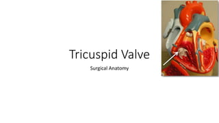
The document details the unique anatomical features of the tricuspid valve, including its anatomy, dynamic function during ventricular contraction, and variations in the number of leaflets and papillary muscles. It emphasizes the complex processes involved in optimal coaptation of the valve leaflets, particularly the importance of the annulus shape and papillary muscle interactions. Additionally, it discusses echocardiographic imaging techniques to assess tricuspid valve anatomy and function, highlighting the variability in individual anatomies and the challenges of surgical repair.


