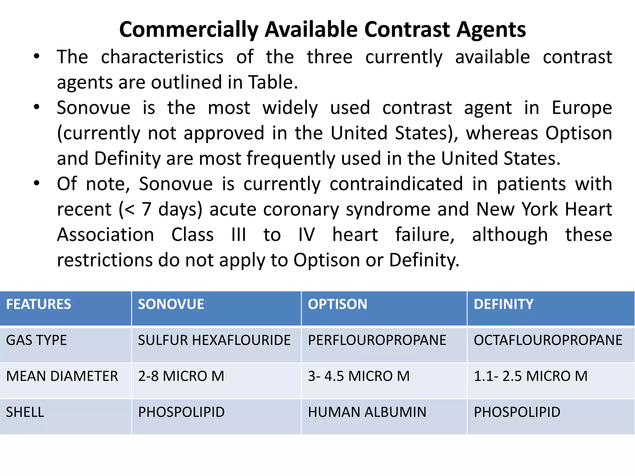
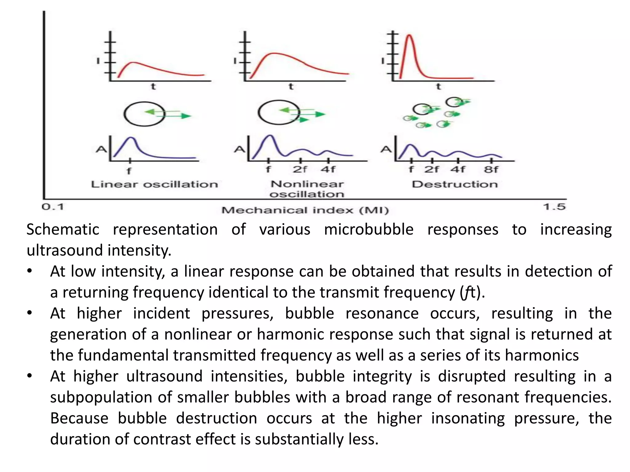
Our concepts of heart disease are based on the enormous reservoir of physiologic and anatomic knowledge derived from the past 70 years' of experience in the cardiac catheterization laboratory.
As Andre Cournand remarked in his Nobel lecture of December 11, 1956, the cardiac catheter was the key in the lock.
By turning this key, Cournand and his colleagues led us into a new era in the understanding of normal and disordered cardiac function in huma
Speckle tracking echocardiography (STE) is an echocardiographic imaging technique that analyzes the motion of tissues in the heart by using the naturally occurring speckle pattern in the myocardium or blood when imaged by ultrasound.
Acute scrotum is a general term referring to an emergency condition affecting the contents or the wall of the scrotum.
There are a number of conditions that present acutely, predominantly with pain and/or swelling
A careful and detailed history and examination, and in some cases, investigations allow differentiation between these diagnoses. A prompt diagnosis is essential as the patient may require urgent surgical intervention
Testicular torsion refers to twisting of the spermatic cord, causing ischaemia of the testicle.
Testicular torsion results from inadequate fixation of the testis to the tunica vaginalis producing ischemia from reduced arterial inflow and venous outflow obstruction.
The prevalence of testicular torsion in adult patients hospitalized with acute scrotal pain is approximately 25 to 50 percent
New Directions in Targeted Therapeutic Approaches for Older Adults With Mantl...i3 Health
i3 Health is pleased to make the speaker slides from this activity available for use as a non-accredited self-study or teaching resource.
This slide deck presented by Dr. Kami Maddocks, Professor-Clinical in the Division of Hematology and
Associate Division Director for Ambulatory Operations
The Ohio State University Comprehensive Cancer Center, will provide insight into new directions in targeted therapeutic approaches for older adults with mantle cell lymphoma.
STATEMENT OF NEED
Mantle cell lymphoma (MCL) is a rare, aggressive B-cell non-Hodgkin lymphoma (NHL) accounting for 5% to 7% of all lymphomas. Its prognosis ranges from indolent disease that does not require treatment for years to very aggressive disease, which is associated with poor survival (Silkenstedt et al, 2021). Typically, MCL is diagnosed at advanced stage and in older patients who cannot tolerate intensive therapy (NCCN, 2022). Although recent advances have slightly increased remission rates, recurrence and relapse remain very common, leading to a median overall survival between 3 and 6 years (LLS, 2021). Though there are several effective options, progress is still needed towards establishing an accepted frontline approach for MCL (Castellino et al, 2022). Treatment selection and management of MCL are complicated by the heterogeneity of prognosis, advanced age and comorbidities of patients, and lack of an established standard approach for treatment, making it vital that clinicians be familiar with the latest research and advances in this area. In this activity chaired by Michael Wang, MD, Professor in the Department of Lymphoma & Myeloma at MD Anderson Cancer Center, expert faculty will discuss prognostic factors informing treatment, the promising results of recent trials in new therapeutic approaches, and the implications of treatment resistance in therapeutic selection for MCL.
Target Audience
Hematology/oncology fellows, attending faculty, and other health care professionals involved in the treatment of patients with mantle cell lymphoma (MCL).
Learning Objectives
1.) Identify clinical and biological prognostic factors that can guide treatment decision making for older adults with MCL
2.) Evaluate emerging data on targeted therapeutic approaches for treatment-naive and relapsed/refractory MCL and their applicability to older adults
3.) Assess mechanisms of resistance to targeted therapies for MCL and their implications for treatment selection
HOT NEW PRODUCT! BIG SALES FAST SHIPPING NOW FROM CHINA!! EU KU DB BK substit...GL Anaacs
Contact us if you are interested:
Email / Skype : kefaya1771@gmail.com
Threema: PXHY5PDH
New BATCH Ku !!! MUCH IN DEMAND FAST SALE EVERY BATCH HAPPY GOOD EFFECT BIG BATCH !
Contact me on Threema or skype to start big business!!
Hot-sale products:
NEW HOT EUTYLONE WHITE CRYSTAL!!
5cl-adba precursor (semi finished )
5cl-adba raw materials
ADBB precursor (semi finished )
ADBB raw materials
APVP powder
5fadb/4f-adb
Jwh018 / Jwh210
Eutylone crystal
Protonitazene (hydrochloride) CAS: 119276-01-6
Flubrotizolam CAS: 57801-95-3
Metonitazene CAS: 14680-51-4
Payment terms: Western Union,MoneyGram,Bitcoin or USDT.
Deliver Time: Usually 7-15days
Shipping method: FedEx, TNT, DHL,UPS etc.Our deliveries are 100% safe, fast, reliable and discreet.
Samples will be sent for your evaluation!If you are interested in, please contact me, let's talk details.
We specializes in exporting high quality Research chemical, medical intermediate, Pharmaceutical chemicals and so on. Products are exported to USA, Canada, France, Korea, Japan,Russia, Southeast Asia and other countries.
Recomendações da OMS sobre cuidados maternos e neonatais para uma experiência pós-natal positiva.
Em consonância com os ODS – Objetivos do Desenvolvimento Sustentável e a Estratégia Global para a Saúde das Mulheres, Crianças e Adolescentes, e aplicando uma abordagem baseada nos direitos humanos, os esforços de cuidados pós-natais devem expandir-se para além da cobertura e da simples sobrevivência, de modo a incluir cuidados de qualidade.
Estas diretrizes visam melhorar a qualidade dos cuidados pós-natais essenciais e de rotina prestados às mulheres e aos recém-nascidos, com o objetivo final de melhorar a saúde e o bem-estar materno e neonatal.
Uma “experiência pós-natal positiva” é um resultado importante para todas as mulheres que dão à luz e para os seus recém-nascidos, estabelecendo as bases para a melhoria da saúde e do bem-estar a curto e longo prazo. Uma experiência pós-natal positiva é definida como aquela em que as mulheres, pessoas que gestam, os recém-nascidos, os casais, os pais, os cuidadores e as famílias recebem informação consistente, garantia e apoio de profissionais de saúde motivados; e onde um sistema de saúde flexível e com recursos reconheça as necessidades das mulheres e dos bebês e respeite o seu contexto cultural.
Estas diretrizes consolidadas apresentam algumas recomendações novas e já bem fundamentadas sobre cuidados pós-natais de rotina para mulheres e neonatos que recebem cuidados no pós-parto em unidades de saúde ou na comunidade, independentemente dos recursos disponíveis.
É fornecido um conjunto abrangente de recomendações para cuidados durante o período puerperal, com ênfase nos cuidados essenciais que todas as mulheres e recém-nascidos devem receber, e com a devida atenção à qualidade dos cuidados; isto é, a entrega e a experiência do cuidado recebido. Estas diretrizes atualizam e ampliam as recomendações da OMS de 2014 sobre cuidados pós-natais da mãe e do recém-nascido e complementam as atuais diretrizes da OMS sobre a gestão de complicações pós-natais.
O estabelecimento da amamentação e o manejo das principais intercorrências é contemplada.
Recomendamos muito.
Vamos discutir essas recomendações no nosso curso de pós-graduação em Aleitamento no Instituto Ciclos.
Esta publicação só está disponível em inglês até o momento.
Prof. Marcus Renato de Carvalho
www.agostodourado.com
Title: Sense of Taste
Presenter: Dr. Faiza, Assistant Professor of Physiology
Qualifications:
MBBS (Best Graduate, AIMC Lahore)
FCPS Physiology
ICMT, CHPE, DHPE (STMU)
MPH (GC University, Faisalabad)
MBA (Virtual University of Pakistan)
Learning Objectives:
Describe the structure and function of taste buds.
Describe the relationship between the taste threshold and taste index of common substances.
Explain the chemical basis and signal transduction of taste perception for each type of primary taste sensation.
Recognize different abnormalities of taste perception and their causes.
Key Topics:
Significance of Taste Sensation:
Differentiation between pleasant and harmful food
Influence on behavior
Selection of food based on metabolic needs
Receptors of Taste:
Taste buds on the tongue
Influence of sense of smell, texture of food, and pain stimulation (e.g., by pepper)
Primary and Secondary Taste Sensations:
Primary taste sensations: Sweet, Sour, Salty, Bitter, Umami
Chemical basis and signal transduction mechanisms for each taste
Taste Threshold and Index:
Taste threshold values for Sweet (sucrose), Salty (NaCl), Sour (HCl), and Bitter (Quinine)
Taste index relationship: Inversely proportional to taste threshold
Taste Blindness:
Inability to taste certain substances, particularly thiourea compounds
Example: Phenylthiocarbamide
Structure and Function of Taste Buds:
Composition: Epithelial cells, Sustentacular/Supporting cells, Taste cells, Basal cells
Features: Taste pores, Taste hairs/microvilli, and Taste nerve fibers
Location of Taste Buds:
Found in papillae of the tongue (Fungiform, Circumvallate, Foliate)
Also present on the palate, tonsillar pillars, epiglottis, and proximal esophagus
Mechanism of Taste Stimulation:
Interaction of taste substances with receptors on microvilli
Signal transduction pathways for Umami, Sweet, Bitter, Sour, and Salty tastes
Taste Sensitivity and Adaptation:
Decrease in sensitivity with age
Rapid adaptation of taste sensation
Role of Saliva in Taste:
Dissolution of tastants to reach receptors
Washing away the stimulus
Taste Preferences and Aversions:
Mechanisms behind taste preference and aversion
Influence of receptors and neural pathways
Impact of Sensory Nerve Damage:
Degeneration of taste buds if the sensory nerve fiber is cut
Abnormalities of Taste Detection:
Conditions: Ageusia, Hypogeusia, Dysgeusia (parageusia)
Causes: Nerve damage, neurological disorders, infections, poor oral hygiene, adverse drug effects, deficiencies, aging, tobacco use, altered neurotransmitter levels
Neurotransmitters and Taste Threshold:
Effects of serotonin (5-HT) and norepinephrine (NE) on taste sensitivity
Supertasters:
25% of the population with heightened sensitivity to taste, especially bitterness
Increased number of fungiform papillae
Title: Sense of Smell
Presenter: Dr. Faiza, Assistant Professor of Physiology
Qualifications:
MBBS (Best Graduate, AIMC Lahore)
FCPS Physiology
ICMT, CHPE, DHPE (STMU)
MPH (GC University, Faisalabad)
MBA (Virtual University of Pakistan)
Learning Objectives:
Describe the primary categories of smells and the concept of odor blindness.
Explain the structure and location of the olfactory membrane and mucosa, including the types and roles of cells involved in olfaction.
Describe the pathway and mechanisms of olfactory signal transmission from the olfactory receptors to the brain.
Illustrate the biochemical cascade triggered by odorant binding to olfactory receptors, including the role of G-proteins and second messengers in generating an action potential.
Identify different types of olfactory disorders such as anosmia, hyposmia, hyperosmia, and dysosmia, including their potential causes.
Key Topics:
Olfactory Genes:
3% of the human genome accounts for olfactory genes.
400 genes for odorant receptors.
Olfactory Membrane:
Located in the superior part of the nasal cavity.
Medially: Folds downward along the superior septum.
Laterally: Folds over the superior turbinate and upper surface of the middle turbinate.
Total surface area: 5-10 square centimeters.
Olfactory Mucosa:
Olfactory Cells: Bipolar nerve cells derived from the CNS (100 million), with 4-25 olfactory cilia per cell.
Sustentacular Cells: Produce mucus and maintain ionic and molecular environment.
Basal Cells: Replace worn-out olfactory cells with an average lifespan of 1-2 months.
Bowman’s Gland: Secretes mucus.
Stimulation of Olfactory Cells:
Odorant dissolves in mucus and attaches to receptors on olfactory cilia.
Involves a cascade effect through G-proteins and second messengers, leading to depolarization and action potential generation in the olfactory nerve.
Quality of a Good Odorant:
Small (3-20 Carbon atoms), volatile, water-soluble, and lipid-soluble.
Facilitated by odorant-binding proteins in mucus.
Membrane Potential and Action Potential:
Resting membrane potential: -55mV.
Action potential frequency in the olfactory nerve increases with odorant strength.
Adaptation Towards the Sense of Smell:
Rapid adaptation within the first second, with further slow adaptation.
Psychological adaptation greater than receptor adaptation, involving feedback inhibition from the central nervous system.
Primary Sensations of Smell:
Camphoraceous, Musky, Floral, Pepperminty, Ethereal, Pungent, Putrid.
Odor Detection Threshold:
Examples: Hydrogen sulfide (0.0005 ppm), Methyl-mercaptan (0.002 ppm).
Some toxic substances are odorless at lethal concentrations.
Characteristics of Smell:
Odor blindness for single substances due to lack of appropriate receptor protein.
Behavioral and emotional influences of smell.
Transmission of Olfactory Signals:
From olfactory cells to glomeruli in the olfactory bulb, involving lateral inhibition.
Primitive, less old, and new olfactory systems with different path
Ozempic: Preoperative Management of Patients on GLP-1 Receptor Agonists Saeid Safari
Preoperative Management of Patients on GLP-1 Receptor Agonists like Ozempic and Semiglutide
ASA GUIDELINE
NYSORA Guideline
2 Case Reports of Gastric Ultrasound
- Video recording of this lecture in English language: https://youtu.be/lK81BzxMqdo
- Video recording of this lecture in Arabic language: https://youtu.be/Ve4P0COk9OI
- Link to download the book free: https://nephrotube.blogspot.com/p/nephrotube-nephrology-books.html
- Link to NephroTube website: www.NephroTube.com
- Link to NephroTube social media accounts: https://nephrotube.blogspot.com/p/join-nephrotube-on-social-media.html
TEST BANK for Operations Management, 14th Edition by William J. Stevenson, Ve...kevinkariuki227
TEST BANK for Operations Management, 14th Edition by William J. Stevenson, Verified Chapters 1 - 19, Complete Newest Version.pdf
Lung Cancer: Artificial Intelligence, Synergetics, Complex System Analysis, S...Oleg Kshivets
RESULTS: Overall life span (LS) was 2252.1±1742.5 days and cumulative 5-year survival (5YS) reached 73.2%, 10 years – 64.8%, 20 years – 42.5%. 513 LCP lived more than 5 years (LS=3124.6±1525.6 days), 148 LCP – more than 10 years (LS=5054.4±1504.1 days).199 LCP died because of LC (LS=562.7±374.5 days). 5YS of LCP after bi/lobectomies was significantly superior in comparison with LCP after pneumonectomies (78.1% vs.63.7%, P=0.00001 by log-rank test). AT significantly improved 5YS (66.3% vs. 34.8%) (P=0.00000 by log-rank test) only for LCP with N1-2. Cox modeling displayed that 5YS of LCP significantly depended on: phase transition (PT) early-invasive LC in terms of synergetics, PT N0—N12, cell ratio factors (ratio between cancer cells- CC and blood cells subpopulations), G1-3, histology, glucose, AT, blood cell circuit, prothrombin index, heparin tolerance, recalcification time (P=0.000-0.038). Neural networks, genetic algorithm selection and bootstrap simulation revealed relationships between 5YS and PT early-invasive LC (rank=1), PT N0—N12 (rank=2), thrombocytes/CC (3), erythrocytes/CC (4), eosinophils/CC (5), healthy cells/CC (6), lymphocytes/CC (7), segmented neutrophils/CC (8), stick neutrophils/CC (9), monocytes/CC (10); leucocytes/CC (11). Correct prediction of 5YS was 100% by neural networks computing (area under ROC curve=1.0; error=0.0).
CONCLUSIONS: 5YS of LCP after radical procedures significantly depended on: 1) PT early-invasive cancer; 2) PT N0--N12; 3) cell ratio factors; 4) blood cell circuit; 5) biochemical factors; 6) hemostasis system; 7) AT; 8) LC characteristics; 9) LC cell dynamics; 10) surgery type: lobectomy/pneumonectomy; 11) anthropometric data. Optimal diagnosis and treatment strategies for LC are: 1) screening and early detection of LC; 2) availability of experienced thoracic surgeons because of complexity of radical procedures; 3) aggressive en block surgery and adequate lymph node dissection for completeness; 4) precise prediction; 5) adjuvant chemoimmunoradiotherapy for LCP with unfavorable prognosis.
The prostate is an exocrine gland of the male mammalian reproductive system
It is a walnut-sized gland that forms part of the male reproductive system and is located in front of the rectum and just below the urinary bladder
Function is to store and secrete a clear, slightly alkaline fluid that constitutes 10-30% of the volume of the seminal fluid that along with the spermatozoa, constitutes semen
A healthy human prostate measures (4cm-vertical, by 3cm-horizontal, 2cm ant-post ).
It surrounds the urethra just below the urinary bladder. It has anterior, median, posterior and two lateral lobes
It’s work is regulated by androgens which are responsible for male sex characteristics
Generalised disease of the prostate due to hormonal derangement which leads to non malignant enlargement of the gland (increase in the number of epithelial cells and stromal tissue)to cause compression of the urethra leading to symptoms (LUTS
Tom Selleck Health: A Comprehensive Look at the Iconic Actor’s Wellness Journeygreendigital
Tom Selleck, an enduring figure in Hollywood. has captivated audiences for decades with his rugged charm, iconic moustache. and memorable roles in television and film. From his breakout role as Thomas Magnum in Magnum P.I. to his current portrayal of Frank Reagan in Blue Bloods. Selleck's career has spanned over 50 years. But beyond his professional achievements. fans have often been curious about Tom Selleck Health. especially as he has aged in the public eye.
Follow us on: Pinterest
Introduction
Many have been interested in Tom Selleck health. not only because of his enduring presence on screen but also because of the challenges. and lifestyle choices he has faced and made over the years. This article delves into the various aspects of Tom Selleck health. exploring his fitness regimen, diet, mental health. and the challenges he has encountered as he ages. We'll look at how he maintains his well-being. the health issues he has faced, and his approach to ageing .
Early Life and Career
Childhood and Athletic Beginnings
Tom Selleck was born on January 29, 1945, in Detroit, Michigan, and grew up in Sherman Oaks, California. From an early age, he was involved in sports, particularly basketball. which played a significant role in his physical development. His athletic pursuits continued into college. where he attended the University of Southern California (USC) on a basketball scholarship. This early involvement in sports laid a strong foundation for his physical health and disciplined lifestyle.
Transition to Acting
Selleck's transition from an athlete to an actor came with its physical demands. His first significant role in "Magnum P.I." required him to perform various stunts and maintain a fit appearance. This role, which he played from 1980 to 1988. necessitated a rigorous fitness routine to meet the show's demands. setting the stage for his long-term commitment to health and wellness.
Fitness Regimen
Workout Routine
Tom Selleck health and fitness regimen has evolved. adapting to his changing roles and age. During his "Magnum, P.I." days. Selleck's workouts were intense and focused on building and maintaining muscle mass. His routine included weightlifting, cardiovascular exercises. and specific training for the stunts he performed on the show.
Selleck adjusted his fitness routine as he aged to suit his body's needs. Today, his workouts focus on maintaining flexibility, strength, and cardiovascular health. He incorporates low-impact exercises such as swimming, walking, and light weightlifting. This balanced approach helps him stay fit without putting undue strain on his joints and muscles.
Importance of Flexibility and Mobility
In recent years, Selleck has emphasized the importance of flexibility and mobility in his fitness regimen. Understanding the natural decline in muscle mass and joint flexibility with age. he includes stretching and yoga in his routine. These practices help prevent injuries, improve posture, and maintain mobilit
2. INTRODUCTION
Contrast echocardiography is a widely used, well tolerated,
non invasive technique employing ultrasound contrast
agents in order to improve image quality.
Contrast echocardiography exploits the ultrasound
properties of micro bubbles, acoustically active gas filled
microspheres, which remains within the intravascular
space and allow the simultaneous assessment of global and
regional myocardial structure, function and perfusion.
3. Red blood cells are poor reflectors of ultrasound during
conventional echocardiography because of small difference
in acoustic impedance between them and surrounding
plasma. Only with aggregation of RBC’s, as seen in low flow
rates do they become echogenic.
Ultrasound contrast agents increase the ultrasound
backscatter intensity even with normal blood flow.
These micro bubble contrast agents behave similar to RBC’s.
When injected intravenously, they remain entirely within the
vascular compartment.
They do not cross the endothelium into the cell intestitium
and should not be confused with iodine based contrast used
in CT or angiography or the gadolinium based contrast agents
used in CMR imaging.
4. Manufacture UCAs consists of micro bubbles
encapsulating an acoustically active, high
molecular gas within an outer shell.
They have distinct ultrasound characteristics
compared with surrounding tissue and enhance
the ultrasound signal, allowing clearer LV
endocardial border detection, improved structural
and functional assessment of LV and assessment
of myocardial perfusion.
5. The first generation of UCAs consists of an
outer shell of denatured albumin
encapsulating air.
Although safe, these micro bubbles were
relatively large by today’s standards, their
passage through the pulmonary capillaries was
inconsistent and the air within the micro
bubble dissipated quickly due to high
solubility, resulting in rapid deterioration of LV
border enhancement
Thus these agents had limited clinical use.
6. • Second generation UCAs consists of a thicker
outer shell of albumin or phospholipids and an
inert gas.(nitrogen or fluorocarbon)
• These micro bubbles are smaller in size to
RBC and range from 1.1 mm to 1.8 mm in
diameter.
• They are small enough to pass through the
smallest blood vessels of the body, the
pulmonary capillaries, in order to reach the
left side of the heart.
• The shells are flexible in order to facilitate
transit across the pulmonary capillary beds.
7. The phospholipids shells are also negatively
charged, which prevent them from aggregating
and occluding the microvasculature.
All the commercially available contrast agents
contain a fluorocarbon gas of high density and
low solubility compared with air which diffuses
out of the slowly, thereby prolonging their
duration of action.
These gases are biologically inert and are
exhaled via their lungs, whereas the shells
undergo hepatic metabolism.
8. The structure of second generation ultrasound contrast agents.
The outer shell consists of albumin or phospholipids and
contains an inner fluorocarbon gas of high density and low
solubility.
9. FEATURES SONOVUE OPTISON DEFINITY
GAS TYPE SULFUR HEXAFLOURIDE PERFLOUROPROPANE OCTAFLOUROPROPANE
MEAN DIAMETER 2-8 MICRO M 3- 4.5 MICRO M 1.1- 2.5 MICRO M
SHELL PHOSPOLIPID HUMAN ALBUMIN PHOSPOLIPID
Commercially Available Contrast Agents
• The characteristics of the three currently available contrast
agents are outlined in Table.
• Sonovue is the most widely used contrast agent in Europe
(currently not approved in the United States), whereas Optison
and Definity are most frequently used in the United States.
• Of note, Sonovue is currently contraindicated in patients with
recent (< 7 days) acute coronary syndrome and New York Heart
Association Class III to IV heart failure, although these
restrictions do not apply to Optison or Definity.
12. Two-syringes and one three-way stopcock apparatus are used for
preparation of agitated saline contrast for intravenous injection.
The total volume in the syringe on the left is approximately 10 mL, which
consists initially of 9.5 mL of saline and 0.5 mL of air.
The contrast is prepared by forcefully injecting the solution from one
syringe to the other through the three-way stopcock.
Turbulence within the stopcock results in the creation of a large number of
microbubbles that are suitable for intravenous injection.
For opacification of right heart structures, a typical intravenous “dose” of
contrast prepared in this manner ranges from 1.0 to 5.0 mL.
13. CONTRAST ADMINISTRATION
UCAs are administered intravenously either by a bolus injection
or continuous infusion..
Bolus Injection
• A slow bolus injection of approximately 0.2 mL of contrast is
followed by a 2-mL saline flush over 10 seconds.
• This results in a rapid increase in signal intensity until a
plateau is reached and there is then a gradual decay in
contrast concentration, the rate of which varies in each
patient.
• Bolus injections are acceptable for studies in which the aim
is to improve endocardial border delineation (EBD), perform
left ventricular opacification (LVO) or enhance Doppler
signals.
• It is best avoided during perfusion studies; however, a
continuous infusion is preferable to ensure a steady-state
concentration of contrast with in the myocardial capillaries.
14. Continuous Infusion
• An infusion provides a steady-state concentration of
microbubbles and reduces the likelihood of artifacts.
• The contrast effect during the course of the study is
consistent and the study is more reproducible.
• Because there is a steady state of contrast concentration, it
allows the assessment of myocardial perfusion both
qualitatively and by quantification of myocardial blood flow
(MBF) and coronary flow reserve (CFR).
•
• Continuous infusions can also be used for EBD or LVO
purposes but are mandatory for perfusion imaging.
16. ULTRASOUND INTERACTION WITH CONTRAST AGENTS
Microbubbles interact with the ultrasound beam in a variety of ways :
• direct reflection at the fundamental transmitted frequency
• resonance with creation of reflected harmonic frequencies.
The frequency at which a bubble has maximal reflectance is
related to bubble diameter.
For any ultrasound frequency, the amplitude of reflection from a
microbubble decreases as the bubble diameter decreases.
All bubbles have a diameter at which reflectance is maximal (the
resonant diameter). Below the resonant diameter of the bubble,
the amplitude of reflection again diminishes with the cube of the
diameter.
It is a fortuitous occurrence that bubbles having a diameter that
allows transpulmonary passage have excellent reflectance when
interacting with clinically relevant transmission frequencies.
17. Interaction of microbubbles with the ultrasound beam has
three phases.
Fundamental frequency
Harmonic frequency
Stimulated acoustic emission.
FUNDAMENTAL FREQUENCY:
• In its simplest form, ultrasound interacts with a
microbubble by pure reflection of the ultrasound beam
at its fundamental (i.e., transmitted) frequency.
• Maximal reflection from the microbubble is dependent
on the relationship of the frequency and diameter.
18. HARMONIC FREQUENCY:
• At higher ultrasound imaging intensities (typically
≥0.3 MI), microbubbles are not pure reflectors but
begin to resonate.
• A resonating bubble will reflect ultrasound not only
at the fundamental insonating frequency (ft) but also
at harmonics of that frequency.
• In this instance, a microbubble insonated with a 2-
MHz interrogating beam will reflect back the 2-MHz
fundamental frequency but also resonate, creating
reflected frequencies at 4, 8 and 16 MHz. Each of
these subsequent harmonic frequencies doubles in
frequency and diminishes in amplitude.
19. • In routine clinical practice, only the first harmonic (i.e.,
twice the fundamental frequency) is typically used for
anatomic imaging.
• Contrast-specific imaging often relies on either multiple
harmonic frequencies or subharmonics of the first
harmonic (i.e., four and eight times the fundamental
frequency). This provides a more contrast-specific
signal.
20. STIMULATED ACOUSTIC EMISSION:
• At increasing ultrasound energy levels, the bubbles are physically
destroyed by the insonating beam.
• The process of destruction results in the creation of
subpopulations of bubbles of variable diameters.
• The highly variable diameter subpopulations result in a broad
range of reflected frequencies.
• By this destructive bubble technique, a large amount of acoustic
energy is generated both as reflected ultrasound and as multiple
detectable Doppler shifts.
• This final phenomenon in which microbubbles are destroyed,
thereby creating detectable ultrasound targets, is referred to as
stimulated acoustic emission.
• This phenomenon can be maximized by the use of a microbubble
with a fragile shell and containing nitrogen, or another rapidly
diffusing gas, resulting in a rapid loss of the contrast effect after
shell disruption.
21. Schematic representation of various microbubble responses to increasing
ultrasound intensity.
• At low intensity, a linear response can be obtained that results in detection of
a returning frequency identical to the transmit frequency (ft).
• At higher incident pressures, bubble resonance occurs, resulting in the
generation of a nonlinear or harmonic response such that signal is returned at
the fundamental transmitted frequency as well as a series of its harmonics
• At higher ultrasound intensities, bubble integrity is disrupted resulting in a
subpopulation of smaller bubbles with a broad range of resonant frequencies.
Because bubble destruction occurs at the higher insonating pressure, the
duration of contrast effect is substantially less.
22. Detection Methods
Machine Settings
• All current manufacturers provide dedicated contrast-specific
presets to account for sensitivity of ultrasound contrast agents in
a high-intensity field.
• Many of them have, as optional addons, contrast-specific
modalities suitable for detection of low intensity contrast in the
myocardium.
• The users should be aware of the specific nature of the contrast
presets, which are proprietary and vary from manufacturer to
manufacturer and from platform to platform.
23. Routine B-mode scanning
• The simplest method for contrast detection is routine B-mode
ultrasound.
• Microbubbles are intense reflectors of ultrasound and the
amount of reflected energy is substantially greater than that of
the surrounding tissue or blood.
• Because of this, routine B-mode scanning is highly sensitive for
the detection of isolated microbubble targets.
• This routine imaging technology is sufficient for detection of
intracardiac shunts such as atrial septal defect using agitated
saline.
• When used with newer perfluorocarbon-based agents,
detection is markedly facilitated by the use of harmonic and
other advanced imaging algorithms
24. Intermittent Imaging
• It was recognized in the mid-1990s that the routine interrogating
ultrasound beam destroyed ultrasound targets.
• This was a fortuitous observation made when investigators
recognized the absence of contrast effect in the left ventricular
cavity or myocardium during continuous imaging. After brief
interruption of scanning, contrast was again detectable without
reinjection of the agent.
• This led to the technique of intermittent imaging in which
ultrasound interrogation is triggered to the electrocardiogram.
• In between triggered imaging, no ultrasound energy is delivered,
allowing time for restitution of the contrast effect and its
subsequent detection when imaging is resumed. Obviously, with
intermittent imaging, the ability to analyze wall motion is lost,
and this imaging technique is typically used for evaluation of
myocardial perfusion.
25. Low Mechanical Index Imaging
• Mechanical index (MI) is a measure of the power of an ultrasound
beam and is defined as peak negative acoustic pressure/ft, where
ft is the transmitted frequency.
• The mechanical index is a unitless number directly proportional to
the power of the ultrasound beam being delivered.
• Typically, structural imaging without contrast enhancement is
undertaken at a mechanical index of 0.9 to 1.5.
• This degree of ultrasound delivery disrupts microbubbles and
reduces the ability to use them clinically.
26. • As such, a mechanical index of ≥0.3 is typically employed for
optimal detection of ultrasound within the left ventricular cavity
or myocardium.
• By imaging at a low mechanical index, contrast within the left
ventricular cavity is not destroyed, and because imaging is
continuous rather than intermittent, wall motion analysis can be
undertaken in real time with boundaries enhanced by the
opacified left ventricular blood pool.
• Low mechanical index imaging is also necessary when detecting
very low concentrations of ultrasound contrast such as for
myocardial perfusion.
• For myocardial perfusion imaging, intermittent high mechanical
index imaging is often undertaken to purposefully destroy
contrast in the blood pool to create a repeated bolus effect from
which time appearance curves can be created.
27. CONTRAST ARTIFACTS
Contrast artifacts can be divided into two broad categories:
• those due to the agent and its interaction with the ultrasound
beam.
• physiologic artifacts, both of which may interfere with
interpretation.
These include:
Attenuation
Shadowing
Apical destruction
28. ATTENUATION:
• As contrast agents are very potent reflectors of ultrasound, their
presence in high concentration results in nearly complete
attenuation of ultrasound penetration. This phenomenon is
particularly prominent when using the newer, more highly
reflective perfluorocarbon-based agents.
• Attenuation occurs when there is an abnormally high
concentration of ultrasound targets in the near field, beyond
which the ultrasound beam cannot penetrate (Figs.). This results
in detection only of the initial layer of contrast-enhanced blood,
with all areas of the heart behind this area being shadowed.
29.
30.
31. • Attenuation is common during bolus injections of
perfluorocarbon-based agents. It can be avoided by delaying
scanning until later in the infusion protocol, after the peak
contrast effect has declined, or preferably by the use of a smaller
bolus or lower concentration of the ultrasound agent.
• Clinically, the attenuation phenomenon is most problematic when
imaging the basal lateral wall in an apical four-chamber view. This
region is often an area of contrast dropout which should not be
confused for the ventricular boundary, either for wall motion
analysis or for volumetric determination. Similarly, this area of
greatest attenuation can be remarkably problematic for assessing
myocardial perfusion.
32. • The amount of microbubble destruction is directly related to the
intensity of the insonating ultrasound beam. Although the
microbubbles generated by agitated saline are resistant to the
destruction of the ultrasound beam, the newer generation of
agents are highly sensitive to ultrasound disruption.
• At a mechanical index used for typical anatomic imaging (0.9-
1.4), microbubbles will be rapidly destroyed in the blood pool,
resulting in a dramatic reduction in the ultrasound contrast.
• By reducing the transmit intensity to a mechanical index (<0.3),
this phenomenon is reduced and the ultrasound contrast effect
is preserved (Fig. ).
• Inadvertent imaging at an inappropriately high mechanical index
results in the destruction of contrast, predominantly in the near
field, and the appearance of a contrast defect in that region.
33. Apical four-chamber view recorded in a patient demonstrating the impact of mechanical
index on contrast appearance. A: Image was recorded with a mechanical index of 0.3 and
reveals smooth opacification of all four cardiac chambers. B: Image was recorded 10
seconds later with a mechanical index of 1.0. Note the complete lack of contrast in the
near field and the swirling nature of the partial filling in the far field.
34. SHADOWING
• Another well-recognized artifact is that created by shadowing
from a papillary muscle when imaging in the four-chamber view.
• The shadow created at the proximal boundary of the contrast with
the papillary muscle extends toward the left atrium in a straight
line. This shadow can be confused with the lateral endocardial
border (Fig.).
35. Apical four-chamber view demonstrates a papillary muscle shadow. A: Image was recorded in
diastole. Note the location of the papillary muscle (black arrows) and the faint shadow behind
it. Also note the true location and thickness of the lateral wall (white arrows). B: Image was
recorded in systole and demonstrates a more exaggerated papillary muscle shadow. Mistaking
the papillary muscle shadow for the lateral wall will result in dramatic underestimation of the
size of the left ventricle. PAP, papillary muscle.
36. Apical four-chamber view recorded after intravenous injection of a perfluorocarbon-based
contrast agent in a patient with an apical aneurysm and focal calcification in the apex. Note
the two distinct shadows arising from the apex in the otherwise smooth homogeneous
filling of the left ventricular cavity. The dotted lines represent the true cavity boundary.
APICAL DESTRUCTION:
If a patient has areas of dense fibrosis or calcification between the transducer and the
blood pool, a shadow will occur behind the echo reflective focal area mimicking a
contrast-free area.
37. Hemodynamic artifacts include
- competitive flow
- marginated flow.
• Because contrast is contained within the bloodstream, its
appearance will parallel that of the blood flow.
• If there is competing flow from another vessel that is not contrast
enhanced, a negative contrast effect will occur.
• This is often seen after intravenous injection of saline contrast for
evaluating an atrial septal defect.
38. • In this instance, superior vena caval flow (assuming an
arm injection) enters the right atrium as a bolus that
merges with the non-contrast enhanced flow from the
inferior vena cava.
• This creates a swirling matrix of contrast and
nonenhanced blood, which is often maximal along the
interatrial septum.
• This effect may be accentuated in a high-flow state in
which there is greater than usual inferior vena caval
flow such as is seen in chronic hepatic disease or
pregnancy.
• On occasion, this effect has been confused with a
pathologic shunt at the atrial level.
39. Apical four-chamber view recorded in a patient after injection of agitated saline into an
upper extremity vein. Note the area of absent contrast effect (large arrow) along the most
superior portion of the atrial septum, which is due to competitive flow from non-contrast-
enhanced inferior vena caval blood flow. Such an area of absent contrast could be confused
with a true negative contrast effect due to an atrial septal defect. This position of the atrial
septum is noted by the smaller arrow.
40. A similar Phenomenon occurs when a prominent eustachian valve marginates superior
vena caval flow in the atrium and may either mimic or mask the presence of an atrial
shunt.
42. CLINICAL APPLICATIONS FOR AGITATED SALINE CONTRAST
SHUNT DETECTION —
• The first clinical use of contrast echocardiography was for
detection of right-to-left shunts.
• Agitated saline is well-suited for this purpose because
microbubbles of air formed from agitating saline persist long
enough to opacify the right heart chambers and diffuse into the
lungs when traveling through the pulmonary circulation.
• Therefore, microbubbles will not gain access to the left heart
chambers unless a right-to-left intracardiac or extracardiac shunt
is present.
• This technique is used most often for the detection of atrial septal
defects, although it can also be used to detect ventricular septal
defects and arteriovenous shunts in the pulmonary vasculature.
43. • The appearance of bubbles in the left heart early (within three to
five beats) after right chamber opacification suggests an
intracardiac shunt.
• Later appearance of bubbles in the left heart (5-15 cycles)
suggests pulmonary arteriovenous shunting.
• Microbubble contrast agents such as Optison, Definity, and
Lumason that traverse the pulmonary vasculature are NOT
designed for shunt detection.
44. Apical four-chamber view recorded in a patient with an atrial septal defect after
intravenous injection of contrast agent.
There is opacification of the right atrium and the right ventricle and the significant
amount of contrast appearing in the left atrium, consistent with a right-to left shunt at
the atrium level, subsequently confirmed to be a secundum atrial defect
45. • Transesophageal echocardiogram recorded in a longitudinal view concentrating on the
atrial septum. Agitated saline has been injected into an upper extremity vein and has
completely filled the right atrium.
• There is small negative contrast effect (arrow) arising from the atrial septum and
projecting into the contrast-enhanced right atrium.
• This effect occurs due to flow of noncontrast- enhanced blood from the left atrium
through a small (4 mm) secundum atrial septal defect into the contrast-filled right
atrium.
46. DOPPLER SIGNAL ENHANCEMENT —
Agitated saline (and other ultrasound contrast agents) may be used
to enhance tricuspid Doppler signals for use in assessment of
transvalvular velocity to estimate right ventricular systolic pressure
• Because the ultrasound contrast agent interacts with all forms of
Doppler imaging, caution should be exercised when color flow
imaging is employed.
• The addition of even very low concentrations of ultrasound
contrast to the blood pool results in a substantially greater color
flow area than would be recorded without contrast.
• Because the color flow jet area is used to estimate regurgitation
severity, the increase in jet area caused by interaction with
contrast will result in systematic overestimation of regurgitation
severity.
• As such, Contrast agents should not be used in conjunction with
color Doppler in clinical practice.
47. Contrast enhancement of a faint tricuspid regurgitation jet by agitated saline injected into an
upper extremity vein.
A: In the spectral images, there is faint tricuspid regurgitation signal from which it is not
possible to ascertain the complete spectral profile or maximal velocity.
B: The spectral profiles were recorded after enhancement of the jet with agitated saline.
There is the substantially more robust signal and the ability to identify the maximal velocity.
48. Apical four-chamber view recorded in a
patient with mild tricuspid regurgitation
before and after injection of a
perfluorocarbon-based contrast agent.
A:The relatively disorganized tricuspid
regurgitation jet consistent with mild
regurgitation.
B: The dramatic increase in the size and
intensity of the color flow signal jet when
intracavitary contrast is present.
49. DIAGNOSIS OF PERSISTENT LEFT SUPERIOR VENA CAVA —
• This venous anomaly is usually detected incidentally.
• A persistent left SVC is suspected when a dilated coronary sinus is
detected in the absence of a cause for elevated right atrial
pressure.
• A persistent left superior vena cava drains directly into the
coronary sinus leading to a characteristic sequence of contrast
appearance: following injection of contrast into a left arm vein,
contrast appears in the coronary sinus before appearing in the
right atrium.
50. • Associated cardiovascular anomalies are present in a minority
of patients with persistent left SVC.
• Associated venous anomalies include absence of the
innominate vein and more rarely absence of the right superior
vena cava or drainage of the left SVC into the left atrium.
• The presence of a left SVC may complicate transvenous
placement of pulmonary artery (Swan-Ganz) catheters,
pacemaker or implantable cardioverter-defibrillator leads, as
well as retrograde cardioplegia
51.
52. CLINICAL APPLICATIONS FOR MICROBUBBLE CONTRAST AGENTS
ENDOCARDIAL BORDER DEFINITION —
The second generation microbubble contrast agents (Definity,
Optison, and Lumason, which are available in most countries)
traverse the pulmonary vasculature and are indicated for LV
opacification and LV endocardial border definition in patients with
technically suboptimal echocardiograms.
In the 2011 ACCF/ASE/AHA Appropriate Use Criteria for
Echocardiography, the use of contrast is considered appropriate
when >2 contiguous LV segments are not seen on noncontrast
images.
53. left ventricular opacification after intravenous injection of a perfluorocarbon based contrast
agent.
Top left: A baseline apical four-chamber view. Note the poor visualization of the apex and
lateral wall. The other three panels were recorded after intravenous injection of a
perfluorocarbon-based contrast agent. Note the excellent delineation of the left ventricular
cavity and the ability to fully identify the apex and lateral walls.
54. REST ECHOCARDIOGRAPHY —
Suboptimal images limit the interpretation in approximately 5 to 20
percent of echocardiographic studies, thereby impairing the
assessment of segmental and global LV systolic function.
Contrast opacification of the LV cavity enhances border detection,
decreasing the variability in the interpretation of regional wall motion
abnormalities, LV volumes and remodeling, and the ejection fraction
(EF).
Although Albunex was the first agent to produce LV opacification from
an intravenous injection, it produced LV opacification only in 64 to 81
percent of the time.
Second generation agents have been more successful, achieving LV
opacification in 90 percent of cases in which baseline images are
suboptimal.
55. 2008 American Society of Echocardiography (ASE) guidelines :
Difficult to image patients presenting for rest echocardiography with
reduced image quality
To enable improved endocardial visualization and assessment of
LV structure and function when ≥2 contiguous segments are not
seen on non-contrast images
To reduce variability and increase accuracy in LV volume and LVEF
measurements by 2D echocardiography
To increase the confidence of the interpreting clinician in LV
functional, structure, and volume assessments.
56. In all patients presenting for rest echocardiographic assessment of LV
systolic function (not solely difficult to image patients)
• To reduce variability in LV volume measurements through 2D
echocardiography
To confirm or exclude the echocardiographic diagnosis of the
following LV structural abnormalities, when nonenhanced images are
suboptimal for definitive diagnosis:
• Apical variant of hypertrophic cardiomyopathy
• LV noncompaction
• Apical thrombus
• Complications of myocardial infarction, such as LV aneurysm,
pseudoaneurysm, and myocardial rupture
57. Apical view recorded in a patient with a vague echo density on noncontrast
imaging. After intravenous injection of a perfluorocarbon-based agent, a distinct
spherical filling defect is noted in the apex, consistent with a pedunculated apical
thrombus (arrows).
58. Apical four-chamber view recorded in a patient with an apical variant of hypertrophic
cardiomyopathy.
A: Recorded with standard B-mode imaging, from which pathologic thickening of the apex is
not appreciable. After injection of contrast for left ventricle opacification
(B), the pathologic thickness of the apical left ventricular walls (arrows) can be appreciated.
59. Apical four-chamber view recorded in a patient with ventricular noncompaction.
A: without contrast, note the irregular thickening of the apex and lateral wall. After
injection of a contrast agent for left ventricular opacification
(B), note the contrast in the multiple sinusoids in the apex and lateral wall
(arrows).
60. A: Apical four-chamber view recorded in a patient with a dilated cardiomyopathy and a vague
echo density in the left ventricular apex (arrows) noted on a non-contrast-enhanced image.
Note the position of the anatomic apex (downward-pointing arrow).
B: Image was recorded after injection of a perfluorocarbon-based contrast agent and
demonstrates complete opacification of the left ventricular cavity. Note that, with contrast,
the entire left ventricular cavity is filled, confirming that the vague echo density in the apex
was not a true mural thrombus.
61. Off-axis apical view recorded in
a patient with a small apical
pseudoaneurysm.
A: Note the nearly spherical
echo-free space at the left
ventricular apex. Contrast has
already opacified the body of
the left and right ventricles.
B: Frame recorded one cardiac
cycle later. Note the
appearance of a small amount
of contrast (arrow) within the
cavity, confirming its
communication with the left
ventricular cavity.
62. Myocardial Perfusion Imaging
• Because myocardial ischemia and infarction affect
both myocardial perfusion and contractility, it would
be ideal to assess both these features simultaneously
.
• Myocardial contrast perfusion echocardiography
provides that capability.
• If the amount of microbubbles in the myocardium is
sufficient, they can be destroyed with a high
mechanical index (>1.5) of ultrasound.
63. • If the myocardium is normal, the
microbubbles are replenished within five to
seven cardiac cycles (or 5 seconds).
• How ever, if the myocardium has no or
decreased perfusion, the microbubbles are
not replenished as normally and the areas of
myocardium affected appear dark or patchy.
• The destruction and replenishment of
microbubbles can be imaged in real time,
which also allows a visual assessment of
myocardial contractility.
64. • A myocardial perfusion defect is more
sensitive for detecting myocardial ischemia
than regional wall motion analysis in patients
who have chest pain syndrome.
• If well-trained personnel are available in the
emergency department, myocardial contrast
perfusion echocardiography can be a very
helpful diagnostic tool to screen the large
population of patients who have chest pain
syndrome.
65. The process of performing real-time myocardial perfusion imaging.
A: A high mechanical index of 1.7 is applied to the heart, destroying all the microbubbles
in the myocardium.
B: Immediately after the destruction, no microbubbles are present (black areas indicated
by arrows).
C:Myocardium w ith normal perfusion is replenished with the microbubbles within 5 to
10 cardiac cycles. LV, left ventricle
66. Contrast myocardial perfusion.
A: Imaging shows patchy defect in the lateral wall of the left ventricle (LV) (arrows).
B: Apical long-axis view shows a dark endocardial area between the two arrows
consistent with myocardial infarction of the apical septum. RV, right ventricle; VS,
ventricular septum.
67. • Myocardial contrast perfusion imaging can be
combined with pharmacologic stress testing with
either dobutamine or a vasodilator such as
dipyridamole or adenosine.
• A perfusion defect demonstrated with myocardial
contrast perfusion echocardiography with a
vasodilator correlates well with a perfusion
defect detected with nuclear imaging.
68. • Many technical and clinical issues still need to be
resolved before myocardial contrast perfusion
echocardiography becomes routine clinical practice.
• This technique has not been standardized regarding the
amount of myocardial contrast used, the mode of
intravenous administration (bolus or continuous
infusion), and other technical aspects.
• Also, contrast imaging depends on the concentration of
microbubbles in the myocardium, and the drop-out area
in the lateral wall or artifactually darkened areas
decrease the specificity of myocardial contrast imaging.
• How ever, considering the steady progress that has been
made with this important technique, its use will soon be
routine and newer contrast agents will be available.
69. • Another important feature of myocardial contrast
echocardiography is to display readily interpretable
images on the screen using parametric data.
• Several attempts have been made to display
myocardial perfusion defects more clearly.
• Contrast echocardiography also has been used in
combination with color kinesis to demonstrate the
degree of myocardial thickening.
• This technique undoubtedly will be combined with
three-dimensional echocardiography to provide a
more robust quantitative method for measuring LV
volume and assessing wall motion with stress
echocardiography.
70. • Myocardial contrast perfusion echocardiography
has been an essential tool for performing alcohol
ablation of the septal coronary artery in patients
with obstructive hypertrophic cardiomyopathy.
• Because the myocardial distribution of the septal
perforator artery varies, it is critical to know
during the procedure the amount of myocardium
that would be damaged by the injection of
alcohol into a septal perforator.