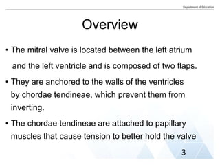
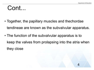
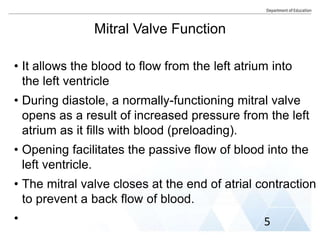
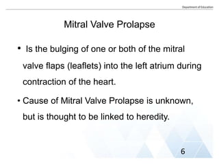
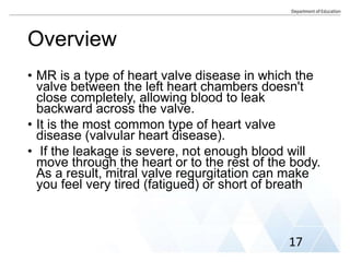
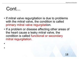
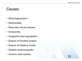
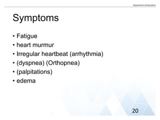
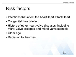
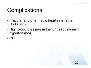
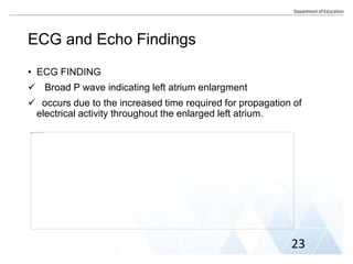
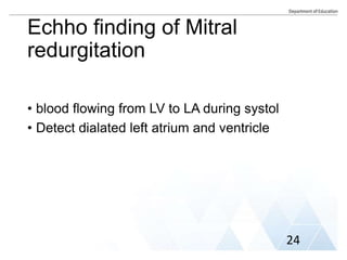
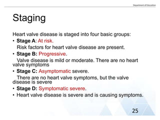
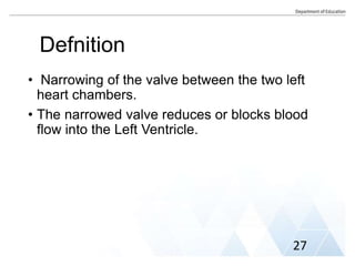
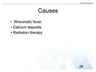
SCHOOL OF NURSING
DEPARTMENT OF MEDICAL SURGICAL NURSING
GROUP ASSIGNMENT
GROUP MEMBERS
1. Menber Yilma. GSRC/4257/
2. Nardos.Denekew GSRC/4259/15
3. Tewodros Ayele GSRC/4263/15
Submited to :- Instructor Tekalgn. ( Msc.CVPN)
June 30/2023
AA, Ethiopia
Acknowledgement
We would ike to.express.our deep gratitude to
Instructor Tekalgn for his commitment and
guiding.us.to be.much familiar and skilled in
Cardiovascular nursing care.
Out line
• Mitral Valve Prolaps
• Mitral Valve Regurgitation
• Mitral Valve Stenosis
• AorticValve regurgitation
• Aortic Valve Stenosis
• Nursing Mngement of Valvular heart disese.
Mitral Valve
1
Heart Chambers and Valves
2
Overview
• The mitral valve is located between the left atrium
and the left ventricle and is composed of two flaps.
• They are anchored to the walls of the ventricles
by chordae tendineae, which prevent them from
inverting.
• The chordae tendineae are attached to papillary
muscles that cause tension to better hold the valve
3
Cont...
• Together, the papillary muscles and thechordae
tendineae are known as the subvalvular apparatus.
• The function of the subvalvular apparatus is to
keep the valves from prolapsing into the atria when
they close
4
Mitral Valve Function
• It allows the blood to flow from the left atrium into
the left ventricle
• During diastole, a normally-functioning mitral valve
opens as a result of increased pressure from the left
atrium as it fills with blood (preloading).
• Opening facilitates the passive flow of blood into the
left ventricle.
• The mitral valve closes at the end of atrial contraction
to prevent a back flow of blood.
• 5
Mitral Valve Prolapse
• Is the bulging of one or both of the mitral
valve flaps (leaflets) into the left atrium during
contraction of the heart.
• Cause of Mitral Valve Prolapse is unknown,
but is thought to be linked to heredity.
6
Sign and Symptoms
Includes
• Chest pain
• Mitral regurgitation
• Irregular heartbeat (arrhythmia)
• Dizziness or lightheadedness
• Shortness of breath, especially during exercis
when lying flat
• Fatigue
• Syncope
• Palpitation 7
Risk Factors
• Men older than 50 years old
• HTN
• Graves' disease.
• Marfan syndrome.
• Muscular dystrophy.
• Scoliosis.
•
•
8
Complications
• Mitral Valve regurgitaion
.
• Heart rhythm problems (arrhythmias).
9
10
Mitral Valve Prolapse ECG Finding
• inversion of T wave
• Enlarged LA
• Atrial Fibrilation
11
12
Mitral Valve Echo finding
• To confirm mitral valve prolapse and severity
• Thickend leaflets greater than 5mm.
• Leaflet displacement >2mm
13
Mitral Valve Prolapse treatment
• Exercise
• Pain relievers
• Relaxation and stress reduction techniques
• Avoidance of caffeine and other stimulants
• Beta-blockers
•
• 14
Follow-Up of Mitral Valve Prolapse
• Most people with mitral valve prolapse should
see a cardiologist every 2 to 3 years. They do
not need regular echocardiograms.
• People with mitral valve prolapse and
moderate or severe mitral regurgitation should
see a doctor and undergo echocardiography
every 6 to 12 months.
• Echocardiography and a doctor's visit are also
recommended if a person develops new
symptoms, or if the symptoms change.
15
Mitral Valve Regurgitation
16
Overview
• MR is a type of heart valve disease in which the
valve between the left heart chambers doesn't
close completely, allowing blood to leak
backward across the valve.
• It is the most common type of heart valve
disease (valvular heart disease).
• If the leakage is severe, not enough blood will
move through the heart or to the rest of the body.
As a result, mitral valve regurgitation can make
you feel very tired (fatigued) or short of breath
17
Cont...
• If mitral valve regurgitation is due to problems
with the mitral valve, the condition is called
primary mitral valve regurgitation.
• If a problem or disease affecting other areas of
the heart cause a leaky mitral valve, the
condition is called functional or secondary
mitral regurgitation.
•
•
18
Causes
• Mitral.Degenaration
• Mitral prolaps
• Rheumatic valvular disease
• Endocarditis
• Congenital mitral regurgitation
• Rupture of Chordate tendane
• Rupture os Papillary muscle
• Dialated cardiomayopathy
• Ischemic heart disease
19
Symptoms
• Fatigue
• heart murmur
• Irregular heartbeat (arrhythmia)
• (dyspnea) (Orthopnea)
• (palpitations)
• edema
20
Risk factors
• Infections that affect the heartHeart attackHeart
• Congenital heart defect
• History of other heart valve diseases, including
mitral valve prolapse and mitral valve stenosis
• Older age
• Radiation to the chest
21
Complications
• Irregular and often rapid heart rate (atrial
fibrillation).
• High blood pressure in the lungs (pulmonary
hypertension).
• CHF
22
ECG and Echo Findings
• ECG FINDING
 Broad P wave indicating left atrium enlargment
 occurs due to the increased time required for propagation of
electrical activity throughout the enlarged left atrium.
23
Echho finding of Mitral
redurgitation
• blood flowing from LV to LA during systol
• Detect dialated left atrium and ventricle
24
Staging
Heart valve disease is staged into four basic groups:
• Stage A: At risk.
Risk factors for heart valve disease are present.
• Stage B: Progressive.
Valve disease is mild or moderate. There are no heart
valve symptoms
• Stage C: Asymptomatic severe.
There are no heart valve symptoms, but the valve
disease is severe
• Stage D: Symptomatic severe.
• Heart valve disease is severe and is causing symptoms.
25
Mitral Stenosis
26
Defnition
• Narrowing of the valve between the two left
heart chambers.
• The narrowed valve reduces or blocks blood
flow into the Left Ventricle.
27
Causes
• Rheumatic fever.
• Calcium deposits
• Radiation therapy
28
• Shortness of breath,
especially with activity
• Orthopnea
• Fatigue, especially
during increased activity
• Swollen feet or legs
• Sensations of a fast,
fluttering or pounding
heartbeat
• Dizziness or fainting
• Irregular heart sound,
also called a heart
murmur
• Fluid buildup in the
lungsI
• rregular heart rhythms
• Chest discomfort or
chest pain
• hemaptosis
Symptoms
29
Risk factors
• Untreated strep infections
• Aging
• Radiation Therapy
30
Complications
• Irregulr heart beat
• Blood clot
• High blood pressure in the lung arteries
• Right side heart failure
31
ECG findings
• There is atrial fibrillation.
• No P waves are visible.
• The rhythm is irregularl.
• There is the suggestion of right ventricular
hypertrophy.
• Right axis deviation and deep S waves in the
lateral leads.
32
33
Echo Finding
• Reduced mitral valve area <1.5mm➋
• Thickened Calcified leaflets
• Increased mean diastolic preasure gradient
across the mitrl valve
• Right Ventricle dialtion
• Left Atrium Enlargment
• Evidences of Pulmonary hypertension
34
Treatment
• provide prophylaxis for infective endocarditis
• reduce symptoms of pulmonary congestion
(eg, orthopnea, paroxysmal nocturnal dyspnea)
• control the ventricular rate if atrial fibrillation is
present, and
• prevent thromboembolic complications
35
Aortic valve regurgitation
36
Defnition
• a condition that occurs when aortic valve
doesn't close tightly.
• As a result, some of the blood pumped out of
theft ventricl leaks backward.
37
Symptoms
• Shortness of breath with exercise or when you lie down
• Fatigue and weakness, especially when you increase
your activity leve
• lHeart murmur
• Irregular pulse (arrhythmia)
• Lightheadedness or fainting
• Chest pain (angina),
• discomfort or tightness, often increasing during exercise
• Palpitations
• Leg swelling
38
Causes
Causes of aortic valve regurgitation include
• Congenital heart valve disease.
• Narrowing of the aortic valve (aortic stenosis).
• Inflammation of the lining of the heart's
chambers and valves (endocarditis)
• Rheumatic fever
39
Risk factors
• Older age
• Congenital heart disease
• History of infections that can affect the heart
• Conditions that can affect the heart, such as
Marfan syndrome
• Valve conditions, such as aortic valve stenosis
• High blood pressure
40
Complications
• Fainting
• feeling lightheaded
• Heart failure
• Heart rhythm problems (arrhythmias)
• Death
41
ECG finding
• Left Ventricular hypertrophic
• T wave inversion
42
Echo Finding
• Dialated LV
43
Treatment
• Antibiotics
• Aortic valve repair
• Aortic valve replacement
• BP lowering medications
• Anticuagulant
• Blood thiner
44
Aortic valve stenosis
45
Defnition
• Narrowing of the Aortic valve resulting in
Obstruction of blood flow from the left ventricle
to aorta during systole.
46
Symptoms
Symptoms of aortic valve stenosis may include
• An irregular heart sound (heart murmur) heard
through a stethoscope
• Chest pain (angina) or tightness with
activityFeeling faint or dizzy or fainting with
activity
• Shortness of breath, especially with
activityFatigue, especially during times of
increased activityRapid, fluttering heartbeat
• palpitations
• Thrill
• Weight lose
47
Causes
• CHF
• Calcium buildup on the valve (aortic valve
calcification)
• Fibrosis
• Rheumatic fever
• Congenital Bicusped aortic valve
• Degerarative disease
48
Risk factors
Risk factors of aortic valve stenosis include:
• Older age
• Congenital heart defects, such as a bicuspid aortic
valve
• Chronic kidney disease
• Having heart disease risk factors, such as diabetes,
high cholesterol and high blood pressure
• History of Rheumatic fever and infective endocarditis
• History of radiation therapy to the chest
49
Complications
Aortic valve stenosis can cause complications,
including:
• Heart failure
• Stroke
• Blood clots
• Bleeding
• Irregular heart rhythms (arrhythmias)
• Infections that affect the heart, such as endocarditis
• HTN
• Death
50
ECG finding
• LVH
• LBBB
• Complete heart block
51
52
• Aortic valve with no
cusp motion (may be
unreliable in congenital
or rheumatic valvular
stenosis)
• A decrease in the
maximal aortic cusp
separation (< 8 mm in
the adult)
• The presence of
unexplained LV
hypertrophy
Echo Finding
53
NursingManagement of
Valvular heart Diseases
54
• Assess mental status (Restlessness, severe anxiety,
and confusion).
• Check vital signs (heart rate and blood pressure).
• Assess heart sounds, noting gallops, S3, S4.
• Assess manually peripheral pulses (with weak rate,
rhythm indicated low cardiac output).
• Assess lung sounds and determine any occurrence
of Paroxysmal Nocturnal Dyspnea (PND) or
orthopnea.
• Monitor central venous, right arterial pressure [RAP],
pulmonary arterial pressure(PAP) Routinely
55
• Assess skin color and temperature (Cold, clammy skin
is secondary to a compensatory increase in
sympathetic nervous system stimulation and low
cardiac output and desaturation).
• Carefully maintain intake output and daily check
weight.
• Administer medication as prescribed, noting response,
and watching for side effects and toxicity.
• Administer stool softeners as needed(straining for a
bowel movement further impairs cardiac output).
• Explain the drug regimen, purpose, dose, and side
effects.
• Maintain adequate ventilation and perfusion (Place the
patient in semi- to high-Fowler’s position or supine
position).
56
• Administer O2 as ordered.
• Assess response to increased activity and help the
patient in daily activities.
• Maintain physical and emotional rest (restrict activity
and provide a quiet and relaxed environment)
• Monitor sleep patterns; administer a sedative
• If invasive adjunct therapies are indicated (e.g.,
intra-aortic balloon pump, pacemaker), maintain
within the prescribed protocol, and prepare the
patient.
• Explain diet restrictions (fluid, sodium).
57
References
1. https://www.mayoclinic.org/diseases-
conditions/mitral-valve-regurgitation/symptoms-
causes/syc-20350178
2. https://www.mayoclinic.org/diseases-
conditions/mitral-valve-stenosis/diagnosis-
treatment/drc-20353165
3. https://www.amboss.com/us/knowledge/mitral-
valve-stenosis
4. https://www.mayoclinic.org/diseases-
conditions/aortic-valve-regurgitation/diagnosis-
treatment/drc-20353135
58
Cont...
5 . https://www.mayoclinic.org/diseases-
conditions/aortic-stenosis/diagnosis-
treatment/drc-20353145
59
THANK YOU!
60

Mitral and Aortic Valves Final .pptx

  • 1.
    SCHOOL OF NURSING DEPARTMENTOF MEDICAL SURGICAL NURSING GROUP ASSIGNMENT GROUP MEMBERS 1. Menber Yilma. GSRC/4257/ 2. Nardos.Denekew GSRC/4259/15 3. Tewodros Ayele GSRC/4263/15 Submited to :- Instructor Tekalgn. ( Msc.CVPN) June 30/2023 AA, Ethiopia
  • 2.
    Acknowledgement We would iketo.express.our deep gratitude to Instructor Tekalgn for his commitment and guiding.us.to be.much familiar and skilled in Cardiovascular nursing care.
  • 3.
    Out line • MitralValve Prolaps • Mitral Valve Regurgitation • Mitral Valve Stenosis • AorticValve regurgitation • Aortic Valve Stenosis • Nursing Mngement of Valvular heart disese.
  • 4.
  • 5.
  • 6.
    Overview • The mitralvalve is located between the left atrium and the left ventricle and is composed of two flaps. • They are anchored to the walls of the ventricles by chordae tendineae, which prevent them from inverting. • The chordae tendineae are attached to papillary muscles that cause tension to better hold the valve 3
  • 7.
    Cont... • Together, thepapillary muscles and thechordae tendineae are known as the subvalvular apparatus. • The function of the subvalvular apparatus is to keep the valves from prolapsing into the atria when they close 4
  • 8.
    Mitral Valve Function •It allows the blood to flow from the left atrium into the left ventricle • During diastole, a normally-functioning mitral valve opens as a result of increased pressure from the left atrium as it fills with blood (preloading). • Opening facilitates the passive flow of blood into the left ventricle. • The mitral valve closes at the end of atrial contraction to prevent a back flow of blood. • 5
  • 9.
    Mitral Valve Prolapse •Is the bulging of one or both of the mitral valve flaps (leaflets) into the left atrium during contraction of the heart. • Cause of Mitral Valve Prolapse is unknown, but is thought to be linked to heredity. 6
  • 10.
    Sign and Symptoms Includes •Chest pain • Mitral regurgitation • Irregular heartbeat (arrhythmia) • Dizziness or lightheadedness • Shortness of breath, especially during exercis when lying flat • Fatigue • Syncope • Palpitation 7
  • 11.
    Risk Factors • Menolder than 50 years old • HTN • Graves' disease. • Marfan syndrome. • Muscular dystrophy. • Scoliosis. • • 8
  • 12.
    Complications • Mitral Valveregurgitaion . • Heart rhythm problems (arrhythmias). 9
  • 13.
  • 14.
    Mitral Valve ProlapseECG Finding • inversion of T wave • Enlarged LA • Atrial Fibrilation 11
  • 15.
  • 16.
    Mitral Valve Echofinding • To confirm mitral valve prolapse and severity • Thickend leaflets greater than 5mm. • Leaflet displacement >2mm 13
  • 17.
    Mitral Valve Prolapsetreatment • Exercise • Pain relievers • Relaxation and stress reduction techniques • Avoidance of caffeine and other stimulants • Beta-blockers • • 14
  • 18.
    Follow-Up of MitralValve Prolapse • Most people with mitral valve prolapse should see a cardiologist every 2 to 3 years. They do not need regular echocardiograms. • People with mitral valve prolapse and moderate or severe mitral regurgitation should see a doctor and undergo echocardiography every 6 to 12 months. • Echocardiography and a doctor's visit are also recommended if a person develops new symptoms, or if the symptoms change. 15
  • 19.
  • 20.
    Overview • MR isa type of heart valve disease in which the valve between the left heart chambers doesn't close completely, allowing blood to leak backward across the valve. • It is the most common type of heart valve disease (valvular heart disease). • If the leakage is severe, not enough blood will move through the heart or to the rest of the body. As a result, mitral valve regurgitation can make you feel very tired (fatigued) or short of breath 17
  • 21.
    Cont... • If mitralvalve regurgitation is due to problems with the mitral valve, the condition is called primary mitral valve regurgitation. • If a problem or disease affecting other areas of the heart cause a leaky mitral valve, the condition is called functional or secondary mitral regurgitation. • • 18
  • 22.
    Causes • Mitral.Degenaration • Mitralprolaps • Rheumatic valvular disease • Endocarditis • Congenital mitral regurgitation • Rupture of Chordate tendane • Rupture os Papillary muscle • Dialated cardiomayopathy • Ischemic heart disease 19
  • 23.
    Symptoms • Fatigue • heartmurmur • Irregular heartbeat (arrhythmia) • (dyspnea) (Orthopnea) • (palpitations) • edema 20
  • 24.
    Risk factors • Infectionsthat affect the heartHeart attackHeart • Congenital heart defect • History of other heart valve diseases, including mitral valve prolapse and mitral valve stenosis • Older age • Radiation to the chest 21
  • 25.
    Complications • Irregular andoften rapid heart rate (atrial fibrillation). • High blood pressure in the lungs (pulmonary hypertension). • CHF 22
  • 26.
    ECG and EchoFindings • ECG FINDING  Broad P wave indicating left atrium enlargment  occurs due to the increased time required for propagation of electrical activity throughout the enlarged left atrium. 23
  • 27.
    Echho finding ofMitral redurgitation • blood flowing from LV to LA during systol • Detect dialated left atrium and ventricle 24
  • 28.
    Staging Heart valve diseaseis staged into four basic groups: • Stage A: At risk. Risk factors for heart valve disease are present. • Stage B: Progressive. Valve disease is mild or moderate. There are no heart valve symptoms • Stage C: Asymptomatic severe. There are no heart valve symptoms, but the valve disease is severe • Stage D: Symptomatic severe. • Heart valve disease is severe and is causing symptoms. 25
  • 29.
  • 30.
    Defnition • Narrowing ofthe valve between the two left heart chambers. • The narrowed valve reduces or blocks blood flow into the Left Ventricle. 27
  • 31.
    Causes • Rheumatic fever. •Calcium deposits • Radiation therapy 28
  • 32.
    • Shortness ofbreath, especially with activity • Orthopnea • Fatigue, especially during increased activity • Swollen feet or legs • Sensations of a fast, fluttering or pounding heartbeat • Dizziness or fainting • Irregular heart sound, also called a heart murmur • Fluid buildup in the lungsI • rregular heart rhythms • Chest discomfort or chest pain • hemaptosis Symptoms 29
  • 33.
    Risk factors • Untreatedstrep infections • Aging • Radiation Therapy 30
  • 34.
    Complications • Irregulr heartbeat • Blood clot • High blood pressure in the lung arteries • Right side heart failure 31
  • 35.
    ECG findings • Thereis atrial fibrillation. • No P waves are visible. • The rhythm is irregularl. • There is the suggestion of right ventricular hypertrophy. • Right axis deviation and deep S waves in the lateral leads. 32
  • 36.
  • 37.
    Echo Finding • Reducedmitral valve area <1.5mm➋ • Thickened Calcified leaflets • Increased mean diastolic preasure gradient across the mitrl valve • Right Ventricle dialtion • Left Atrium Enlargment • Evidences of Pulmonary hypertension 34
  • 38.
    Treatment • provide prophylaxisfor infective endocarditis • reduce symptoms of pulmonary congestion (eg, orthopnea, paroxysmal nocturnal dyspnea) • control the ventricular rate if atrial fibrillation is present, and • prevent thromboembolic complications 35
  • 39.
  • 40.
    Defnition • a conditionthat occurs when aortic valve doesn't close tightly. • As a result, some of the blood pumped out of theft ventricl leaks backward. 37
  • 41.
    Symptoms • Shortness ofbreath with exercise or when you lie down • Fatigue and weakness, especially when you increase your activity leve • lHeart murmur • Irregular pulse (arrhythmia) • Lightheadedness or fainting • Chest pain (angina), • discomfort or tightness, often increasing during exercise • Palpitations • Leg swelling 38
  • 42.
    Causes Causes of aorticvalve regurgitation include • Congenital heart valve disease. • Narrowing of the aortic valve (aortic stenosis). • Inflammation of the lining of the heart's chambers and valves (endocarditis) • Rheumatic fever 39
  • 43.
    Risk factors • Olderage • Congenital heart disease • History of infections that can affect the heart • Conditions that can affect the heart, such as Marfan syndrome • Valve conditions, such as aortic valve stenosis • High blood pressure 40
  • 44.
    Complications • Fainting • feelinglightheaded • Heart failure • Heart rhythm problems (arrhythmias) • Death 41
  • 45.
    ECG finding • LeftVentricular hypertrophic • T wave inversion 42
  • 46.
  • 47.
    Treatment • Antibiotics • Aorticvalve repair • Aortic valve replacement • BP lowering medications • Anticuagulant • Blood thiner 44
  • 48.
  • 49.
    Defnition • Narrowing ofthe Aortic valve resulting in Obstruction of blood flow from the left ventricle to aorta during systole. 46
  • 50.
    Symptoms Symptoms of aorticvalve stenosis may include • An irregular heart sound (heart murmur) heard through a stethoscope • Chest pain (angina) or tightness with activityFeeling faint or dizzy or fainting with activity • Shortness of breath, especially with activityFatigue, especially during times of increased activityRapid, fluttering heartbeat • palpitations • Thrill • Weight lose 47
  • 51.
    Causes • CHF • Calciumbuildup on the valve (aortic valve calcification) • Fibrosis • Rheumatic fever • Congenital Bicusped aortic valve • Degerarative disease 48
  • 52.
    Risk factors Risk factorsof aortic valve stenosis include: • Older age • Congenital heart defects, such as a bicuspid aortic valve • Chronic kidney disease • Having heart disease risk factors, such as diabetes, high cholesterol and high blood pressure • History of Rheumatic fever and infective endocarditis • History of radiation therapy to the chest 49
  • 53.
    Complications Aortic valve stenosiscan cause complications, including: • Heart failure • Stroke • Blood clots • Bleeding • Irregular heart rhythms (arrhythmias) • Infections that affect the heart, such as endocarditis • HTN • Death 50
  • 54.
    ECG finding • LVH •LBBB • Complete heart block 51
  • 55.
  • 56.
    • Aortic valvewith no cusp motion (may be unreliable in congenital or rheumatic valvular stenosis) • A decrease in the maximal aortic cusp separation (< 8 mm in the adult) • The presence of unexplained LV hypertrophy Echo Finding 53
  • 57.
  • 58.
    • Assess mentalstatus (Restlessness, severe anxiety, and confusion). • Check vital signs (heart rate and blood pressure). • Assess heart sounds, noting gallops, S3, S4. • Assess manually peripheral pulses (with weak rate, rhythm indicated low cardiac output). • Assess lung sounds and determine any occurrence of Paroxysmal Nocturnal Dyspnea (PND) or orthopnea. • Monitor central venous, right arterial pressure [RAP], pulmonary arterial pressure(PAP) Routinely 55
  • 59.
    • Assess skincolor and temperature (Cold, clammy skin is secondary to a compensatory increase in sympathetic nervous system stimulation and low cardiac output and desaturation). • Carefully maintain intake output and daily check weight. • Administer medication as prescribed, noting response, and watching for side effects and toxicity. • Administer stool softeners as needed(straining for a bowel movement further impairs cardiac output). • Explain the drug regimen, purpose, dose, and side effects. • Maintain adequate ventilation and perfusion (Place the patient in semi- to high-Fowler’s position or supine position). 56
  • 60.
    • Administer O2as ordered. • Assess response to increased activity and help the patient in daily activities. • Maintain physical and emotional rest (restrict activity and provide a quiet and relaxed environment) • Monitor sleep patterns; administer a sedative • If invasive adjunct therapies are indicated (e.g., intra-aortic balloon pump, pacemaker), maintain within the prescribed protocol, and prepare the patient. • Explain diet restrictions (fluid, sodium). 57
  • 61.
    References 1. https://www.mayoclinic.org/diseases- conditions/mitral-valve-regurgitation/symptoms- causes/syc-20350178 2. https://www.mayoclinic.org/diseases- conditions/mitral-valve-stenosis/diagnosis- treatment/drc-20353165 3.https://www.amboss.com/us/knowledge/mitral- valve-stenosis 4. https://www.mayoclinic.org/diseases- conditions/aortic-valve-regurgitation/diagnosis- treatment/drc-20353135 58
  • 62.
  • 63.