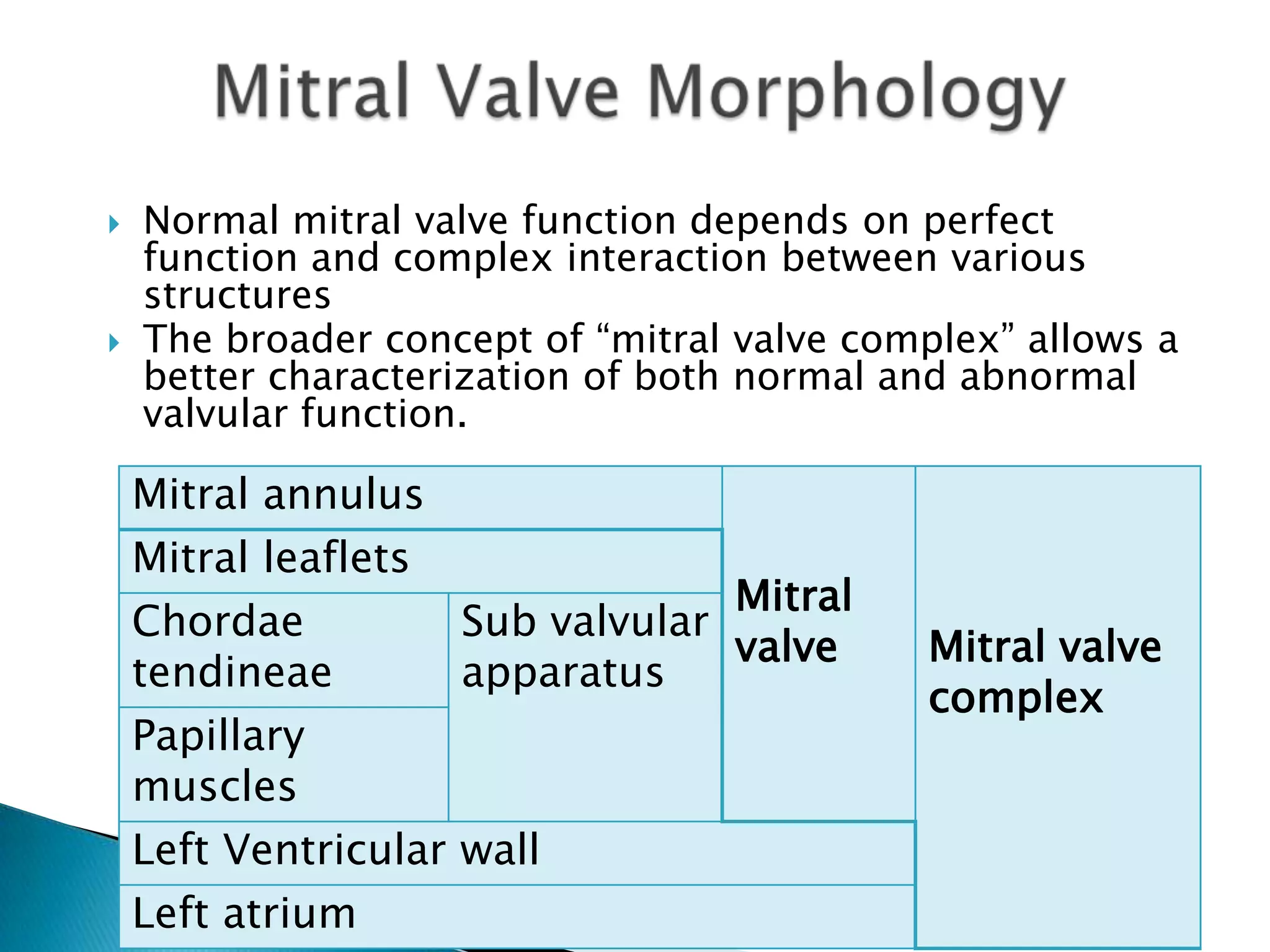
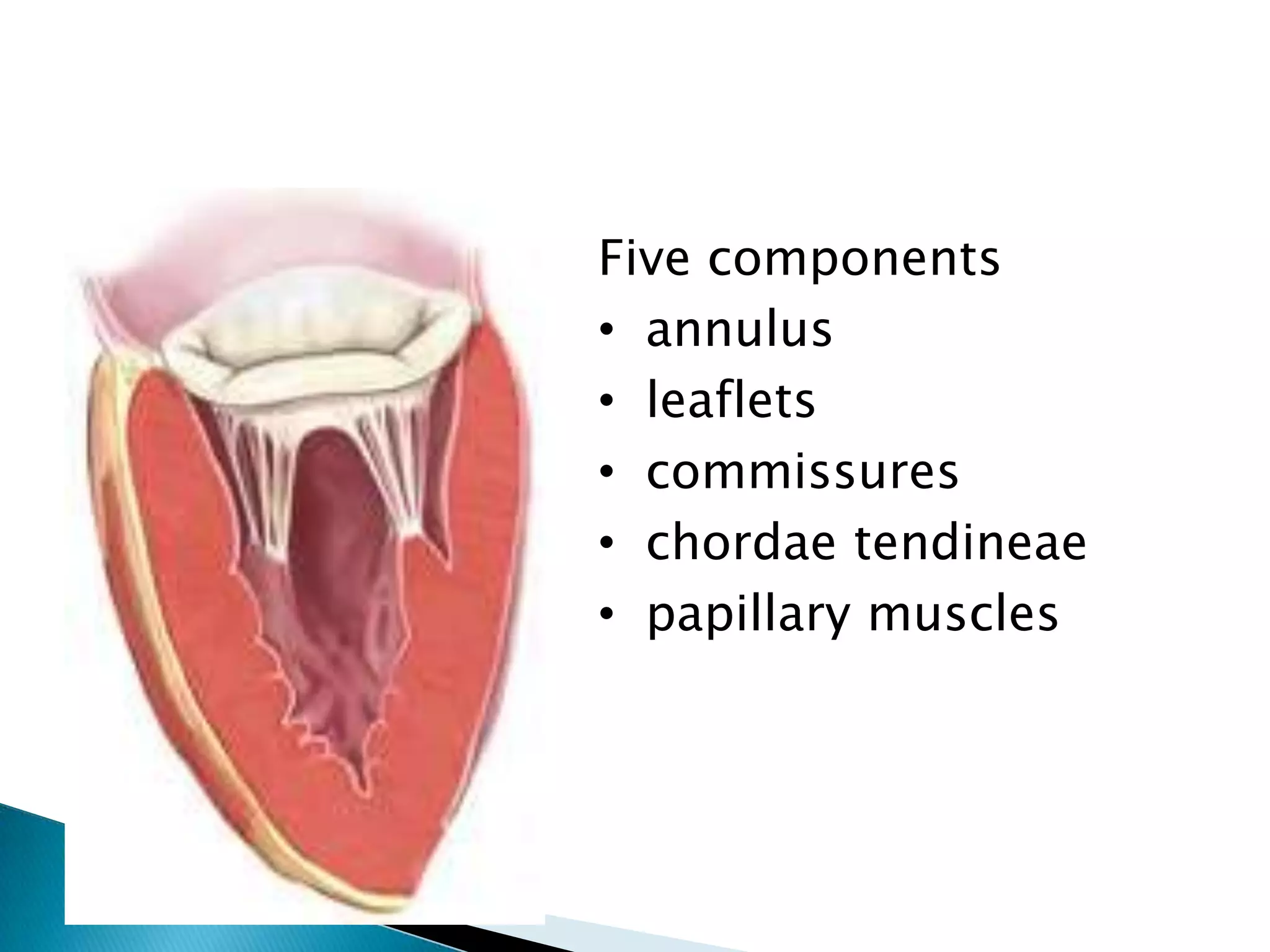
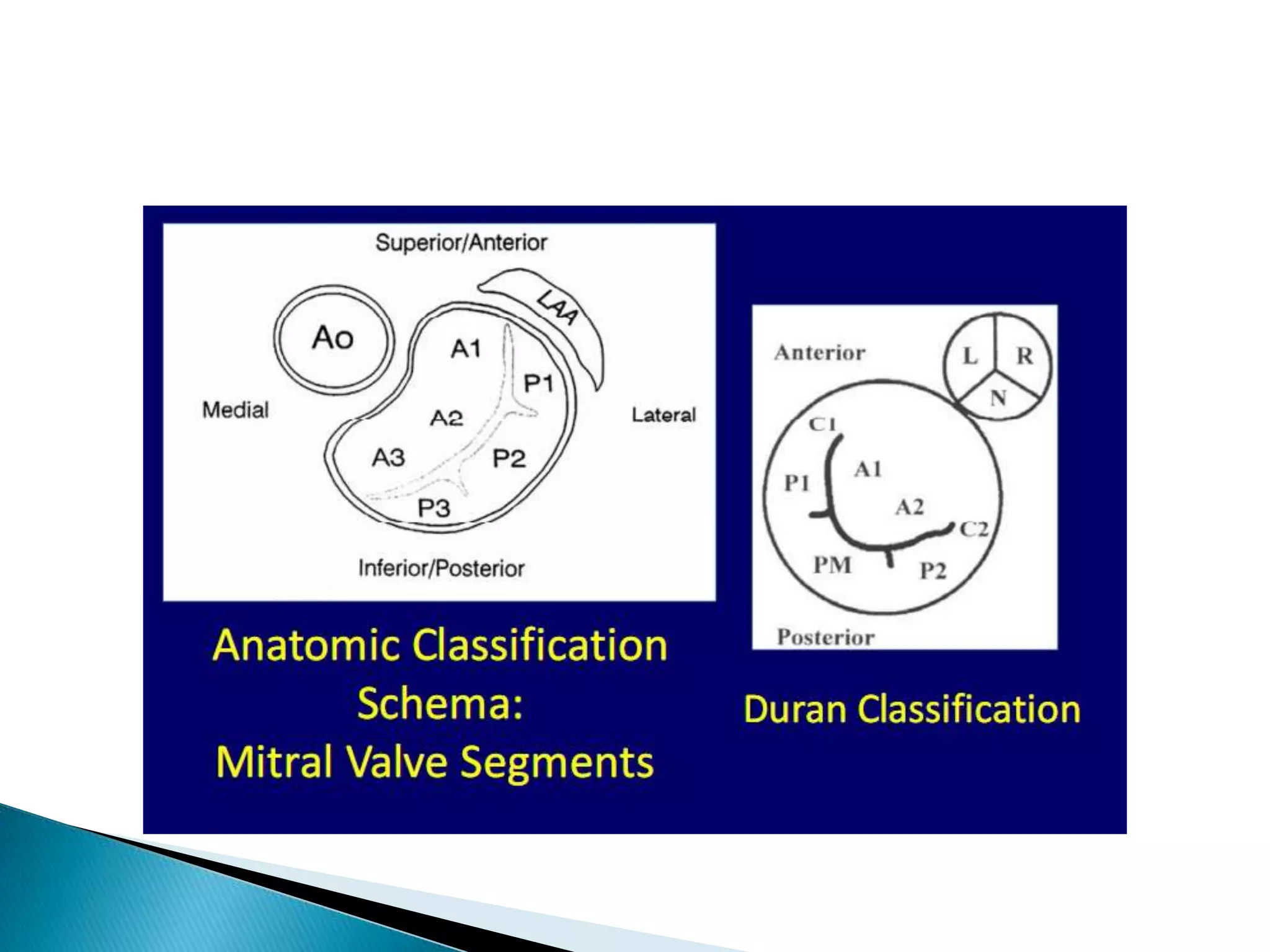
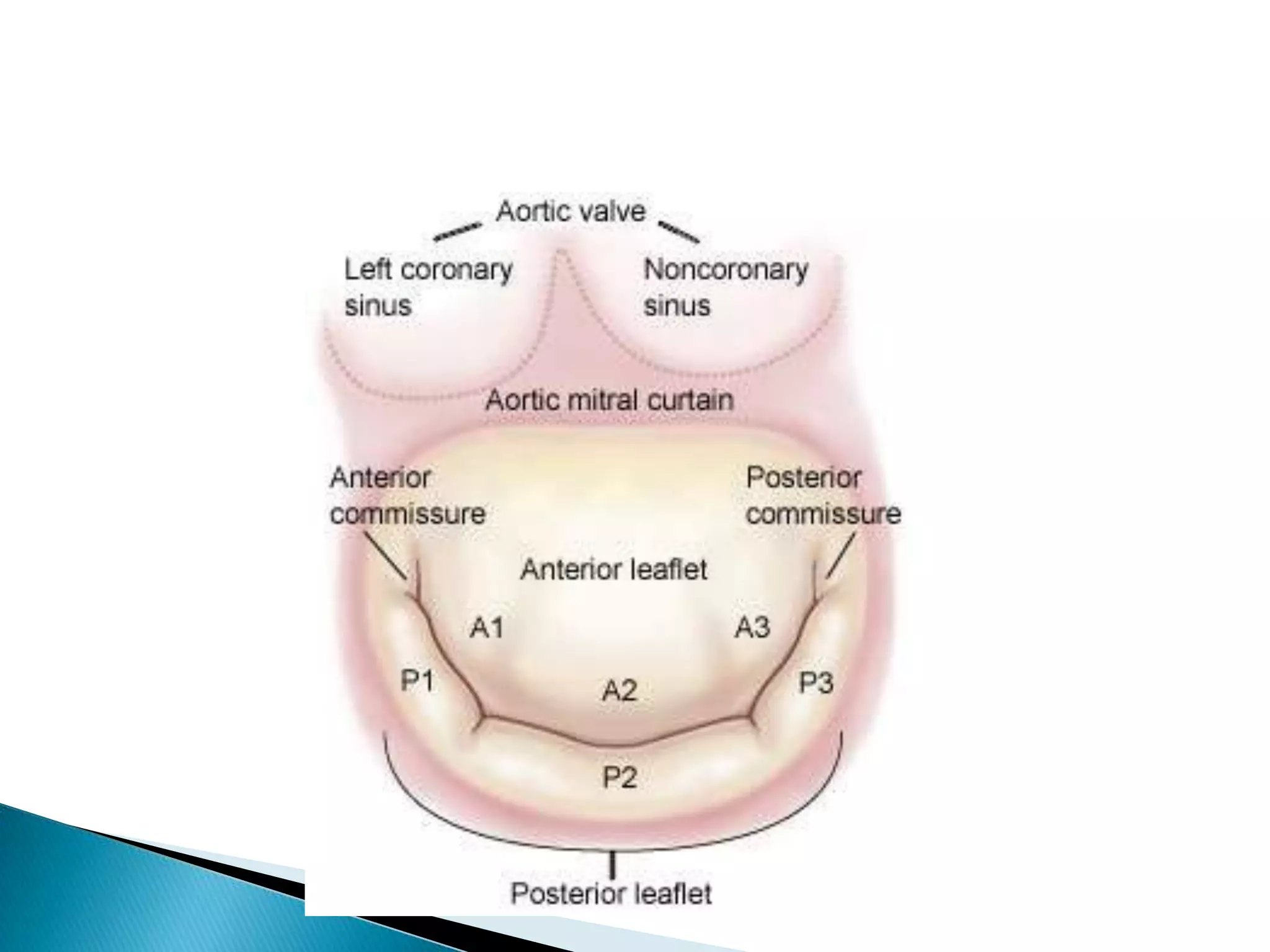
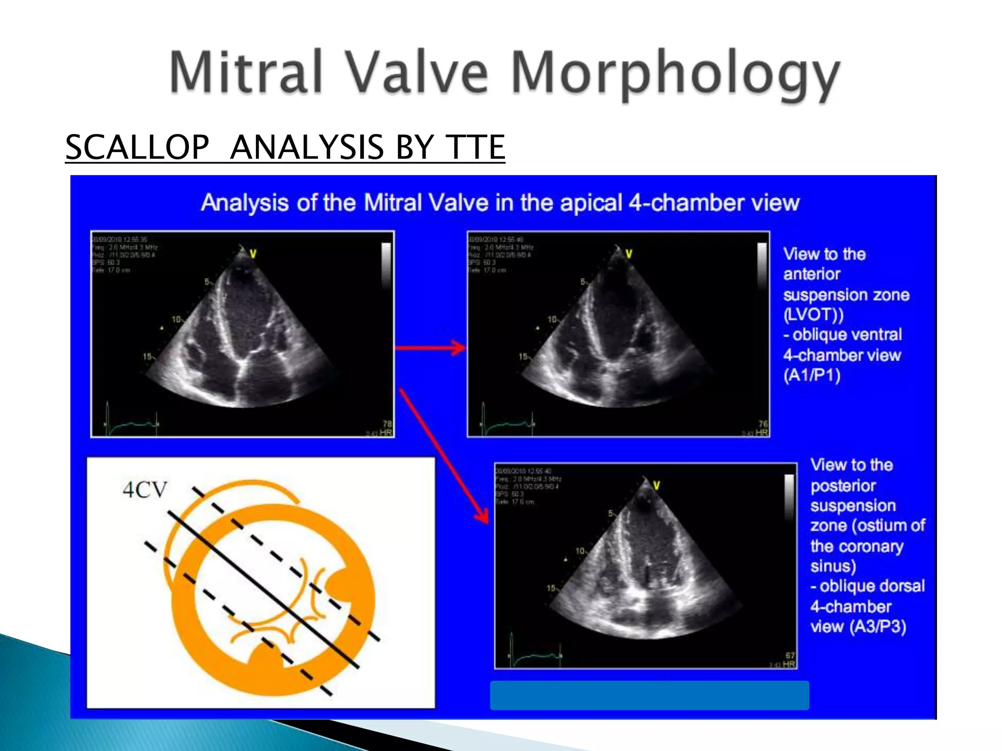
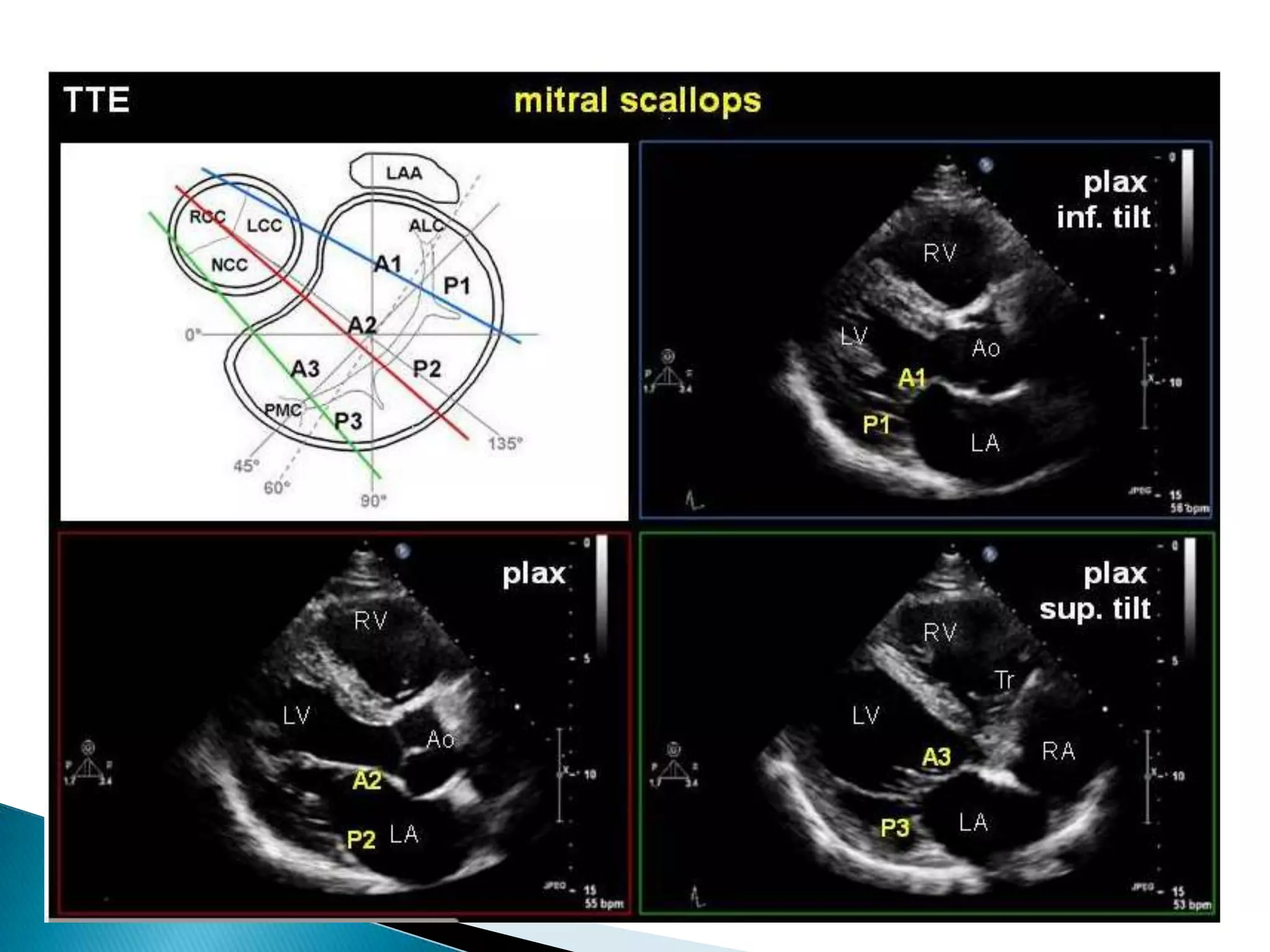
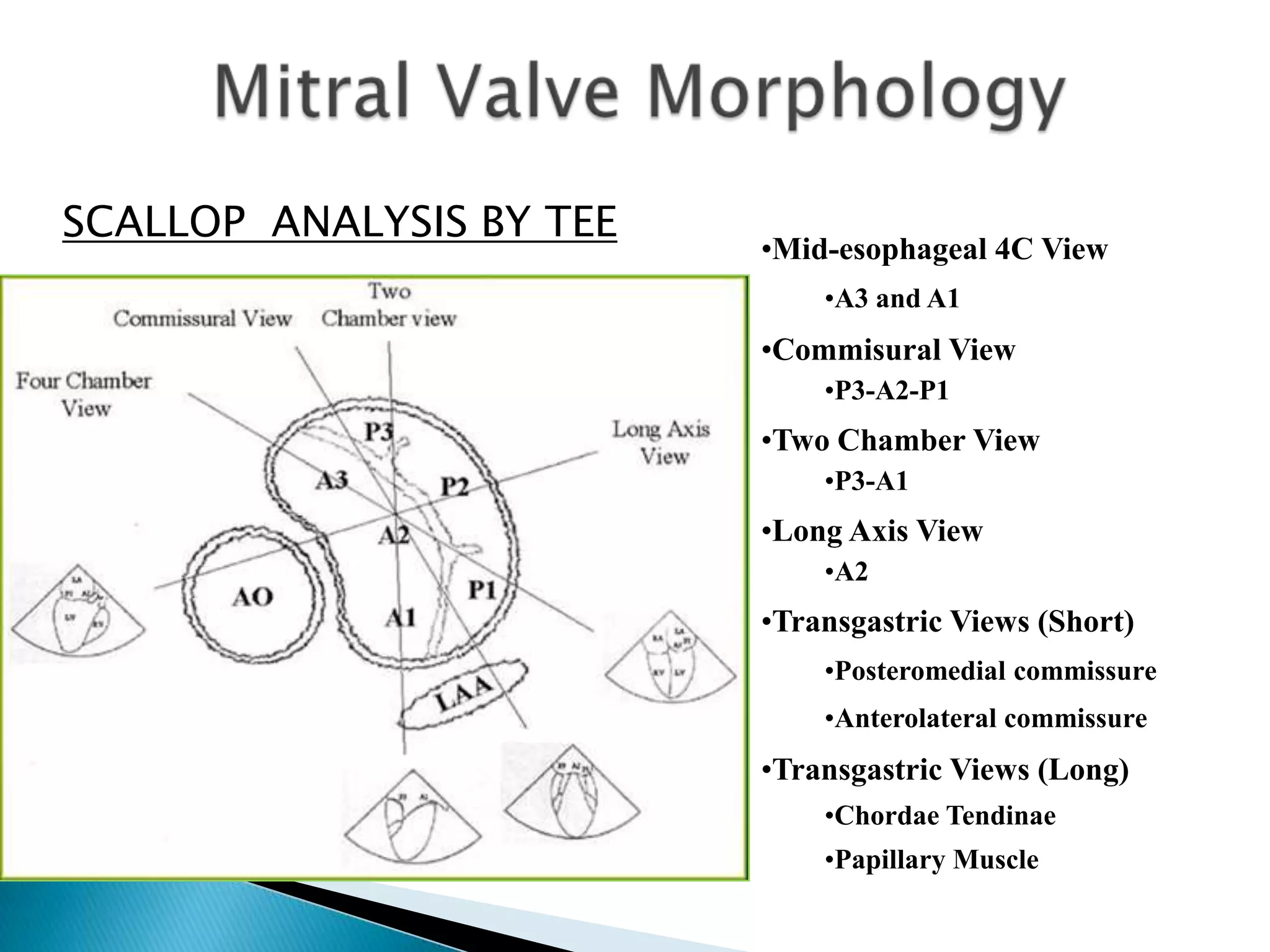
SlideShare a Scribd company logo
Dr. K.V.Siva krishna
 The mitral valve was the first of the four
cardiac valves to be evaluated with
echocardiography.
 This was due to the relatively high prevalence
of rheumatic heart disease and the relatively
large excursion of the mitral valve leaflets,
which made them an easier target for early
M-mode techniques
The mitral valve has a triple function:
 it regulates blood flow towards the left
ventricle during diastole at low pressure
gradient while preventing systolic backflow
towards the left atrium
 it contributes to the formation of the left
ventricular outflow tract during systole and
 its integrity is essential to maintain normal
size, geometry and function of the left
ventricle.
 The mitral valve, located between the left
atrium and left ventricle, is a functional
complex that relies on normal morphology,
geometrical relations and function of all its
constituents: the left atrium, the mitral
annulus, the mitral leaflets, the subvalvular
apparatus (tendinous chords and papillary
muscles) and the left ventricle.
 It is important to recognize that the leaflets
of the mitral valve constitute only a portion of
the mitral valve apparatus and that diseases
resulting in mitral dysfunction often are
caused by abnormalities in the overall
apparatus rather than in the actual leaflets.
 Normal mitral valve function depends on perfect
function and complex interaction between various
structures
 The broader concept of “mitral valve complex” allows a
better characterization of both normal and abnormal
valvular function.
Mitral annulus
Mitral
valve Mitral valve
complex
Mitral leaflets
Chordae
tendineae
Sub valvular
apparatus
Papillary
muscles
Left Ventricular wall
Left atrium
Five components
• annulus
• leaflets
• commissures
• chordae tendineae
• papillary muscles
 MITRAL ANNULUS
 The mitral annulus constitutes the anatomical
junction between the LV and the LA, and serves as
insertion site for the leaflet tissue.
 It is oval and saddle shaped.
 The mitral annulus is a fibrotic ring that consists of
an anterior part and a posterior part
 The anterior portion of the mitral annulus is attached to
the fibrous trigones and is generally more developed than
the posterior annulus.
 The aortic-mitral curtain is a fibrous structure that
connects the anterior mitral annulus intimately with the
aortic valve annulus (at the level of the left and non-
coronary cusps)
 The annulus is deficient anteriorly at the level of the aorto
mitral curtain.
• The posterior part of the mitral annulus is not
enforced and rather discontinuous (making it prone
to dilatation)
• Both parts of annulus may dilate in pathological
conditions
• The antero-posterior diameter forms the minor axis
and the inter-commissural distance refers to the
major axis
 identifying the mechanism of valvular
insufficiency,
 is useful to determine the type of mitral valve
intervention in case of dysfunction and
 is of interest to size mitral valve prosthesis or
annuloplasty ring.
 MITRAL ANNULUS
 The anterior-posterior diameter can be measured
using real-time 3D or by conventional 2D in the
parasternal long-axis view.
 Conventionally, a parasternal long axis transthoracic
view has been advocated for measuring minor
annular diameter
 More appropriate measurement of the minor
axis (antero-posterior diameter) of the mitral
annulus can be performed at end-systole
during transthoracic echocardiography in the
apical long axis view (3-chamber view) or its
transoesophageal equivalent, found at mid-
oesophageal level with 135° tilt of the probe
 The major axis (inter-commissural
diameter) of the mitral annulus is found at a
bicommissural 2-chamber transthoracic
echocardiographic view (when P1-A2-P3
mitral leaflet scallops are visualized) or a
mid-oesophageal bicommissural view at 45-
60° during transoesophageal
echocardiography
 MITRAL ANNULUS
 The orifice at the level of the left AV junction is ovoidal
with a longer intercommissural (IC) and a shorter
septal-to-lateral axis (SL).
 Body-weight-corrected data are: 0.39-0.59 mm/kg for
the IC and 0.32-0.48 mm/kg for the SL diameters
respectively .
 However, dimensions are underestimated “in vivo” by
2D ECHO as compared by 3D ECHO and with MRI.
 Annular dilatation is present when
 the ratio annulus/anterior leaflet during diastole is
>1.3
 the diameter is ≥ 35 mm
• MITRAL ANNULUS
• The annulus depicts complex modifications during
the cardiac cycle
• Annular flexion - a 23-40% variation in the annular
circumference between the systolic and diastolic
configuration
• Excursion Or Annular Descent - movement in apical-
to-basal direction
• The Rotation represents the torque movement while
the complex 3D modifications in shape are called,
Folding of the annulus.
• All such modifications are reduced or disappear with
the use of rigid annuloplasty rings, postoperative
fibrosis or extensive reduction of the posterior
 MITRAL ANNULUS
 Changes to be
observed are
 Mitral annulus
diameter
 Mitral annular
motion
 Calcification
 Perivalvular Leak
 (Prosthetic Valves)
 The mitral valve consists of an anterior and
posterior leaflet that converge at
the posteromedial and anterolateral
commissures
 Line of contact between leaf lets is termed
as coaptation line
 Region of leaf let overlap is called zone of
apposition
 The largest part of the atrial floor is formed
by the anterior mitral valve leaflet.
 Normal leaflets are thin and pliable structures
with a thickness <5 mm
 Normal mitral valve area is 4 to 5 cm2
 MITRAL VALVULAR LEAFLETS
Leaflets Commisure Coaptation
line
Zone of
apposition
 Posterior mitral leaf let
 The posterior leaflet has a quadrangular shape
 Attached to approximately two-thirds of the
annular circumference.
 The posterior leaflet typically has two well defined
indentations which divide the Leaflet into three
individual scallops as P1,P2,P3.
 The P1 scallop corresponds to the external
anterolateral portion of the posteror leaflet.
close to the anterior commissure and the left
atrium (LA) appendage.
 The P2 scallop medium and more developed.
 The P3 scallop is internal and close to posterior
comissure and tricuspid annulus
 Anterior mitral leaf let
 Anterior leaf let has semicircular shape
 Is in continuity with non coronary cusp of aortic valve
 The free edge of leaf let is not having any indentation
but divided into three segments A1 A2 A3
corresponding to posterior leaf let
 By Echocardiography,
 The presence and the extent of inadequate tissue
 Of excess leaflet tissue and the precise localization
of the leaflet lesions should be analysed.
 Describing the mitral valve segmentation is
particularly useful to precisely define the
anatomical lesions and the prolapsing segments in
patients with degenerative MR
 TEE still remains the recommended approach
 TTE predict accurately valve reparability.
TTE -
PSAX
TEE – TRANSGASTRIC
VIEW IN 0
SCALLOP ANALYSIS
SCALLOP ANALYSIS BY TTE
SCALLOP ANALYSIS BY
TTE
SCALLOP ANALYSIS BY TTE
SCALLOP ANALYSIS BY TEE
•Mid-esophageal 4C View
•A3 and A1
•Commisural View
•P3-A2-P1
•Two Chamber View
•P3-A1
•Long Axis View
•A2
•Transgastric Views (Short)
•Posteromedial commissure
•Anterolateral commissure
•Transgastric Views (Long)
•Chordae Tendinae
•Papillary Muscle
ME 5CV - Anteriorly DirectedSCALLOP ANALYSIS BY TEE
ME 4CV - Neutrally DirectedSCALLOP ANALYSIS BY TEE
ME 4CV - Posteroirly DirectedSCALLOP ANALYSIS BY TEE
SCALLOP ANALYSIS BY TEE
 TEE is probably the method of choice
 Multiple views are available which permit to
precisely determine the localization and the
extent of prolapse.
 The 'en face' view seen from the LA
perspective is identical to the surgical view
in the operating room.
 This view allows to perfectly analysing the
extent of commissural fusion in rheumatic
MR.
 Chordae tendinae
 There are three sets of chordae arising from the
papillary muscles. They are classified according to
their site of insertion between the free margin and the
base of leaflets.
 Marginal chordae (primary chordae) are inserted on
the free margin of the Leaflets and function to prevent
prolapse of the leaflet margin.
 Intermediate chordae (secondary chordae) insert on
the ventricular surface of the leaflets and relieve
valvular tissue of excess tension. Often two large
secondary or „strut‟ chordae can be individualized.
They may be important in preserving ventricular
shape and function.
 Basal chordae (tertiary chordae) are limited to the
posterior leaflet and connect the leaflet base and
mitral annulus to the papillary muscle.
Chordae tendineae
 Commisural chordae arise from ALPM &PMPM
and branch in a fan like fashion and insert
onto both commisures .
 These chordae divide about 3 times before
their final attachments leading to around 120
chordal attachment to both leaflets .
 Chordae tendineae
Thickening
Fusion
Calcification
Elongation
Rupture
 Papillary muscles
 The papillary muscles of the LV are three types
1. Completely tethered papillary muscle: In this
type papillary muscle was fully adherent to the
subjacent ventricular myocardium and
protruded very little into the ventricular cavity
with few trabecular attachments.
2. Finger like papillary muscle: in this type one third
or more of the body of the papillary muscle protruded
freely into the ventricular cavity with very few or no
trabecular attachments.
3. Mixed type papillary muscle: This papillary muscle
had part of the body protruding freely into the
ventricular cavity but also with considerable
trabecular attachments and tethering.
 The posteromedial PM gives chords to the
medial half of both leaflets (i.e.
posteromedial commissure, P3, A3 and half
of P2 and A2). Similarly, the anterolateral PM
chords attach to the lateral half of the MV
leaflets (i.e. anterolateral commissure, A1,P1
and half of P2 and A2)
 Papillary muscles
TTE - PSAX
VIEW
 Papillary muscles
TTE - PLAX
VIEW
TTE – APICAL 4C
VIEW
 Real-time 3-dimensional echocardiography
of the mitral valve allows easy identification
of different anatomical segments of the mitral
valve, including both commissures
 The `en face` view of the mitral valve can be
constructed and refers to exposure of the
mitral valve from the atrial perspective,
similar to the surgeons view during mitral
valve surgery.
 Thank you

More Related Content

What's hot

Lv systolic function
Lv systolic functionLv systolic function
Lv systolic function
Sruthi Meenaxshi
 
Echocardiographic Evaluation of Hypertrophic Cardiomyopathy
Echocardiographic Evaluation of Hypertrophic CardiomyopathyEchocardiographic Evaluation of Hypertrophic Cardiomyopathy
Echocardiographic Evaluation of Hypertrophic Cardiomyopathy
Hatem Soliman Aboumarie
 
Ebstein's anomaly echocardiogram
Ebstein's anomaly echocardiogramEbstein's anomaly echocardiogram
Ebstein's anomaly echocardiogram
Malleswara rao Dangeti
 
Echocardiographic assesment of systolic and diastolic dysfunction
Echocardiographic assesment of systolic and diastolic dysfunctionEchocardiographic assesment of systolic and diastolic dysfunction
Echocardiographic assesment of systolic and diastolic dysfunction
Malleswara rao Dangeti
 
Echocardiography of Mitral regurgitation
Echocardiography of Mitral regurgitationEchocardiography of Mitral regurgitation
Echocardiography of Mitral regurgitation
Dr. Muhammad AzAm Shah
 
ECHOCARDIOGRAPHIC EVALUATION OF MITRAL VALVE DISEASE -MITRAL REGURGITATION
ECHOCARDIOGRAPHIC EVALUATION OF MITRAL VALVE DISEASE -MITRAL REGURGITATIONECHOCARDIOGRAPHIC EVALUATION OF MITRAL VALVE DISEASE -MITRAL REGURGITATION
ECHOCARDIOGRAPHIC EVALUATION OF MITRAL VALVE DISEASE -MITRAL REGURGITATION
Praveen Nagula
 
Echo views
Echo viewsEcho views
Echo views
nmonty02
 
Echocardiography assessment of Aortic Regurgitation severity
Echocardiography assessment of Aortic Regurgitation severityEchocardiography assessment of Aortic Regurgitation severity
Echocardiography assessment of Aortic Regurgitation severity
PRAVEEN GUPTA
 
Echo assesment of rv function
Echo assesment of rv function Echo assesment of rv function
Echo assesment of rv function
Nizam Uddin
 
Aortic stenosis Echo
Aortic stenosis Echo Aortic stenosis Echo
Aortic stenosis Echo
madhusiva03
 
Mitral stenosis - Echocardiography
Mitral stenosis - EchocardiographyMitral stenosis - Echocardiography
Mitral stenosis - Echocardiography
Ankur Gupta
 
Echo assessment of cardiomyopathy pdf
Echo assessment of cardiomyopathy pdfEcho assessment of cardiomyopathy pdf
Echo assessment of cardiomyopathy pdf
Nizam Uddin
 
Echo assessment of Aortic Regurgitation
Echo assessment of Aortic RegurgitationEcho assessment of Aortic Regurgitation
Echo assessment of Aortic Regurgitation
Mashiul Alam
 
PBMV:Tips and Tricks
PBMV:Tips and TricksPBMV:Tips and Tricks
PBMV:Tips and Tricks
Ramachandra Barik
 
Echo assessment of RV function
Echo assessment of RV functionEcho assessment of RV function
Echo assessment of RV function
Dr. Md. Ahasanul Kabir Shahin
 
Echocardiography of Aortic stenosis
Echocardiography of Aortic stenosis Echocardiography of Aortic stenosis
Echocardiography of Aortic stenosis
Dr. Muhammad AzAm Shah
 
MVP Mitral Valve Prolapse - Echocardiographic Evaluation
 MVP Mitral Valve  Prolapse - Echocardiographic Evaluation MVP Mitral Valve  Prolapse - Echocardiographic Evaluation
MVP Mitral Valve Prolapse - Echocardiographic Evaluation
Praveen Nagula
 
Right Ventricle Echocardiography
Right Ventricle EchocardiographyRight Ventricle Echocardiography
Right Ventricle Echocardiography
Dr. Muhammad AzAm Shah
 

What's hot (20)

Lv systolic function
Lv systolic functionLv systolic function
Lv systolic function
 
Echocardiographic Evaluation of Hypertrophic Cardiomyopathy
Echocardiographic Evaluation of Hypertrophic CardiomyopathyEchocardiographic Evaluation of Hypertrophic Cardiomyopathy
Echocardiographic Evaluation of Hypertrophic Cardiomyopathy
 
Ebstein's anomaly echocardiogram
Ebstein's anomaly echocardiogramEbstein's anomaly echocardiogram
Ebstein's anomaly echocardiogram
 
Echocardiographic assesment of systolic and diastolic dysfunction
Echocardiographic assesment of systolic and diastolic dysfunctionEchocardiographic assesment of systolic and diastolic dysfunction
Echocardiographic assesment of systolic and diastolic dysfunction
 
Echocardiography of Mitral regurgitation
Echocardiography of Mitral regurgitationEchocardiography of Mitral regurgitation
Echocardiography of Mitral regurgitation
 
ECHOCARDIOGRAPHIC EVALUATION OF MITRAL VALVE DISEASE -MITRAL REGURGITATION
ECHOCARDIOGRAPHIC EVALUATION OF MITRAL VALVE DISEASE -MITRAL REGURGITATIONECHOCARDIOGRAPHIC EVALUATION OF MITRAL VALVE DISEASE -MITRAL REGURGITATION
ECHOCARDIOGRAPHIC EVALUATION OF MITRAL VALVE DISEASE -MITRAL REGURGITATION
 
Echo views
Echo viewsEcho views
Echo views
 
Echocardiography assessment of Aortic Regurgitation severity
Echocardiography assessment of Aortic Regurgitation severityEchocardiography assessment of Aortic Regurgitation severity
Echocardiography assessment of Aortic Regurgitation severity
 
Echo assesment of rv function
Echo assesment of rv function Echo assesment of rv function
Echo assesment of rv function
 
Aortic stenosis Echo
Aortic stenosis Echo Aortic stenosis Echo
Aortic stenosis Echo
 
Echo assessment of mitral regurgitation
Echo assessment of mitral regurgitationEcho assessment of mitral regurgitation
Echo assessment of mitral regurgitation
 
PTMC/PBMC
PTMC/PBMCPTMC/PBMC
PTMC/PBMC
 
Mitral stenosis - Echocardiography
Mitral stenosis - EchocardiographyMitral stenosis - Echocardiography
Mitral stenosis - Echocardiography
 
Echo assessment of cardiomyopathy pdf
Echo assessment of cardiomyopathy pdfEcho assessment of cardiomyopathy pdf
Echo assessment of cardiomyopathy pdf
 
Echo assessment of Aortic Regurgitation
Echo assessment of Aortic RegurgitationEcho assessment of Aortic Regurgitation
Echo assessment of Aortic Regurgitation
 
PBMV:Tips and Tricks
PBMV:Tips and TricksPBMV:Tips and Tricks
PBMV:Tips and Tricks
 
Echo assessment of RV function
Echo assessment of RV functionEcho assessment of RV function
Echo assessment of RV function
 
Echocardiography of Aortic stenosis
Echocardiography of Aortic stenosis Echocardiography of Aortic stenosis
Echocardiography of Aortic stenosis
 
MVP Mitral Valve Prolapse - Echocardiographic Evaluation
 MVP Mitral Valve  Prolapse - Echocardiographic Evaluation MVP Mitral Valve  Prolapse - Echocardiographic Evaluation
MVP Mitral Valve Prolapse - Echocardiographic Evaluation
 
Right Ventricle Echocardiography
Right Ventricle EchocardiographyRight Ventricle Echocardiography
Right Ventricle Echocardiography
 

Similar to Anatomy of mitral valve echo evaluation

Mitral valve anatomy
Mitral valve anatomyMitral valve anatomy
Mitral valve anatomy
DR NIKUNJ SHEKHADA
 
Anatomy of heart
Anatomy of heartAnatomy of heart
Anatomy of heart
nmonty02
 
Mitral valve anatomy.pptx
Mitral valve anatomy.pptxMitral valve anatomy.pptx
Mitral valve anatomy.pptx
YasserKamal16
 
Mitral valve surgical anatomy DR NIKUNJ R SHEKHADA (MBBS ,MS GRN SURG , DNB C...
Mitral valve surgical anatomy DR NIKUNJ R SHEKHADA (MBBS ,MS GRN SURG , DNB C...Mitral valve surgical anatomy DR NIKUNJ R SHEKHADA (MBBS ,MS GRN SURG , DNB C...
Mitral valve surgical anatomy DR NIKUNJ R SHEKHADA (MBBS ,MS GRN SURG , DNB C...
DR NIKUNJ SHEKHADA
 
Clinical Anatomy 2009 Anatomia de la valvula mitral.pptx
Clinical Anatomy 2009 Anatomia de la valvula mitral.pptxClinical Anatomy 2009 Anatomia de la valvula mitral.pptx
Clinical Anatomy 2009 Anatomia de la valvula mitral.pptx
Juan Diego
 
anatomy of mitral valve
anatomy of mitral valve  anatomy of mitral valve
anatomy of mitral valve
India CTVS
 
Anatomy of cardiac structures & conducting system in
Anatomy of cardiac structures & conducting system inAnatomy of cardiac structures & conducting system in
Anatomy of cardiac structures & conducting system in
Ramachandra Barik
 
Anatomy tricuspid valve DR NIKUNJ R SHEKHADA (MBBS,MS GEN SURG ,DNB CTS SR)
Anatomy tricuspid valve DR NIKUNJ R SHEKHADA (MBBS,MS GEN SURG ,DNB CTS SR)Anatomy tricuspid valve DR NIKUNJ R SHEKHADA (MBBS,MS GEN SURG ,DNB CTS SR)
Anatomy tricuspid valve DR NIKUNJ R SHEKHADA (MBBS,MS GEN SURG ,DNB CTS SR)
DR NIKUNJ SHEKHADA
 
Presentation1.pptx, radiological anatomy of the chest.
Presentation1.pptx, radiological anatomy of the chest.Presentation1.pptx, radiological anatomy of the chest.
Presentation1.pptx, radiological anatomy of the chest.Abdellah Nazeer
 
Aortic root anatomy
Aortic root anatomyAortic root anatomy
Aortic root anatomy
Mohamed Gabr
 
Mitral valve anatomy - ppt by kunwar sidharth
Mitral valve    anatomy - ppt by kunwar sidharthMitral valve    anatomy - ppt by kunwar sidharth
Mitral valve anatomy - ppt by kunwar sidharth
kunwar sidharth
 
Mitral valve
Mitral valveMitral valve
Mitral valve
Uphar Gupta
 
Surgery 5th year, 9th, 10th &11th lectures (Dr. Ahmed Al-Azzawi)
Surgery 5th year, 9th, 10th &11th lectures (Dr. Ahmed Al-Azzawi)Surgery 5th year, 9th, 10th &11th lectures (Dr. Ahmed Al-Azzawi)
Surgery 5th year, 9th, 10th &11th lectures (Dr. Ahmed Al-Azzawi)
College of Medicine, Sulaymaniyah
 
Anatomy of cardiac structures & conducting system in
Anatomy of cardiac structures & conducting system inAnatomy of cardiac structures & conducting system in
Anatomy of cardiac structures & conducting system inGopi Krishna Rayidi
 
Mitral regurgitation
Mitral regurgitationMitral regurgitation
Mitral regurgitation
Ramachandra Barik
 
Morphological anatomy of heart
Morphological anatomy of heartMorphological anatomy of heart
Morphological anatomy of heart
drbashyal85
 
ECHO - share.ppt
ECHO  - share.pptECHO  - share.ppt
ECHO - share.ppt
DrQamarmalik1
 
Variations In Branching Pattern Of Coeliac Trunk
Variations In Branching Pattern Of Coeliac TrunkVariations In Branching Pattern Of Coeliac Trunk
Variations In Branching Pattern Of Coeliac Trunk
iosrjce
 
rad ana of chest.pdf
rad ana of chest.pdfrad ana of chest.pdf
rad ana of chest.pdf
rohanjohnjacob
 
Management of Small Aortic Root
Management of Small Aortic RootManagement of Small Aortic Root
Management of Small Aortic Root
DR.NABAJYOTI HAZARIKA
 

Similar to Anatomy of mitral valve echo evaluation (20)

Mitral valve anatomy
Mitral valve anatomyMitral valve anatomy
Mitral valve anatomy
 
Anatomy of heart
Anatomy of heartAnatomy of heart
Anatomy of heart
 
Mitral valve anatomy.pptx
Mitral valve anatomy.pptxMitral valve anatomy.pptx
Mitral valve anatomy.pptx
 
Mitral valve surgical anatomy DR NIKUNJ R SHEKHADA (MBBS ,MS GRN SURG , DNB C...
Mitral valve surgical anatomy DR NIKUNJ R SHEKHADA (MBBS ,MS GRN SURG , DNB C...Mitral valve surgical anatomy DR NIKUNJ R SHEKHADA (MBBS ,MS GRN SURG , DNB C...
Mitral valve surgical anatomy DR NIKUNJ R SHEKHADA (MBBS ,MS GRN SURG , DNB C...
 
Clinical Anatomy 2009 Anatomia de la valvula mitral.pptx
Clinical Anatomy 2009 Anatomia de la valvula mitral.pptxClinical Anatomy 2009 Anatomia de la valvula mitral.pptx
Clinical Anatomy 2009 Anatomia de la valvula mitral.pptx
 
anatomy of mitral valve
anatomy of mitral valve  anatomy of mitral valve
anatomy of mitral valve
 
Anatomy of cardiac structures & conducting system in
Anatomy of cardiac structures & conducting system inAnatomy of cardiac structures & conducting system in
Anatomy of cardiac structures & conducting system in
 
Anatomy tricuspid valve DR NIKUNJ R SHEKHADA (MBBS,MS GEN SURG ,DNB CTS SR)
Anatomy tricuspid valve DR NIKUNJ R SHEKHADA (MBBS,MS GEN SURG ,DNB CTS SR)Anatomy tricuspid valve DR NIKUNJ R SHEKHADA (MBBS,MS GEN SURG ,DNB CTS SR)
Anatomy tricuspid valve DR NIKUNJ R SHEKHADA (MBBS,MS GEN SURG ,DNB CTS SR)
 
Presentation1.pptx, radiological anatomy of the chest.
Presentation1.pptx, radiological anatomy of the chest.Presentation1.pptx, radiological anatomy of the chest.
Presentation1.pptx, radiological anatomy of the chest.
 
Aortic root anatomy
Aortic root anatomyAortic root anatomy
Aortic root anatomy
 
Mitral valve anatomy - ppt by kunwar sidharth
Mitral valve    anatomy - ppt by kunwar sidharthMitral valve    anatomy - ppt by kunwar sidharth
Mitral valve anatomy - ppt by kunwar sidharth
 
Mitral valve
Mitral valveMitral valve
Mitral valve
 
Surgery 5th year, 9th, 10th &11th lectures (Dr. Ahmed Al-Azzawi)
Surgery 5th year, 9th, 10th &11th lectures (Dr. Ahmed Al-Azzawi)Surgery 5th year, 9th, 10th &11th lectures (Dr. Ahmed Al-Azzawi)
Surgery 5th year, 9th, 10th &11th lectures (Dr. Ahmed Al-Azzawi)
 
Anatomy of cardiac structures & conducting system in
Anatomy of cardiac structures & conducting system inAnatomy of cardiac structures & conducting system in
Anatomy of cardiac structures & conducting system in
 
Mitral regurgitation
Mitral regurgitationMitral regurgitation
Mitral regurgitation
 
Morphological anatomy of heart
Morphological anatomy of heartMorphological anatomy of heart
Morphological anatomy of heart
 
ECHO - share.ppt
ECHO  - share.pptECHO  - share.ppt
ECHO - share.ppt
 
Variations In Branching Pattern Of Coeliac Trunk
Variations In Branching Pattern Of Coeliac TrunkVariations In Branching Pattern Of Coeliac Trunk
Variations In Branching Pattern Of Coeliac Trunk
 
rad ana of chest.pdf
rad ana of chest.pdfrad ana of chest.pdf
rad ana of chest.pdf
 
Management of Small Aortic Root
Management of Small Aortic RootManagement of Small Aortic Root
Management of Small Aortic Root
 

More from madhusiva03

Cardiac transplantation
Cardiac transplantationCardiac transplantation
Cardiac transplantation
madhusiva03
 
Pulmonary arterial hypertension in congenital heart disease
Pulmonary arterial hypertension in congenital heart diseasePulmonary arterial hypertension in congenital heart disease
Pulmonary arterial hypertension in congenital heart disease
madhusiva03
 
Land mark trials 2015
Land mark trials 2015Land mark trials 2015
Land mark trials 2015
madhusiva03
 
Fetal Echocardiography
Fetal EchocardiographyFetal Echocardiography
Fetal Echocardiography
madhusiva03
 
The hibernating myocardium
The hibernating myocardiumThe hibernating myocardium
The hibernating myocardiummadhusiva03
 
Dynamic auscultation
Dynamic auscultationDynamic auscultation
Dynamic auscultationmadhusiva03
 

More from madhusiva03 (6)

Cardiac transplantation
Cardiac transplantationCardiac transplantation
Cardiac transplantation
 
Pulmonary arterial hypertension in congenital heart disease
Pulmonary arterial hypertension in congenital heart diseasePulmonary arterial hypertension in congenital heart disease
Pulmonary arterial hypertension in congenital heart disease
 
Land mark trials 2015
Land mark trials 2015Land mark trials 2015
Land mark trials 2015
 
Fetal Echocardiography
Fetal EchocardiographyFetal Echocardiography
Fetal Echocardiography
 
The hibernating myocardium
The hibernating myocardiumThe hibernating myocardium
The hibernating myocardium
 
Dynamic auscultation
Dynamic auscultationDynamic auscultation
Dynamic auscultation
 

Recently uploaded

Physiology of Chemical Sensation of smell.pdf
Physiology of Chemical Sensation of smell.pdfPhysiology of Chemical Sensation of smell.pdf
Physiology of Chemical Sensation of smell.pdf
MedicoseAcademics
 
Basavarajeeyam - Ayurvedic heritage book of Andhra pradesh
Basavarajeeyam - Ayurvedic heritage book of Andhra pradeshBasavarajeeyam - Ayurvedic heritage book of Andhra pradesh
Basavarajeeyam - Ayurvedic heritage book of Andhra pradesh
Dr. Madduru Muni Haritha
 
How STIs Influence the Development of Pelvic Inflammatory Disease.pptx
How STIs Influence the Development of Pelvic Inflammatory Disease.pptxHow STIs Influence the Development of Pelvic Inflammatory Disease.pptx
How STIs Influence the Development of Pelvic Inflammatory Disease.pptx
FFragrant
 
Vision-1.pptx, Eye structure, basics of optics
Vision-1.pptx, Eye structure, basics of opticsVision-1.pptx, Eye structure, basics of optics
Vision-1.pptx, Eye structure, basics of optics
Sai Sailesh Kumar Goothy
 
263778731218 Abortion Clinic /Pills In Harare ,
263778731218 Abortion Clinic /Pills In Harare ,263778731218 Abortion Clinic /Pills In Harare ,
263778731218 Abortion Clinic /Pills In Harare ,
sisternakatoto
 
Sex determination from mandible pelvis and skull
Sex determination from mandible pelvis and skullSex determination from mandible pelvis and skull
Sex determination from mandible pelvis and skull
ShashankRoodkee
 
Flu Vaccine Alert in Bangalore Karnataka
Flu Vaccine Alert in Bangalore KarnatakaFlu Vaccine Alert in Bangalore Karnataka
Flu Vaccine Alert in Bangalore Karnataka
addon Scans
 
planning for change nursing Management ppt
planning for change nursing Management pptplanning for change nursing Management ppt
planning for change nursing Management ppt
Thangamjayarani
 
Hemodialysis: Chapter 3, Dialysis Water Unit - Dr.Gawad
Hemodialysis: Chapter 3, Dialysis Water Unit - Dr.GawadHemodialysis: Chapter 3, Dialysis Water Unit - Dr.Gawad
Hemodialysis: Chapter 3, Dialysis Water Unit - Dr.Gawad
NephroTube - Dr.Gawad
 
Light House Retreats: Plant Medicine Retreat Europe
Light House Retreats: Plant Medicine Retreat EuropeLight House Retreats: Plant Medicine Retreat Europe
Light House Retreats: Plant Medicine Retreat Europe
Lighthouse Retreat
 
Effective-Soaps-for-Fungal-Skin-Infections.pptx
Effective-Soaps-for-Fungal-Skin-Infections.pptxEffective-Soaps-for-Fungal-Skin-Infections.pptx
Effective-Soaps-for-Fungal-Skin-Infections.pptx
SwisschemDerma
 
Ophthalmology Clinical Tests for OSCE exam
Ophthalmology Clinical Tests for OSCE examOphthalmology Clinical Tests for OSCE exam
Ophthalmology Clinical Tests for OSCE exam
KafrELShiekh University
 
BRACHYTHERAPY OVERVIEW AND APPLICATORS
BRACHYTHERAPY OVERVIEW  AND  APPLICATORSBRACHYTHERAPY OVERVIEW  AND  APPLICATORS
BRACHYTHERAPY OVERVIEW AND APPLICATORS
Krishan Murari
 
Dehradun #ℂall #gIRLS Oyo Hotel 9719300533 #ℂall #gIRL in Dehradun
Dehradun #ℂall #gIRLS Oyo Hotel 9719300533 #ℂall #gIRL in DehradunDehradun #ℂall #gIRLS Oyo Hotel 9719300533 #ℂall #gIRL in Dehradun
Dehradun #ℂall #gIRLS Oyo Hotel 9719300533 #ℂall #gIRL in Dehradun
chandankumarsmartiso
 
Pharynx and Clinical Correlations BY Dr.Rabia Inam Gandapore.pptx
Pharynx and Clinical Correlations BY Dr.Rabia Inam Gandapore.pptxPharynx and Clinical Correlations BY Dr.Rabia Inam Gandapore.pptx
Pharynx and Clinical Correlations BY Dr.Rabia Inam Gandapore.pptx
Dr. Rabia Inam Gandapore
 
New Drug Discovery and Development .....
New Drug Discovery and Development .....New Drug Discovery and Development .....
New Drug Discovery and Development .....
NEHA GUPTA
 
Physiology of Special Chemical Sensation of Taste
Physiology of Special Chemical Sensation of TastePhysiology of Special Chemical Sensation of Taste
Physiology of Special Chemical Sensation of Taste
MedicoseAcademics
 
TEST BANK for Operations Management, 14th Edition by William J. Stevenson, Ve...
TEST BANK for Operations Management, 14th Edition by William J. Stevenson, Ve...TEST BANK for Operations Management, 14th Edition by William J. Stevenson, Ve...
TEST BANK for Operations Management, 14th Edition by William J. Stevenson, Ve...
kevinkariuki227
 
Tom Selleck Health: A Comprehensive Look at the Iconic Actor’s Wellness Journey
Tom Selleck Health: A Comprehensive Look at the Iconic Actor’s Wellness JourneyTom Selleck Health: A Comprehensive Look at the Iconic Actor’s Wellness Journey
Tom Selleck Health: A Comprehensive Look at the Iconic Actor’s Wellness Journey
greendigital
 
A Classical Text Review on Basavarajeeyam
A Classical Text Review on BasavarajeeyamA Classical Text Review on Basavarajeeyam
A Classical Text Review on Basavarajeeyam
Dr. Jyothirmai Paindla
 

Recently uploaded (20)

Physiology of Chemical Sensation of smell.pdf
Physiology of Chemical Sensation of smell.pdfPhysiology of Chemical Sensation of smell.pdf
Physiology of Chemical Sensation of smell.pdf
 
Basavarajeeyam - Ayurvedic heritage book of Andhra pradesh
Basavarajeeyam - Ayurvedic heritage book of Andhra pradeshBasavarajeeyam - Ayurvedic heritage book of Andhra pradesh
Basavarajeeyam - Ayurvedic heritage book of Andhra pradesh
 
How STIs Influence the Development of Pelvic Inflammatory Disease.pptx
How STIs Influence the Development of Pelvic Inflammatory Disease.pptxHow STIs Influence the Development of Pelvic Inflammatory Disease.pptx
How STIs Influence the Development of Pelvic Inflammatory Disease.pptx
 
Vision-1.pptx, Eye structure, basics of optics
Vision-1.pptx, Eye structure, basics of opticsVision-1.pptx, Eye structure, basics of optics
Vision-1.pptx, Eye structure, basics of optics
 
263778731218 Abortion Clinic /Pills In Harare ,
263778731218 Abortion Clinic /Pills In Harare ,263778731218 Abortion Clinic /Pills In Harare ,
263778731218 Abortion Clinic /Pills In Harare ,
 
Sex determination from mandible pelvis and skull
Sex determination from mandible pelvis and skullSex determination from mandible pelvis and skull
Sex determination from mandible pelvis and skull
 
Flu Vaccine Alert in Bangalore Karnataka
Flu Vaccine Alert in Bangalore KarnatakaFlu Vaccine Alert in Bangalore Karnataka
Flu Vaccine Alert in Bangalore Karnataka
 
planning for change nursing Management ppt
planning for change nursing Management pptplanning for change nursing Management ppt
planning for change nursing Management ppt
 
Hemodialysis: Chapter 3, Dialysis Water Unit - Dr.Gawad
Hemodialysis: Chapter 3, Dialysis Water Unit - Dr.GawadHemodialysis: Chapter 3, Dialysis Water Unit - Dr.Gawad
Hemodialysis: Chapter 3, Dialysis Water Unit - Dr.Gawad
 
Light House Retreats: Plant Medicine Retreat Europe
Light House Retreats: Plant Medicine Retreat EuropeLight House Retreats: Plant Medicine Retreat Europe
Light House Retreats: Plant Medicine Retreat Europe
 
Effective-Soaps-for-Fungal-Skin-Infections.pptx
Effective-Soaps-for-Fungal-Skin-Infections.pptxEffective-Soaps-for-Fungal-Skin-Infections.pptx
Effective-Soaps-for-Fungal-Skin-Infections.pptx
 
Ophthalmology Clinical Tests for OSCE exam
Ophthalmology Clinical Tests for OSCE examOphthalmology Clinical Tests for OSCE exam
Ophthalmology Clinical Tests for OSCE exam
 
BRACHYTHERAPY OVERVIEW AND APPLICATORS
BRACHYTHERAPY OVERVIEW  AND  APPLICATORSBRACHYTHERAPY OVERVIEW  AND  APPLICATORS
BRACHYTHERAPY OVERVIEW AND APPLICATORS
 
Dehradun #ℂall #gIRLS Oyo Hotel 9719300533 #ℂall #gIRL in Dehradun
Dehradun #ℂall #gIRLS Oyo Hotel 9719300533 #ℂall #gIRL in DehradunDehradun #ℂall #gIRLS Oyo Hotel 9719300533 #ℂall #gIRL in Dehradun
Dehradun #ℂall #gIRLS Oyo Hotel 9719300533 #ℂall #gIRL in Dehradun
 
Pharynx and Clinical Correlations BY Dr.Rabia Inam Gandapore.pptx
Pharynx and Clinical Correlations BY Dr.Rabia Inam Gandapore.pptxPharynx and Clinical Correlations BY Dr.Rabia Inam Gandapore.pptx
Pharynx and Clinical Correlations BY Dr.Rabia Inam Gandapore.pptx
 
New Drug Discovery and Development .....
New Drug Discovery and Development .....New Drug Discovery and Development .....
New Drug Discovery and Development .....
 
Physiology of Special Chemical Sensation of Taste
Physiology of Special Chemical Sensation of TastePhysiology of Special Chemical Sensation of Taste
Physiology of Special Chemical Sensation of Taste
 
TEST BANK for Operations Management, 14th Edition by William J. Stevenson, Ve...
TEST BANK for Operations Management, 14th Edition by William J. Stevenson, Ve...TEST BANK for Operations Management, 14th Edition by William J. Stevenson, Ve...
TEST BANK for Operations Management, 14th Edition by William J. Stevenson, Ve...
 
Tom Selleck Health: A Comprehensive Look at the Iconic Actor’s Wellness Journey
Tom Selleck Health: A Comprehensive Look at the Iconic Actor’s Wellness JourneyTom Selleck Health: A Comprehensive Look at the Iconic Actor’s Wellness Journey
Tom Selleck Health: A Comprehensive Look at the Iconic Actor’s Wellness Journey
 
A Classical Text Review on Basavarajeeyam
A Classical Text Review on BasavarajeeyamA Classical Text Review on Basavarajeeyam
A Classical Text Review on Basavarajeeyam
 

Anatomy of mitral valve echo evaluation

  • 2.
  • 3.  The mitral valve was the first of the four cardiac valves to be evaluated with echocardiography.  This was due to the relatively high prevalence of rheumatic heart disease and the relatively large excursion of the mitral valve leaflets, which made them an easier target for early M-mode techniques
  • 4. The mitral valve has a triple function:  it regulates blood flow towards the left ventricle during diastole at low pressure gradient while preventing systolic backflow towards the left atrium  it contributes to the formation of the left ventricular outflow tract during systole and  its integrity is essential to maintain normal size, geometry and function of the left ventricle.
  • 5.  The mitral valve, located between the left atrium and left ventricle, is a functional complex that relies on normal morphology, geometrical relations and function of all its constituents: the left atrium, the mitral annulus, the mitral leaflets, the subvalvular apparatus (tendinous chords and papillary muscles) and the left ventricle.
  • 6.  It is important to recognize that the leaflets of the mitral valve constitute only a portion of the mitral valve apparatus and that diseases resulting in mitral dysfunction often are caused by abnormalities in the overall apparatus rather than in the actual leaflets.
  • 7.  Normal mitral valve function depends on perfect function and complex interaction between various structures  The broader concept of “mitral valve complex” allows a better characterization of both normal and abnormal valvular function. Mitral annulus Mitral valve Mitral valve complex Mitral leaflets Chordae tendineae Sub valvular apparatus Papillary muscles Left Ventricular wall Left atrium
  • 8. Five components • annulus • leaflets • commissures • chordae tendineae • papillary muscles
  • 9.  MITRAL ANNULUS  The mitral annulus constitutes the anatomical junction between the LV and the LA, and serves as insertion site for the leaflet tissue.  It is oval and saddle shaped.  The mitral annulus is a fibrotic ring that consists of an anterior part and a posterior part
  • 10.
  • 11.  The anterior portion of the mitral annulus is attached to the fibrous trigones and is generally more developed than the posterior annulus.  The aortic-mitral curtain is a fibrous structure that connects the anterior mitral annulus intimately with the aortic valve annulus (at the level of the left and non- coronary cusps)  The annulus is deficient anteriorly at the level of the aorto mitral curtain.
  • 12. • The posterior part of the mitral annulus is not enforced and rather discontinuous (making it prone to dilatation) • Both parts of annulus may dilate in pathological conditions • The antero-posterior diameter forms the minor axis and the inter-commissural distance refers to the major axis
  • 13.  identifying the mechanism of valvular insufficiency,  is useful to determine the type of mitral valve intervention in case of dysfunction and  is of interest to size mitral valve prosthesis or annuloplasty ring.
  • 14.  MITRAL ANNULUS  The anterior-posterior diameter can be measured using real-time 3D or by conventional 2D in the parasternal long-axis view.  Conventionally, a parasternal long axis transthoracic view has been advocated for measuring minor annular diameter
  • 15.
  • 16.  More appropriate measurement of the minor axis (antero-posterior diameter) of the mitral annulus can be performed at end-systole during transthoracic echocardiography in the apical long axis view (3-chamber view) or its transoesophageal equivalent, found at mid- oesophageal level with 135° tilt of the probe
  • 17.  The major axis (inter-commissural diameter) of the mitral annulus is found at a bicommissural 2-chamber transthoracic echocardiographic view (when P1-A2-P3 mitral leaflet scallops are visualized) or a mid-oesophageal bicommissural view at 45- 60° during transoesophageal echocardiography
  • 18.
  • 19.  MITRAL ANNULUS  The orifice at the level of the left AV junction is ovoidal with a longer intercommissural (IC) and a shorter septal-to-lateral axis (SL).  Body-weight-corrected data are: 0.39-0.59 mm/kg for the IC and 0.32-0.48 mm/kg for the SL diameters respectively .  However, dimensions are underestimated “in vivo” by 2D ECHO as compared by 3D ECHO and with MRI.  Annular dilatation is present when  the ratio annulus/anterior leaflet during diastole is >1.3  the diameter is ≥ 35 mm
  • 20. • MITRAL ANNULUS • The annulus depicts complex modifications during the cardiac cycle • Annular flexion - a 23-40% variation in the annular circumference between the systolic and diastolic configuration • Excursion Or Annular Descent - movement in apical- to-basal direction • The Rotation represents the torque movement while the complex 3D modifications in shape are called, Folding of the annulus. • All such modifications are reduced or disappear with the use of rigid annuloplasty rings, postoperative fibrosis or extensive reduction of the posterior
  • 21.  MITRAL ANNULUS  Changes to be observed are  Mitral annulus diameter  Mitral annular motion  Calcification  Perivalvular Leak  (Prosthetic Valves)
  • 22.  The mitral valve consists of an anterior and posterior leaflet that converge at the posteromedial and anterolateral commissures  Line of contact between leaf lets is termed as coaptation line  Region of leaf let overlap is called zone of apposition
  • 23.  The largest part of the atrial floor is formed by the anterior mitral valve leaflet.  Normal leaflets are thin and pliable structures with a thickness <5 mm  Normal mitral valve area is 4 to 5 cm2
  • 24.  MITRAL VALVULAR LEAFLETS Leaflets Commisure Coaptation line Zone of apposition
  • 25.  Posterior mitral leaf let  The posterior leaflet has a quadrangular shape  Attached to approximately two-thirds of the annular circumference.  The posterior leaflet typically has two well defined indentations which divide the Leaflet into three individual scallops as P1,P2,P3.
  • 26.  The P1 scallop corresponds to the external anterolateral portion of the posteror leaflet. close to the anterior commissure and the left atrium (LA) appendage.  The P2 scallop medium and more developed.  The P3 scallop is internal and close to posterior comissure and tricuspid annulus
  • 27.  Anterior mitral leaf let  Anterior leaf let has semicircular shape  Is in continuity with non coronary cusp of aortic valve  The free edge of leaf let is not having any indentation but divided into three segments A1 A2 A3 corresponding to posterior leaf let
  • 28.
  • 29.
  • 30.  By Echocardiography,  The presence and the extent of inadequate tissue  Of excess leaflet tissue and the precise localization of the leaflet lesions should be analysed.  Describing the mitral valve segmentation is particularly useful to precisely define the anatomical lesions and the prolapsing segments in patients with degenerative MR  TEE still remains the recommended approach  TTE predict accurately valve reparability.
  • 31. TTE - PSAX TEE – TRANSGASTRIC VIEW IN 0 SCALLOP ANALYSIS
  • 35.
  • 36. SCALLOP ANALYSIS BY TEE •Mid-esophageal 4C View •A3 and A1 •Commisural View •P3-A2-P1 •Two Chamber View •P3-A1 •Long Axis View •A2 •Transgastric Views (Short) •Posteromedial commissure •Anterolateral commissure •Transgastric Views (Long) •Chordae Tendinae •Papillary Muscle
  • 37. ME 5CV - Anteriorly DirectedSCALLOP ANALYSIS BY TEE
  • 38. ME 4CV - Neutrally DirectedSCALLOP ANALYSIS BY TEE
  • 39. ME 4CV - Posteroirly DirectedSCALLOP ANALYSIS BY TEE
  • 42.
  • 43.  TEE is probably the method of choice  Multiple views are available which permit to precisely determine the localization and the extent of prolapse.  The 'en face' view seen from the LA perspective is identical to the surgical view in the operating room.  This view allows to perfectly analysing the extent of commissural fusion in rheumatic MR.
  • 44.
  • 45.  Chordae tendinae  There are three sets of chordae arising from the papillary muscles. They are classified according to their site of insertion between the free margin and the base of leaflets.  Marginal chordae (primary chordae) are inserted on the free margin of the Leaflets and function to prevent prolapse of the leaflet margin.
  • 46.  Intermediate chordae (secondary chordae) insert on the ventricular surface of the leaflets and relieve valvular tissue of excess tension. Often two large secondary or „strut‟ chordae can be individualized. They may be important in preserving ventricular shape and function.  Basal chordae (tertiary chordae) are limited to the posterior leaflet and connect the leaflet base and mitral annulus to the papillary muscle.
  • 47.
  • 49.
  • 50.  Commisural chordae arise from ALPM &PMPM and branch in a fan like fashion and insert onto both commisures .  These chordae divide about 3 times before their final attachments leading to around 120 chordal attachment to both leaflets .
  • 52.  Papillary muscles  The papillary muscles of the LV are three types 1. Completely tethered papillary muscle: In this type papillary muscle was fully adherent to the subjacent ventricular myocardium and protruded very little into the ventricular cavity with few trabecular attachments.
  • 53. 2. Finger like papillary muscle: in this type one third or more of the body of the papillary muscle protruded freely into the ventricular cavity with very few or no trabecular attachments. 3. Mixed type papillary muscle: This papillary muscle had part of the body protruding freely into the ventricular cavity but also with considerable trabecular attachments and tethering.
  • 54.  The posteromedial PM gives chords to the medial half of both leaflets (i.e. posteromedial commissure, P3, A3 and half of P2 and A2). Similarly, the anterolateral PM chords attach to the lateral half of the MV leaflets (i.e. anterolateral commissure, A1,P1 and half of P2 and A2)
  • 56.  Papillary muscles TTE - PLAX VIEW TTE – APICAL 4C VIEW
  • 57.
  • 58.  Real-time 3-dimensional echocardiography of the mitral valve allows easy identification of different anatomical segments of the mitral valve, including both commissures  The `en face` view of the mitral valve can be constructed and refers to exposure of the mitral valve from the atrial perspective, similar to the surgeons view during mitral valve surgery.
  • 59.