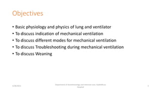
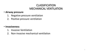
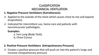
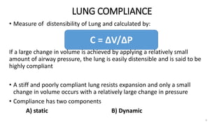
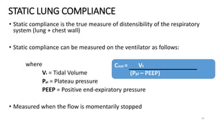
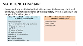
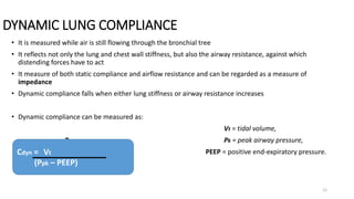
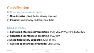
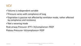
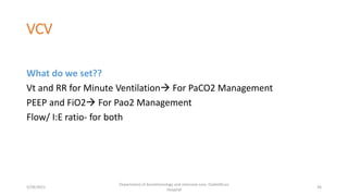
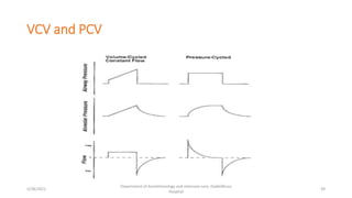
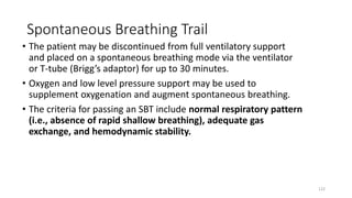
Mechanical Ventilation: Part –I(Basics and Modes)
Presenter:
Dr. Tirtha Raj Bhandari
Anesthesiologist
Dadeldhura Hospital, Dadeldhura
5/28/2021
Department of Anesthesiology and intensive care, Dadeldhura
Hospital
1
Objectives
• Basic physiology and physics of lung and ventilator
• To discuss indication of mechanical ventilation
• To discuss different modes for mechanical ventilation
• To discuss Troubleshooting during mechanical ventilation
• To discuss Weaning
5/28/2021
Department of Anesthesiology and intensive care, Dadeldhura
Hospital
2
CLASSIFICATION
MECHANICAL VENTILATION
3
• Airway pressure
1. Negative pressure ventilation
2. Positive pressure ventilation
• Invasiveness
1. Invasive Ventilation
2. Non-invasive mechanical ventilation
CLASSIFICATION
MECHANICAL VENTILATION
4
1. Negative Pressure Ventilators (Extrathoracic)
 Applied to the outside of the chest which causes chest to rise and expand
(inspiration).
 Indicated for intermittent use, home care and patients with
neuromuscular pathologies.
Examples:
a. lron Lung (Body Tank).
b. Chest Cuirass
2. Positive Pressure Ventilators (lntrapulmonary Pressure)
 Creates a positive pressure that will push air into the patient's lungs and
increase intrapulmonary pressure
AIRWAY RESISTANCE
5
• Occurs as a result of the friction between the air molecules and the walls of
the tracheobronchial tree, and to some extent, as a result of the friction
between the air molecules themselves
• Poiseuille’s law states that the resistance (Raw) to the flow of fluids through a
long and narrow tube is:
►Proportional to the length of the tube (l) and the viscosity of the
fluid (η)
► Inversely proportional to the fourth power of the radius (r).
• This means small changes in the radius can have inordinate effects on airway
resistance
AIRWAY RESISTANCE
6
• Normal Raw ranges from 0.6 to 2.4 cm H2O/L/s. With an endotracheal tube in situ
• The driving pressure across a tube in ventilated patient is a function of the difference
between the pressures at its ends
• In a patient who is not being given positive pressure breaths is dependent upon the
difference between the atmospheric pressure (Patm) and the alveolar pressure (PALV)
(Raw) in a Spontaneous breathing
Patient
AIRWAY RESISTANCE
7
Clinical conditions that increase airway resistance are:
AIRWAY RESISTANCE
8
• The airflow resistance of a patient-ventilator system may be monitored using the pressure-
volume (P-V) loop
• An increased bowing of the P-V
loop suggests an overall increase
in airflow resistance
• When the inspiratory flow exceeds
a patient’s tidal volume and
inspiratory time requirement,
bowing of the inspiratory limb may
result (line A2)
• When the expiratory airflow resistance is increased (e.g., bronchospasm), bowing of the
expiratory limb (line B2) may occur
LUNG COMPLIANCE
9
• Measure of distensibility of Lung and calculated by:
If a large change in volume is achieved by applying a relatively small
amount of airway pressure, the lung is easily distensible and is said to be
highly compliant
• A stiff and poorly compliant lung resists expansion and only a small
change in volume occurs with a relatively large change in pressure
• Compliance has two components
A) static B) Dynamic
C = ∆V/∆P
STATIC LUNG COMPLIANCE
10
• Static compliance is the true measure of distensibility of the respiratory
system (lung + chest wall)
• Static compliance can be measured on the ventilator as follows:
where
Vt = Tidal Volume
Ppl = Plateau pressure
PEEP = Positive end-expiratory pressure
• Measured when the flow is momentarily stopped
Cstat = Vt
(Ppl – PEEP)
STATIC LUNG COMPLIANCE
11
• In mechanically ventilated patient with an essentially normal chest wall
and lungs, the static compliance of the respiratory system is usually in the
range of 70–100 mL/cm H2O
DYNAMIC LUNG COMPLIANCE
12
• It is measured while air is still flowing through the bronchial tree
• It reflects not only the lung and chest wall stiffness, but also the airway resistance, against which
distending forces have to act
• It measure of both static compliance and airflow resistance and can be regarded as a measure of
impedance
• Dynamic compliance falls when either lung stiffness or airway resistance increases
• Dynamic compliance can be measured as:
Vt = tidal volume,
P Pk = peak airway pressure,
PEEP = positive end-expiratory pressure.
Cdyn = Vt
(Ppk – PEEP)
LUNG COMPLIANCE
13
LUNG COMPLIANCE
14
• Static and dynamic compliance in various lung conditions
Alveolar Ventilation & Dead-Space
• The total surface area of the alveolar epithelium is about 72–80 m2
• Approximately 85–95% (about 70 m2) of this is in contact with pulmonary
capillaries: this constitutes the alveolocapillary interface
• The part of the inspired gas that does not come into contact with the
pulmonary capillary bed is termed the dead-space
• Dead space can be: 1)Anatomical
2)Alveolar
3) Physiological
Alveolar Ventilation & Dead-Space
• Minute ventilation is the product of
the tidal volume times the respiratory
rate
• It is inversely proportional to the
PaCO2.
• It also affects the PaO2 in a nonlinear
fashion; however, manipulating the
minute volume for the change in PaO2
is undesirable, as only
• small changes in PaO2 can be brought
about by large alterations in minute
ventilation.
• Such large changes in minute
ventilation can have a profound and
unwanted effect on PaCO2.
EXAMPLE:
• A set tidal volume of 500 mL and a RR of 10 breaths/min results in a minute ventilation of 500
× 10 = 5,000 mL/min
• The same minute ventilation can be produced by a tidal volume of 250 mL delivered at a RR of 20
breaths/min 250 × 20 = 5,000 mL/min
• When dead-space 150ML is taken the alveolar ventilation
In the first example would be (500 – 150) × 10 = 3,500,
In the second example would be (250 – 150) × 20 = 2,000
• The alveolar ventilation in the first instance would vastly exceed the alveolar ventilation in the second
example
• The PaCO2 is inversely proportional not to all of the minute ventilation, but to that part of the ventilation
that is independent of dead-space
Alveolar Ventilation & Dead-Space
Peak Inspiratory Pressure (Ppeak)
• During a ventilator-driven tidal breath, the
airway pressure rises rapidly to a peak which is
called as Peak Inspiratory Pressure
• It is influenced both by airway resistance and
compliance
• It can be high either on account of narrowed
airways or stiff lung
• It is used to deliver the tidal volume by
overcoming non-elastic (airways) and elastic(lung
parenchyma) resistance
Plateau Pressure (Pplateau)
• At the end of inspiration, the airway
pressure falls to a plateau as the air
diffuses out to the periphery of
tracheobronchial tree
• This pressure within the airway during
no airflow period is called the pause
pressure or the plateau pressure
• It is a reflection of the static compliance
• Any condition that stiffens the lung will
increase the pause pressure.
Plateau Pressure (Pplateau)
POSITIVE END-EXPIRATORY
PRESSURE (PEEP)
• PEEP is alveolar pressure above atmospheric pressure that exist at the end of
expiration
• Types of PEEP:
A)Extrinsic PEEP
►PEEP provided by mechanical ventilator
B)Intrinsic PEEP
• Secondary to incomplete expiration
• Also known as auto PEEP
Baseline Pressure PEEP/CPAP
• Physiological effects of positive end-expiratory pressure (PEEP)
Mean Airway Pressure(Pmean)
►Average pressure within the airway over the entire respiratory cycle
►Is significantly affected by:
 PEEP
 Inspiratory time (TI)
►Is affected to a lesser degree by:
 Peak pressure
►Has significant affect on oxygenation
AIRWAY OPENING
PRESSURE
PLEURAL & ALVEOLAR PRESSURE
• Pressure, volume and flow relationship is described by equation of motion
for the respiratory system. (stems from Newton’s third law of motion)
PTR = PE+PR
PTR Transrespiratory pressure(P at the airway opening minus pressure at
the body surface)
PE pressure caused by elastic resistance,
PR Resistance due to airflow
29
Physics
Volume
Compliance
A (PAW)
Flow x resistance +
Physics
PEEP
Compliance
Volume
Resistance
Flow
(Paw)
pressure
Airway 



Only possible to set one of:
Pressure
Flow
Volume
Other variables become a dependent variable
+PEEP
Indications
1) Can not oxygenate (low PaO2/SpO2)- Hypoxemic respiratory failure
2) Can not ventilate (high PCO2)- Hypercarbic Respiratory failure
3) Both
4) Can not protect airway/ secure airway( Low GCS)
5) When clinician/ physician confused, either patient need or not,
needs mechanical ventilation
6) Elective ventilation for GA
5/28/2021
Department of Anesthesiology and intensive care, Dadeldhura
Hospital
31
Goals/Principles of mechanical Ventilation
1) To provide adequate ventilation and oxygenation
2) To achieve adequate lung volume and recruitment
3) To improve lung compliance
4) To reduce work of breathing
5) To limit lung injury
5/28/2021
Department of Anesthesiology and intensive care, Dadeldhura
Hospital
32
Assessment for MV
We should know
1)History and physical examination
2) Laboratory and radiological finding of patient
3) Anatomical size and structure of airway and lungs
4) Disease severity
5/28/2021
Department of Anesthesiology and intensive care, Dadeldhura
Hospital
33
Classification
Both on inferior airway invasion
1) Non- invasive : No inferior airway invasion
2) Invasive: Invasion by endotracheal tube
Based on modes
1) Controlled Mechanical Ventilation: PCV, VCV, PRVC, HFV, CMV, IMV
2) Supported spontaneous breathing: PSV, VSV
3)Mixed Respiratory Support: SIMV/ or +PS
4) Assisted spontaneous breathing: CPAP, APRV
5/28/2021
Department of Anesthesiology and intensive care, Dadeldhura
Hospital
34
VCV
Volume is independent variable
Pressure varies with compliance of lung
Expiration is passive not affected by ventilator mode, rather affected
by compliance and resistance
Not a weaning mode
Peak airway Pressure= R*F+ Vt/compliance+ PEEP
Plateau Pressure= Vt/compliance+ PEEP
5/28/2021
Department of Anesthesiology and intensive care, Dadeldhura
Hospital
35
VCV
What do we set??
Vt and RR for Minute Ventilation For PaCO2 Management
PEEP and FiO2 For Pao2 Management
Flow/ I:E ratio- for both
5/28/2021
Department of Anesthesiology and intensive care, Dadeldhura
Hospital
36
PCV
Pressure is independent variable
Volume is dependent variable Depends on: level of pressure, I-time
also it is the function of compliance and airway resistance. That’s why
volume deliver can vary breath to breath.
Ventilator adjust flow to maintain pressure.
Flow decreases throughout the inspiratory cycle.
Good mode to use if patient has large air leak, the ventilator will
increase the flow to compensate it.
Not a weaning mode
5/28/2021
Department of Anesthesiology and intensive care, Dadeldhura
Hospital
37
PCV
What do we set??
Pressure limit, T-Insp, RR,FIO2,PEEP
P and RR for Minute ventilation PaCO2 management
FiO2 and PEEP for PaO2 management
5/28/2021
Department of Anesthesiology and intensive care, Dadeldhura
Hospital
38
VCV and PCV
5/28/2021
Department of Anesthesiology and intensive care, Dadeldhura
Hospital
39
Pressure Support=PS
Supports each spontaneous breath with supplemental flow to
achieve preset pressure
All breath are triggered by patient
Preset value  PIP, PEEP, FiO2
Patient determine Rate, Ti, I/E ratio, TV
5/28/2021
Department of Anesthesiology and intensive care, Dadeldhura
Hospital
40
PS
Needs intact respiratory drive
Helps to overcome airway resistance/ tube resistance, so that
spontaneous breathing will be easier
Can not be use in patient not having spontaneous breathing(i.e.
muscle relaxant)
5/28/2021
Department of Anesthesiology and intensive care, Dadeldhura
Hospital
41
SIMV= Synchronized intermittent mandatory
ventilation
Preset mechanical breath delivered within interval acc to preset and
wait for spontaneous in between, which it will use as a trigger to
deliver full breath.
If not sensed it will automatically give a breath
Vt on spontaneous breaths depends entirely upon the patient effort
and lung mechanics, can be pressure or volume controlled.
5/28/2021
Department of Anesthesiology and intensive care, Dadeldhura
Hospital
42
SIMV
Tb(time for breathing) = Tm(mandatory)+Ts(spontaneous)
If patient tries to breath during Tm, ventilator gives a fully assisted
breath
If Patient tries to breath during Ts, the ventilator will allow the
patient to take breath
5/28/2021
Department of Anesthesiology and intensive care, Dadeldhura
Hospital
43
5/28/2021
Department of Anesthesiology and intensive care, Dadeldhura
Hospital
44
5/28/2021
Department of Anesthesiology and intensive care, Dadeldhura
Hospital
45
5/28/2021
Department of Anesthesiology and intensive care, Dadeldhura
Hospital
46
SIMV+PS
SIMV+PS= provides assistance for spontaneous breath to overcome tube resistance
5/28/2021
Department of Anesthesiology and intensive care, Dadeldhura
Hospital
47
CPAP
Provides continuous positive pressure throughout the respiratory
cycle
So gives supports to inspiration and resistance to expiration
Can be use both in invasive and non-invasive form.
Very similar to PEEP
5/28/2021
Department of Anesthesiology and intensive care, Dadeldhura
Hospital
48
PEEP
PEEP is a residual pressure above atmospheric pressure maintained
at the end of expiration.
PEEP can be added to any mode
PEEP helps to recruit alveoli, increase FRC , Redistribute pulmonary
edema, Decrease intrapulmonary shunt, Increase PaO2.(GOOD)
PEEP , decreases venous return/CO, increase ICP/intensify cerebral
ischemia/ risk of barotrauma(Bad)
5/28/2021
Department of Anesthesiology and intensive care, Dadeldhura
Hospital
49
CPAP
5/28/2021 Dadeldhura Hospital Dadeldhura 50
CPAP
5/28/2021 Dadeldhura Hospital Dadeldhura 51
CPAP
5/28/2021 Dadeldhura Hospital Dadeldhura 52
BiPAP
5/28/2021 Dadeldhura Hospital Dadeldhura 53
BiPAP
5/28/2021 Dadeldhura Hospital Dadeldhura 54
CPAP Vs BiPAP
5/28/2021 Dadeldhura Hospital Dadeldhura 55
CPAP Vs BiPAP
5/28/2021 Dadeldhura Hospital Dadeldhura 56
5/28/2021 Dadeldhura Hospital Dadeldhura 57
5/28/2021 Dadeldhura Hospital Dadeldhura 58
5/28/2021 Dadeldhura Hospital Dadeldhura 59
5/28/2021 Dadeldhura Hospital Dadeldhura 60
IPAP/EPAP
5/28/2021 Dadeldhura Hospital Dadeldhura 61
5/28/2021 Dadeldhura Hospital Dadeldhura 62
5/28/2021 Dadeldhura Hospital Dadeldhura 63
5/28/2021 Dadeldhura Hospital Dadeldhura 64
5/28/2021 Dadeldhura Hospital Dadeldhura 65
5/28/2021 Dadeldhura Hospital Dadeldhura 66
5/28/2021 Dadeldhura Hospital Dadeldhura 67
5/28/2021 Dadeldhura Hospital Dadeldhura 68
NASAL CPAP FOR NEONATE/INFANT
Nasal prong/Nasopharyngral tube/ET tube can be use for nasal cpap
5/28/2021
Department of Anesthesiology and intensive care, Dadeldhura
Hospital
69
Indication
Disease condition;
1)Retained lung fluid
2)Post-extubation (if risk of airway collapse)
3)Atelectasis
4)Respiratory distress syndrome
5) For administration of controlled concentration of nitric oxide
5/28/2021
Department of Anesthesiology and intensive care, Dadeldhura
Hospital
70
CONTD
Physical findings:
1)Increase WOB ,indicated by increase RR by 30-40%
2)Sub-sternal/suprasternal retraction
3)Grunting and nasal flaring
4)Pale or cyanotic skin color
5)Agitation
5/28/2021
Department of Anesthesiology and intensive care, Dadeldhura
Hospital
71
Contraindication/ Should 'Not Try
C/I:
Choanal atresia
Untreated diaphragmatic hernia
Cleft palate
TEF
Should 'not try:
Cardiovascular instability
Severe ventilatory impairment
Severe hypoxemia
Frequent apnea
High level of sedation
5/28/2021
Department of Anesthesiology and intensive care, Dadeldhura
Hospital
72
How it Helps??
Reduces grunting and tachypnea
Increases FRC and PaO2
Decreases intrapulmonary shunting
Improves lung compliance
Aids in stabilization of floppy infant chest wall
5/28/2021
Department of Anesthesiology and intensive care, Dadeldhura
Hospital
73
CONTD
Improves distribution of ventilation
Reduce WOB
Reduces central and obstructive sleep apnea by mechanically
splinting the upper airway.
Better recruitment and oxygenation
Stimulation of infant/neonate for breathing
5/28/2021
Department of Anesthesiology and intensive care, Dadeldhura
Hospital
74
Mechanical Ventilation:
Part-II(Troubleshooting and Weaning)
Presenter:
Dr.Tirtha Raj Bhandari
Anesthesiologist
Dadeldhura Hospital,dadeldhura
5/28/2021
Department of Anesthesiology and intensive care, Dadeldhura
Hospital
75
Troubleshooting and Weaning
5/28/2021
Department of Anesthesiology and intensive care, Dadeldhura
Hospital
76
Common Troubleshooting
1) Ventilator alarms
2) Hypoxia(Desaturation)
3) Hypotension
4) Patient ventilator dysynchrony
77
Assessment
• Is the chest moving and is it moving symmetrically?
• Is the patient cyanosed?
• What is the arterial saturation?
• Is the patient haemo-dynamically stable?
-Do not forget to observe respiratory pattern and feature of respiratory
distress
78
High Airway Pressure
Why does this matter??
-Risk of barotrauma
-Hypoventilation(premature termination by high airway pressure)
79
Causes of high airway pressure;
80
CONTD
• Ventilator malfunction/setting
• circuits problems
• Endotracheal tube obstruction
• Increase airway resistance
• Decrease compliance of lung
81
Dynamic Compliance Static compliance(lung)
Bronchospasm Obesity
Kinking of tubes Atelectasis, ARDS
Airway obstruction(mucus, secretions) Pneumothorax
Retained secretion
82
Dynamic compliance  PIP
83
Static Compliance Both PIP and PLAT
84
Low Airway Pressure/volume
Why it is important??
-Risk of hypoventilation hypoxia and hypercarbia(Lactic and
respiratory acidosis)  Deasaturation Bradycardia Cardiac Arrest
85
86
Auto-peep
87
Causes of Auto-peep formation
• Inadequate time for expiration, either by flow limitation or
development of resistance in airway or endotracheal tube
• More time for inspiration
• Obstructive airway disease
• High minute ventilation( High Tidal Volume, High Frequency)
• Dysynchrony
88
Pathophysiological Consequences of air
trapping
• Air-trappingDynamic hyperinflation Autopeep
leads following consequences,
1) Increase intra-thoracic pressure
2) Increase work of breathing
3) Decrease preload  Hypotension
4) Worsened V/Q mismatch
5) Increase risk of barotrauma
89
90
91
92
Management of Auto-PEEP
• Depends upon cause
-decrease MV
-allow sufficient time for expiration
-bronchodilator
-use higher size tube
-remove obstruction in airway
-increase sedation
93
Desaturation(Hypoxia)
• Ventilatory Malfunction
• ET tube disconnection(circuit malfunction), cuff leak
• Oxygen failure
• Any causes of hypoxic respiratory failure
• Special consideration: endobronchial intubation,
pneumothorax, collapse of part of lung, pulmonary
edema, bronchospasm and pulmonary embolism
94
Management
• Increase Fio2 to 1.0
• Check whether the chest is moving or not
• Briefly examine chest to determine the cause of desaturation
• If cause is not obvious manually ventilate the patient with
100% oxygen to exclude ventilator malfunction as the cause
• Treat underlying cause
• Alter ventilator settings to improve oxygenation
• Chest x-ray
95
96
97
Hypotension
Immediately after the initiation of mechanical ventilation
hypotension can occur:
Causes:
• Relative Hypovolaemia: reduction in venous return exacerbated
by positive intra-thoracic pressure.
• Drug induced vasodilation and myocardial depression: all
induction agents have some short lived vaso-dilatory myocardial
depressant effects.
• Tension pneumothorax
• Gas trapping ( dyanamic hyperinflation)
Delayed cause may be due to pathological process going on in
patient’s body.
98
Management
• Depends upon causes
-fluid therapy
-decrease positive pressure/peep
-treat air trapping
-chest tube(if pneumothorax)
99
Dysynchrony
100
Dysynchrony
According to its relationship to specific phases of the delivered breath:
• breath initiation/trigger,
• flow delivery and
• breath cycling/termination.
*Asynchrony/dysynchrony index
101
102
Ineffective trigger
Causes: improper setting of sensitivity, weak patient
effort, auto-peep 103
Auto-trigger
Causes: low trigger sensitivity, circuit leak, water in the circuit, cardiac oscillation. 104
Double trigger
Causes: unusually high ventilatory demand, low PaO2/FiO2, longer/too short inspiratory time of patient
105
Flow Dysynchrony
Causes: High respiratory drive, insufficient flow setting 106
Premature cycling
Causes: Increase neural inspiratory time, earlier termination of inspiration by ventilator
107
Delayed cycling
108
Management
• Select the appropriate mode
• Proper trigger setting
• Adjust adequate flow
• Adjust proper cycling
• Clear airway
• Treat auto-peep
• Sedation
• Neuromuscular blockade
109
WEANING
• Weaning is the process of withdrawing mechanical
ventilatory support and transferring the work of
breathing from the ventilator to the patient.
• In most cases, weaning may be accomplished rapidly
from full ventilator support to unassisted spontaneous
breathing.
110
111
112
Formula
• RSBI= RR(f)/Tidal Volume(L)
• P/F ratio: PO2/FiO2:,FiO2=1=100%
5/28/2021
Department of Anesthesiology and intensive care, Dadeldhura
Hospital
113
Formula
5/28/2021
Department of Anesthesiology and intensive care, Dadeldhura
Hospital
114
Formula
5/28/2021
Department of Anesthesiology and intensive care, Dadeldhura
Hospital
115
116
Weaning success
• Weaning success is defined as absence of ventilatory
support 48 hours following the extubation.
• The spontaneous breaths are unassisted by mechanical
ventilation,
• Supplemental oxygen, bronchodilators, pressure
support ventilation, or continuous positive airway
pressure may be used to support and maintain adequate
spontaneous ventilation and oxygenation.
117
Weaning In Progress
• Weaning in progress is an intermediate category (between weaning
success and weaning failure) for patients who are extubated but
continue to receive ventilatory support by noninvasive ventilation
(NIV)
118
Weaning Failure
• Weaning failure is defined as either the failure of
spontaneous breathing trial (SBT) or for reintubation
within 48 hours following extubation.
• Patients who fail the SBT often exhibit the following
clinical signs: tachypnea, tachycardia, hypertension,
hypotension, hypoxemia, acidosis, or arrhythmias.
• Physical signs of SBT failure may include agitation,
distress, diminished mental status, diaphoresis, and
increased work of breathing
119
120
CONTD
• Causes of failure are
1)Increase airway resistance
2)Decrease in compliance of lungs
3)Respiratory muscle fatigue
121
Spontaneous Breathing Trail
• The patient may be discontinued from full ventilatory support
and placed on a spontaneous breathing mode via the ventilator
or T-tube (Brigg’s adaptor) for up to 30 minutes.
• Oxygen and low level pressure support may be used to
supplement oxygenation and augment spontaneous breathing.
• The criteria for passing an SBT include normal respiratory pattern
(i.e., absence of rapid shallow breathing), adequate gas
exchange, and hemodynamic stability.
122
123
Rapid Shallow Breathing Index(RSBI)
124
<100=success of weaning
>100=failure of weaning
125
5/28/2021
Department of Anesthesiology and intensive care, Dadeldhura
Hospital
126

Basics of Mechanical Ventilation

  • 1.
    Mechanical Ventilation: Part–I(Basics and Modes) Presenter: Dr. Tirtha Raj Bhandari Anesthesiologist Dadeldhura Hospital, Dadeldhura 5/28/2021 Department of Anesthesiology and intensive care, Dadeldhura Hospital 1
  • 2.
    Objectives • Basic physiologyand physics of lung and ventilator • To discuss indication of mechanical ventilation • To discuss different modes for mechanical ventilation • To discuss Troubleshooting during mechanical ventilation • To discuss Weaning 5/28/2021 Department of Anesthesiology and intensive care, Dadeldhura Hospital 2
  • 3.
    CLASSIFICATION MECHANICAL VENTILATION 3 • Airwaypressure 1. Negative pressure ventilation 2. Positive pressure ventilation • Invasiveness 1. Invasive Ventilation 2. Non-invasive mechanical ventilation
  • 4.
    CLASSIFICATION MECHANICAL VENTILATION 4 1. NegativePressure Ventilators (Extrathoracic)  Applied to the outside of the chest which causes chest to rise and expand (inspiration).  Indicated for intermittent use, home care and patients with neuromuscular pathologies. Examples: a. lron Lung (Body Tank). b. Chest Cuirass 2. Positive Pressure Ventilators (lntrapulmonary Pressure)  Creates a positive pressure that will push air into the patient's lungs and increase intrapulmonary pressure
  • 5.
    AIRWAY RESISTANCE 5 • Occursas a result of the friction between the air molecules and the walls of the tracheobronchial tree, and to some extent, as a result of the friction between the air molecules themselves • Poiseuille’s law states that the resistance (Raw) to the flow of fluids through a long and narrow tube is: ►Proportional to the length of the tube (l) and the viscosity of the fluid (η) ► Inversely proportional to the fourth power of the radius (r). • This means small changes in the radius can have inordinate effects on airway resistance
  • 6.
    AIRWAY RESISTANCE 6 • NormalRaw ranges from 0.6 to 2.4 cm H2O/L/s. With an endotracheal tube in situ • The driving pressure across a tube in ventilated patient is a function of the difference between the pressures at its ends • In a patient who is not being given positive pressure breaths is dependent upon the difference between the atmospheric pressure (Patm) and the alveolar pressure (PALV) (Raw) in a Spontaneous breathing Patient
  • 7.
    AIRWAY RESISTANCE 7 Clinical conditionsthat increase airway resistance are:
  • 8.
    AIRWAY RESISTANCE 8 • Theairflow resistance of a patient-ventilator system may be monitored using the pressure- volume (P-V) loop • An increased bowing of the P-V loop suggests an overall increase in airflow resistance • When the inspiratory flow exceeds a patient’s tidal volume and inspiratory time requirement, bowing of the inspiratory limb may result (line A2) • When the expiratory airflow resistance is increased (e.g., bronchospasm), bowing of the expiratory limb (line B2) may occur
  • 9.
    LUNG COMPLIANCE 9 • Measureof distensibility of Lung and calculated by: If a large change in volume is achieved by applying a relatively small amount of airway pressure, the lung is easily distensible and is said to be highly compliant • A stiff and poorly compliant lung resists expansion and only a small change in volume occurs with a relatively large change in pressure • Compliance has two components A) static B) Dynamic C = ∆V/∆P
  • 10.
    STATIC LUNG COMPLIANCE 10 •Static compliance is the true measure of distensibility of the respiratory system (lung + chest wall) • Static compliance can be measured on the ventilator as follows: where Vt = Tidal Volume Ppl = Plateau pressure PEEP = Positive end-expiratory pressure • Measured when the flow is momentarily stopped Cstat = Vt (Ppl – PEEP)
  • 11.
    STATIC LUNG COMPLIANCE 11 •In mechanically ventilated patient with an essentially normal chest wall and lungs, the static compliance of the respiratory system is usually in the range of 70–100 mL/cm H2O
  • 12.
    DYNAMIC LUNG COMPLIANCE 12 •It is measured while air is still flowing through the bronchial tree • It reflects not only the lung and chest wall stiffness, but also the airway resistance, against which distending forces have to act • It measure of both static compliance and airflow resistance and can be regarded as a measure of impedance • Dynamic compliance falls when either lung stiffness or airway resistance increases • Dynamic compliance can be measured as: Vt = tidal volume, P Pk = peak airway pressure, PEEP = positive end-expiratory pressure. Cdyn = Vt (Ppk – PEEP)
  • 13.
  • 14.
    LUNG COMPLIANCE 14 • Staticand dynamic compliance in various lung conditions
  • 15.
    Alveolar Ventilation &Dead-Space • The total surface area of the alveolar epithelium is about 72–80 m2 • Approximately 85–95% (about 70 m2) of this is in contact with pulmonary capillaries: this constitutes the alveolocapillary interface • The part of the inspired gas that does not come into contact with the pulmonary capillary bed is termed the dead-space • Dead space can be: 1)Anatomical 2)Alveolar 3) Physiological
  • 16.
  • 17.
    Alveolar Ventilation &Dead-Space • Minute ventilation is the product of the tidal volume times the respiratory rate • It is inversely proportional to the PaCO2. • It also affects the PaO2 in a nonlinear fashion; however, manipulating the minute volume for the change in PaO2 is undesirable, as only • small changes in PaO2 can be brought about by large alterations in minute ventilation. • Such large changes in minute ventilation can have a profound and unwanted effect on PaCO2.
  • 18.
    EXAMPLE: • A settidal volume of 500 mL and a RR of 10 breaths/min results in a minute ventilation of 500 × 10 = 5,000 mL/min • The same minute ventilation can be produced by a tidal volume of 250 mL delivered at a RR of 20 breaths/min 250 × 20 = 5,000 mL/min • When dead-space 150ML is taken the alveolar ventilation In the first example would be (500 – 150) × 10 = 3,500, In the second example would be (250 – 150) × 20 = 2,000 • The alveolar ventilation in the first instance would vastly exceed the alveolar ventilation in the second example • The PaCO2 is inversely proportional not to all of the minute ventilation, but to that part of the ventilation that is independent of dead-space Alveolar Ventilation & Dead-Space
  • 19.
  • 20.
    Peak Inspiratory Pressure(Ppeak) • During a ventilator-driven tidal breath, the airway pressure rises rapidly to a peak which is called as Peak Inspiratory Pressure • It is influenced both by airway resistance and compliance • It can be high either on account of narrowed airways or stiff lung • It is used to deliver the tidal volume by overcoming non-elastic (airways) and elastic(lung parenchyma) resistance
  • 21.
    Plateau Pressure (Pplateau) •At the end of inspiration, the airway pressure falls to a plateau as the air diffuses out to the periphery of tracheobronchial tree • This pressure within the airway during no airflow period is called the pause pressure or the plateau pressure • It is a reflection of the static compliance • Any condition that stiffens the lung will increase the pause pressure.
  • 22.
  • 23.
    POSITIVE END-EXPIRATORY PRESSURE (PEEP) •PEEP is alveolar pressure above atmospheric pressure that exist at the end of expiration • Types of PEEP: A)Extrinsic PEEP ►PEEP provided by mechanical ventilator B)Intrinsic PEEP • Secondary to incomplete expiration • Also known as auto PEEP
  • 24.
    Baseline Pressure PEEP/CPAP •Physiological effects of positive end-expiratory pressure (PEEP)
  • 25.
    Mean Airway Pressure(Pmean) ►Averagepressure within the airway over the entire respiratory cycle ►Is significantly affected by:  PEEP  Inspiratory time (TI) ►Is affected to a lesser degree by:  Peak pressure ►Has significant affect on oxygenation
  • 26.
  • 27.
  • 29.
    • Pressure, volumeand flow relationship is described by equation of motion for the respiratory system. (stems from Newton’s third law of motion) PTR = PE+PR PTR Transrespiratory pressure(P at the airway opening minus pressure at the body surface) PE pressure caused by elastic resistance, PR Resistance due to airflow 29 Physics
  • 30.
    Volume Compliance A (PAW) Flow xresistance + Physics PEEP Compliance Volume Resistance Flow (Paw) pressure Airway     Only possible to set one of: Pressure Flow Volume Other variables become a dependent variable +PEEP
  • 31.
    Indications 1) Can notoxygenate (low PaO2/SpO2)- Hypoxemic respiratory failure 2) Can not ventilate (high PCO2)- Hypercarbic Respiratory failure 3) Both 4) Can not protect airway/ secure airway( Low GCS) 5) When clinician/ physician confused, either patient need or not, needs mechanical ventilation 6) Elective ventilation for GA 5/28/2021 Department of Anesthesiology and intensive care, Dadeldhura Hospital 31
  • 32.
    Goals/Principles of mechanicalVentilation 1) To provide adequate ventilation and oxygenation 2) To achieve adequate lung volume and recruitment 3) To improve lung compliance 4) To reduce work of breathing 5) To limit lung injury 5/28/2021 Department of Anesthesiology and intensive care, Dadeldhura Hospital 32
  • 33.
    Assessment for MV Weshould know 1)History and physical examination 2) Laboratory and radiological finding of patient 3) Anatomical size and structure of airway and lungs 4) Disease severity 5/28/2021 Department of Anesthesiology and intensive care, Dadeldhura Hospital 33
  • 34.
    Classification Both on inferiorairway invasion 1) Non- invasive : No inferior airway invasion 2) Invasive: Invasion by endotracheal tube Based on modes 1) Controlled Mechanical Ventilation: PCV, VCV, PRVC, HFV, CMV, IMV 2) Supported spontaneous breathing: PSV, VSV 3)Mixed Respiratory Support: SIMV/ or +PS 4) Assisted spontaneous breathing: CPAP, APRV 5/28/2021 Department of Anesthesiology and intensive care, Dadeldhura Hospital 34
  • 35.
    VCV Volume is independentvariable Pressure varies with compliance of lung Expiration is passive not affected by ventilator mode, rather affected by compliance and resistance Not a weaning mode Peak airway Pressure= R*F+ Vt/compliance+ PEEP Plateau Pressure= Vt/compliance+ PEEP 5/28/2021 Department of Anesthesiology and intensive care, Dadeldhura Hospital 35
  • 36.
    VCV What do weset?? Vt and RR for Minute Ventilation For PaCO2 Management PEEP and FiO2 For Pao2 Management Flow/ I:E ratio- for both 5/28/2021 Department of Anesthesiology and intensive care, Dadeldhura Hospital 36
  • 37.
    PCV Pressure is independentvariable Volume is dependent variable Depends on: level of pressure, I-time also it is the function of compliance and airway resistance. That’s why volume deliver can vary breath to breath. Ventilator adjust flow to maintain pressure. Flow decreases throughout the inspiratory cycle. Good mode to use if patient has large air leak, the ventilator will increase the flow to compensate it. Not a weaning mode 5/28/2021 Department of Anesthesiology and intensive care, Dadeldhura Hospital 37
  • 38.
    PCV What do weset?? Pressure limit, T-Insp, RR,FIO2,PEEP P and RR for Minute ventilation PaCO2 management FiO2 and PEEP for PaO2 management 5/28/2021 Department of Anesthesiology and intensive care, Dadeldhura Hospital 38
  • 39.
    VCV and PCV 5/28/2021 Departmentof Anesthesiology and intensive care, Dadeldhura Hospital 39
  • 40.
    Pressure Support=PS Supports eachspontaneous breath with supplemental flow to achieve preset pressure All breath are triggered by patient Preset value  PIP, PEEP, FiO2 Patient determine Rate, Ti, I/E ratio, TV 5/28/2021 Department of Anesthesiology and intensive care, Dadeldhura Hospital 40
  • 41.
    PS Needs intact respiratorydrive Helps to overcome airway resistance/ tube resistance, so that spontaneous breathing will be easier Can not be use in patient not having spontaneous breathing(i.e. muscle relaxant) 5/28/2021 Department of Anesthesiology and intensive care, Dadeldhura Hospital 41
  • 42.
    SIMV= Synchronized intermittentmandatory ventilation Preset mechanical breath delivered within interval acc to preset and wait for spontaneous in between, which it will use as a trigger to deliver full breath. If not sensed it will automatically give a breath Vt on spontaneous breaths depends entirely upon the patient effort and lung mechanics, can be pressure or volume controlled. 5/28/2021 Department of Anesthesiology and intensive care, Dadeldhura Hospital 42
  • 43.
    SIMV Tb(time for breathing)= Tm(mandatory)+Ts(spontaneous) If patient tries to breath during Tm, ventilator gives a fully assisted breath If Patient tries to breath during Ts, the ventilator will allow the patient to take breath 5/28/2021 Department of Anesthesiology and intensive care, Dadeldhura Hospital 43
  • 44.
    5/28/2021 Department of Anesthesiologyand intensive care, Dadeldhura Hospital 44
  • 45.
    5/28/2021 Department of Anesthesiologyand intensive care, Dadeldhura Hospital 45
  • 46.
    5/28/2021 Department of Anesthesiologyand intensive care, Dadeldhura Hospital 46
  • 47.
    SIMV+PS SIMV+PS= provides assistancefor spontaneous breath to overcome tube resistance 5/28/2021 Department of Anesthesiology and intensive care, Dadeldhura Hospital 47
  • 48.
    CPAP Provides continuous positivepressure throughout the respiratory cycle So gives supports to inspiration and resistance to expiration Can be use both in invasive and non-invasive form. Very similar to PEEP 5/28/2021 Department of Anesthesiology and intensive care, Dadeldhura Hospital 48
  • 49.
    PEEP PEEP is aresidual pressure above atmospheric pressure maintained at the end of expiration. PEEP can be added to any mode PEEP helps to recruit alveoli, increase FRC , Redistribute pulmonary edema, Decrease intrapulmonary shunt, Increase PaO2.(GOOD) PEEP , decreases venous return/CO, increase ICP/intensify cerebral ischemia/ risk of barotrauma(Bad) 5/28/2021 Department of Anesthesiology and intensive care, Dadeldhura Hospital 49
  • 50.
  • 51.
  • 52.
  • 53.
  • 54.
  • 55.
    CPAP Vs BiPAP 5/28/2021Dadeldhura Hospital Dadeldhura 55
  • 56.
    CPAP Vs BiPAP 5/28/2021Dadeldhura Hospital Dadeldhura 56
  • 57.
  • 58.
  • 59.
  • 60.
  • 61.
  • 62.
  • 63.
  • 64.
  • 65.
  • 66.
  • 67.
  • 68.
  • 69.
    NASAL CPAP FORNEONATE/INFANT Nasal prong/Nasopharyngral tube/ET tube can be use for nasal cpap 5/28/2021 Department of Anesthesiology and intensive care, Dadeldhura Hospital 69
  • 70.
    Indication Disease condition; 1)Retained lungfluid 2)Post-extubation (if risk of airway collapse) 3)Atelectasis 4)Respiratory distress syndrome 5) For administration of controlled concentration of nitric oxide 5/28/2021 Department of Anesthesiology and intensive care, Dadeldhura Hospital 70
  • 71.
    CONTD Physical findings: 1)Increase WOB,indicated by increase RR by 30-40% 2)Sub-sternal/suprasternal retraction 3)Grunting and nasal flaring 4)Pale or cyanotic skin color 5)Agitation 5/28/2021 Department of Anesthesiology and intensive care, Dadeldhura Hospital 71
  • 72.
    Contraindication/ Should 'NotTry C/I: Choanal atresia Untreated diaphragmatic hernia Cleft palate TEF Should 'not try: Cardiovascular instability Severe ventilatory impairment Severe hypoxemia Frequent apnea High level of sedation 5/28/2021 Department of Anesthesiology and intensive care, Dadeldhura Hospital 72
  • 73.
    How it Helps?? Reducesgrunting and tachypnea Increases FRC and PaO2 Decreases intrapulmonary shunting Improves lung compliance Aids in stabilization of floppy infant chest wall 5/28/2021 Department of Anesthesiology and intensive care, Dadeldhura Hospital 73
  • 74.
    CONTD Improves distribution ofventilation Reduce WOB Reduces central and obstructive sleep apnea by mechanically splinting the upper airway. Better recruitment and oxygenation Stimulation of infant/neonate for breathing 5/28/2021 Department of Anesthesiology and intensive care, Dadeldhura Hospital 74
  • 75.
    Mechanical Ventilation: Part-II(Troubleshooting andWeaning) Presenter: Dr.Tirtha Raj Bhandari Anesthesiologist Dadeldhura Hospital,dadeldhura 5/28/2021 Department of Anesthesiology and intensive care, Dadeldhura Hospital 75
  • 76.
    Troubleshooting and Weaning 5/28/2021 Departmentof Anesthesiology and intensive care, Dadeldhura Hospital 76
  • 77.
    Common Troubleshooting 1) Ventilatoralarms 2) Hypoxia(Desaturation) 3) Hypotension 4) Patient ventilator dysynchrony 77
  • 78.
    Assessment • Is thechest moving and is it moving symmetrically? • Is the patient cyanosed? • What is the arterial saturation? • Is the patient haemo-dynamically stable? -Do not forget to observe respiratory pattern and feature of respiratory distress 78
  • 79.
    High Airway Pressure Whydoes this matter?? -Risk of barotrauma -Hypoventilation(premature termination by high airway pressure) 79
  • 80.
    Causes of highairway pressure; 80
  • 81.
    CONTD • Ventilator malfunction/setting •circuits problems • Endotracheal tube obstruction • Increase airway resistance • Decrease compliance of lung 81
  • 82.
    Dynamic Compliance Staticcompliance(lung) Bronchospasm Obesity Kinking of tubes Atelectasis, ARDS Airway obstruction(mucus, secretions) Pneumothorax Retained secretion 82
  • 83.
  • 84.
  • 85.
    Low Airway Pressure/volume Whyit is important?? -Risk of hypoventilation hypoxia and hypercarbia(Lactic and respiratory acidosis)  Deasaturation Bradycardia Cardiac Arrest 85
  • 86.
  • 87.
  • 88.
    Causes of Auto-peepformation • Inadequate time for expiration, either by flow limitation or development of resistance in airway or endotracheal tube • More time for inspiration • Obstructive airway disease • High minute ventilation( High Tidal Volume, High Frequency) • Dysynchrony 88
  • 89.
    Pathophysiological Consequences ofair trapping • Air-trappingDynamic hyperinflation Autopeep leads following consequences, 1) Increase intra-thoracic pressure 2) Increase work of breathing 3) Decrease preload  Hypotension 4) Worsened V/Q mismatch 5) Increase risk of barotrauma 89
  • 90.
  • 91.
  • 92.
  • 93.
    Management of Auto-PEEP •Depends upon cause -decrease MV -allow sufficient time for expiration -bronchodilator -use higher size tube -remove obstruction in airway -increase sedation 93
  • 94.
    Desaturation(Hypoxia) • Ventilatory Malfunction •ET tube disconnection(circuit malfunction), cuff leak • Oxygen failure • Any causes of hypoxic respiratory failure • Special consideration: endobronchial intubation, pneumothorax, collapse of part of lung, pulmonary edema, bronchospasm and pulmonary embolism 94
  • 95.
    Management • Increase Fio2to 1.0 • Check whether the chest is moving or not • Briefly examine chest to determine the cause of desaturation • If cause is not obvious manually ventilate the patient with 100% oxygen to exclude ventilator malfunction as the cause • Treat underlying cause • Alter ventilator settings to improve oxygenation • Chest x-ray 95
  • 96.
  • 97.
  • 98.
    Hypotension Immediately after theinitiation of mechanical ventilation hypotension can occur: Causes: • Relative Hypovolaemia: reduction in venous return exacerbated by positive intra-thoracic pressure. • Drug induced vasodilation and myocardial depression: all induction agents have some short lived vaso-dilatory myocardial depressant effects. • Tension pneumothorax • Gas trapping ( dyanamic hyperinflation) Delayed cause may be due to pathological process going on in patient’s body. 98
  • 99.
    Management • Depends uponcauses -fluid therapy -decrease positive pressure/peep -treat air trapping -chest tube(if pneumothorax) 99
  • 100.
  • 101.
    Dysynchrony According to itsrelationship to specific phases of the delivered breath: • breath initiation/trigger, • flow delivery and • breath cycling/termination. *Asynchrony/dysynchrony index 101
  • 102.
  • 103.
    Ineffective trigger Causes: impropersetting of sensitivity, weak patient effort, auto-peep 103
  • 104.
    Auto-trigger Causes: low triggersensitivity, circuit leak, water in the circuit, cardiac oscillation. 104
  • 105.
    Double trigger Causes: unusuallyhigh ventilatory demand, low PaO2/FiO2, longer/too short inspiratory time of patient 105
  • 106.
    Flow Dysynchrony Causes: Highrespiratory drive, insufficient flow setting 106
  • 107.
    Premature cycling Causes: Increaseneural inspiratory time, earlier termination of inspiration by ventilator 107
  • 108.
  • 109.
    Management • Select theappropriate mode • Proper trigger setting • Adjust adequate flow • Adjust proper cycling • Clear airway • Treat auto-peep • Sedation • Neuromuscular blockade 109
  • 110.
    WEANING • Weaning isthe process of withdrawing mechanical ventilatory support and transferring the work of breathing from the ventilator to the patient. • In most cases, weaning may be accomplished rapidly from full ventilator support to unassisted spontaneous breathing. 110
  • 111.
  • 112.
  • 113.
    Formula • RSBI= RR(f)/TidalVolume(L) • P/F ratio: PO2/FiO2:,FiO2=1=100% 5/28/2021 Department of Anesthesiology and intensive care, Dadeldhura Hospital 113
  • 114.
    Formula 5/28/2021 Department of Anesthesiologyand intensive care, Dadeldhura Hospital 114
  • 115.
    Formula 5/28/2021 Department of Anesthesiologyand intensive care, Dadeldhura Hospital 115
  • 116.
  • 117.
    Weaning success • Weaningsuccess is defined as absence of ventilatory support 48 hours following the extubation. • The spontaneous breaths are unassisted by mechanical ventilation, • Supplemental oxygen, bronchodilators, pressure support ventilation, or continuous positive airway pressure may be used to support and maintain adequate spontaneous ventilation and oxygenation. 117
  • 118.
    Weaning In Progress •Weaning in progress is an intermediate category (between weaning success and weaning failure) for patients who are extubated but continue to receive ventilatory support by noninvasive ventilation (NIV) 118
  • 119.
    Weaning Failure • Weaningfailure is defined as either the failure of spontaneous breathing trial (SBT) or for reintubation within 48 hours following extubation. • Patients who fail the SBT often exhibit the following clinical signs: tachypnea, tachycardia, hypertension, hypotension, hypoxemia, acidosis, or arrhythmias. • Physical signs of SBT failure may include agitation, distress, diminished mental status, diaphoresis, and increased work of breathing 119
  • 120.
  • 121.
    CONTD • Causes offailure are 1)Increase airway resistance 2)Decrease in compliance of lungs 3)Respiratory muscle fatigue 121
  • 122.
    Spontaneous Breathing Trail •The patient may be discontinued from full ventilatory support and placed on a spontaneous breathing mode via the ventilator or T-tube (Brigg’s adaptor) for up to 30 minutes. • Oxygen and low level pressure support may be used to supplement oxygenation and augment spontaneous breathing. • The criteria for passing an SBT include normal respiratory pattern (i.e., absence of rapid shallow breathing), adequate gas exchange, and hemodynamic stability. 122
  • 123.
  • 124.
    Rapid Shallow BreathingIndex(RSBI) 124 <100=success of weaning >100=failure of weaning
  • 125.
  • 126.
    5/28/2021 Department of Anesthesiologyand intensive care, Dadeldhura Hospital 126

Editor's Notes

  • #7 Since airway resistance is directly related to pressure change (the work of breathing), an increase in airway resistance means the patient must exert more energy for ventilation.
  • #9 Increased bowing (from dotted to solid lines) of the pressure–volume loop suggests an increase in airflow resistance. Bowing of inspiratory limb (from A1 to A2) may be caused by excessive inspiratory flow. Bowing of the expiratory limb (from B1 to B2) may be caused by an increase in expiratory flow resistance such as bronchospasm.
  • #11 Increased bowing (from dotted to solid lines) of the pressure–volume loop suggests an increase in airflow resistance. Bowing of inspiratory limb (from A1 to A2) may be caused by excessive inspiratory flow. Bowing of the expiratory limb (from B1 to B2) may be caused by an increase in expiratory flow resistance such as bronchospasm.
  • #13 . Emphysema is an example of high compliance where the gas exchange process is impaired. This condition is due to chronic air trapping, destruction of lung tissues, and enlargement of terminal and respiratory bronchioles.
  • #14 . Emphysema is an example of high compliance where the gas exchange process is impaired. This condition is due to chronic air trapping, destruction of lung tissues, and enlargement of terminal and respiratory bronchioles.
  • #15 . Emphysema is an example of high compliance where the gas exchange process is impaired. This condition is due to chronic air trapping, destruction of lung tissues, and enlargement of terminal and respiratory bronchioles.
  • #30 Inorder to understand how this ventilators work, have to know mechanics of breathing. It deals with force, displacement n rate of change of displacement P=F/A, force is measured as pressure, Disp=Volume, V=AxDisp, Pressure necessray to cause flow of gas into airway and increase lung volume PTR transrespiratory pressure(P at the airway opening minus pressure at the body surface, PE pressure caused by elastic resistance, PR resistance due to airflow
  • #31 The component due to alveolar pressure is equal to volume divided by compliance plus PEEP
  • #34 Symptom; dyspnea, orthopnea, increased cough/ wheez,somnolence Signs: stridor, tachypnea, use of accessory muscle of respiration, retraction, prolonged expiratory phase, Paradoxical abdominal motion on inspiration, cyanosis Lab: Arterial blood gas measurement, pulse oxymetric study Others; chest radiograph, measurement of pulmonary compliance
  • #71 RDS= TTN, MAS, Pulmonary hemorrhage, paralysis of hemodiaphragm, following repair of diaphragm, congenital cardiac anomalies, RSV, apnea of prematurity, (VLBW baby, premature infant risk for RDS)
  • #105 Ventilator triggers unscheduled breath that is not initiated by patient. More than one breath occurring in a single patient effort.
  • #107 Ventilator flow output does not coincide with patient demand.
  • #108  Occurs when patients’ inspiratory time exceeds ventilator set inspiratory time.
  • #109 Occurs when the ventilator set inspiratory time exceeds patients’ neural inspiratory time. Auto peep develops Cause- opposite of premature cycling