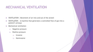
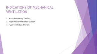
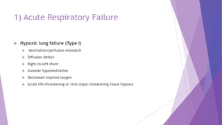
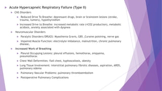
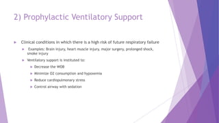
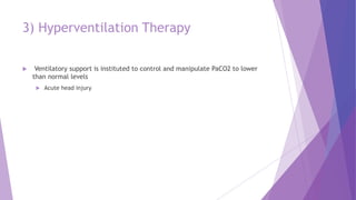
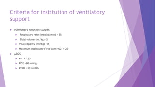
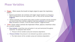
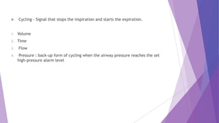
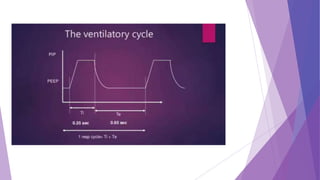
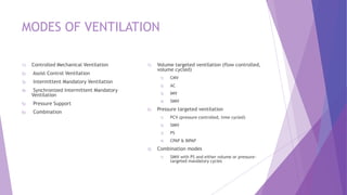
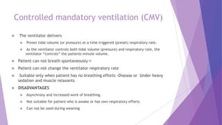
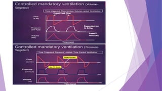
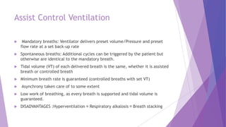
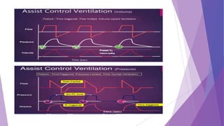
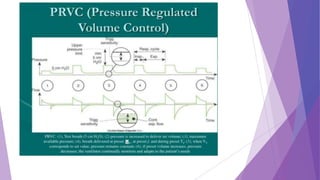
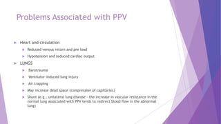
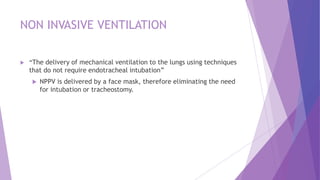
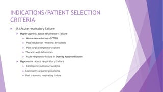
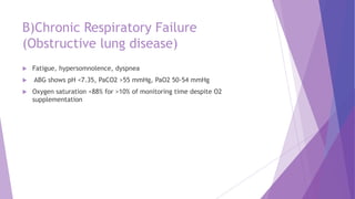
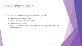
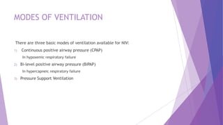
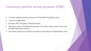
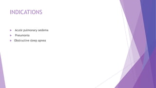
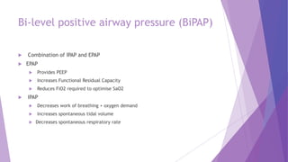
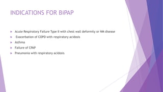
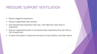
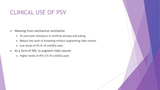
INVASIVE AND NON
INVASIVE VENTILATION
DR M AWAIS IQBAL
PGR-ANESTHESIA ,JHL
MECHANICAL VENTILATION
 VENTILATION : Movement of air into and out of the alveoli
 VENTILATOR : A machine that generates a controlled flow of gas into a
patient’s airways
 Mechanical ventilation
a) Negative pressure
b) Positive pressure
a) Invasive
b) Noninvasive
INDICATIONS OF MECHANICAL
VENTILATION
1) Acute Respiratory Failure
2) Prophylactic Ventilatory Support
3) Hyperventilation Therapy
1) Acute Respiratory Failure
 Hypoxic lung failure (Type I)
 Ventilation/perfusion mismatch
 Diffusion defect
 Right-to-left shunt
 Alveolar hypoventilation
 Decreased inspired oxygen
 Acute life-threatening or vital organ-threatening tissue hypoxia
 Acute Hypercapneic Respiratory Failure (Type II)
 CNS Disorders
 Reduced Drive To Breathe: depressant drugs, brain or brainstem lesions (stroke,
trauma, tumors), hypothyroidism
 Increased Drive to Breathe: increased metabolic rate ( CO2 production), metabolic
acidosis, anxiety associated with dyspnea
 Neuromuscular Disorders
 Paralytic Disorders/DRUGS: Myasthenia Gravis, GBS ,Curaine poisining, nerve gas
 Impaired Muscle Function: electrolyte imbalance, malnutrition, chronic pulmonary
disease,
 Increased Work of Breathing
 Pleural Occupying Lesions: pleural effusions, hemothorax, empyema,
pneumothorax
 Chest Wall Deformities: flail chest, kyphoscoliosis, obesity
 Lung Tissue Involvement: interstitial pulmonary fibrotic diseases, aspiration, ARDS,
pulmonary edema
 Pulmonary Vascular Problems: pulmonary thromboembolism
 Postoperative Pulmonary Complications
2) Prophylactic Ventilatory Support
 Clinical conditions in which there is a high risk of future respiratory failure
 Examples: Brain injury, heart muscle injury, major surgery, prolonged shock,
smoke injury
 Ventilatory support is instituted to:
 Decrease the WOB
 Minimize O2 consumption and hypoxemia
 Reduce cardiopulmonary stress
 Control airway with sedation
3) Hyperventilation Therapy
 Ventilatory support is instituted to control and manipulate PaCO2 to lower
than normal levels
 Acute head injury
Criteria for institution of ventilatory
support
 Pulmonary function studies:
 Respiratory rate (breaths/min) > 35
 Tidal volume (ml/kg) <5
 Vital capacity (ml/kg) <15
 Maximum Inspiratory Force (cm HO2) <-20
 ABGS
 PH <7.25
 PO2 <60 mmHg
 PCO2 >50 mmHG
Basic Ventilator Parameters
 Mode – The way machine ventilates the patient.
 Tidal volume
 Frequency
 FiO2
 PI & Plateau Pressure
 PEEP
 Inspiratory Time
 Expiratory time
 I:E Ratio
Phase Variables
 Trigger - What causes the breath to begin (signal to open the inspiratory
valve)
 Machine (controlled): the ventilator will trigger regular breaths at a frequency
which will depend on the set respiratory rate, ie, they will be ventilator time
triggered.
 Patient (assisted): If the patient does make an effort to breathe and the ventilator
can sense it (by either sensing a negative inspiratory pressure or an inspiratory
flow) and deliver a breath, it will be called a patient- triggered breath.
 Limit- Factor which controls the inspiration inflow ,places a maximum value
on a control variable
 Flow Limited: a fixed flow rate and pattern is set and maintained throughout
inspiration- An adequate tidal volume.
 Pressure will be variable (comp and resistance dependent)
 Pressure limited: the pressure is not allowed to go above a preset limit.
 The tidal volume will be variable (comp and resistance dependent)
 Cycling - Signal that stops the inspiration and starts the expiration.
1. Volume
2. Time
3. Flow
4. Pressure : back-up form of cycling when the airway pressure reaches the set
high-pressure alarm level
MODES OF VENTILATION
1) Controlled Mechanical Ventilation
2) Assist Control Ventilation
3) Intermittent Mandatory Ventilation
4) Synchronized Intermittent Mandatory
Ventilation
5) Pressure Support
6) Combination
1) Volume targeted ventilation (flow controlled,
volume cycled)
1) CMV
2) AC
3) IMV
4) SIMV
2) Pressure targeted ventilation
1) PCV (pressure controlled, time cycled)
2) SIMV
3) PS
4) CPAP & BIPAP
3) Combination modes
1) SIMV with PS and either volume or pressure-
targeted mandatory cycles
Controlled mandatory ventilation (CMV)
 The ventilator delivers
 Preset tidal volume (or pressure) at a time triggered (preset) respiratory rate.
 As the ventilator controls both tidal volume (pressure) and respiratory rate, the
ventilator “controls” the patients minute volume.
 Patient can not breath spontaneously
 Patient can not change the ventilator respiratory rate
 Suitable only when patient has no breathing efforts -Disease or Under heavy
sedation and muscle relaxants
 DISADVANTAGES
 Asynchrony and increased work of breathing.
 Not suitable for patient who is awake or has own respiratory efforts
 Can not be used during weaning
Assist Control Ventilation
 Mandatory breaths: Ventilator delivers preset volume/Pressure and preset
flow rate at a set back-up rate
 Spontaneous breaths: Additional cycles can be triggered by the patient but
otherwise are identical to the mandatory breath.
 Tidal volume (VT) of each delivered breath is the same, whether it is assisted
breath or controlled breath
 Minimum breath rate is guaranteed (controlled breaths with set VT)
 Asynchrony taken care of to some extent
 Low work of breathing, as every breath is supported and tidal volume is
guaranteed.
 DISADVANTAGES :Hyperventilation Respiratory alkalosis Breath stacking
Intermittent Mandatory Ventilation
(IMV)
 Machine breaths are delivered at a set rate (volume or pressure limit)
 Patient is allowed to breath spontaneously from either a demand valve or a
continuous flow of gases but not offering any inspiratory assistance.
 Patient’s capability determines Tidal volume of spontaneously breaths
 Pros: Freedom for natural spontaneous breaths even on machine Lesser
chances of hyperventilation
 Cons: Asynchrony Random chance of breath stacking. Increase work of
breathing Random high airway pressure (barotrauma) and lung volume
(volutrauma) Setting appropriate pressure limit is important to reduce the
risk of barotrauma
Synchronized Intermittent Mandatory
Ventilation
 3 types of breathing:
1) Patient initiated assisted ventilation
If the patient makes a spontaneous inspiratory effort that falls in sync window,
the ventilator is patient triggered to deliver an assisted breath and will count it
as mandatory breath
2) Ventilator generated controlled ventilation
if patient does not make an inspiratory effort then ventilator will deliver a time
triggered mandatory breath. Time triggered mandatory breath
3) Unassisted spontaneous breath.
If the patient breathes between mandatory breaths, the ventilator will allow the
patient to breathe a normal breath by opening the demand (inspiratory) valve but
not offering any inspiratory assistance.
Pressure Support Ventilation
 Pressure (or Pressure above PEEP) is the setting variable
 No mandatory breaths , Applicable on Spontaneous breaths: a preset pressure
assist,
 Flow cycling: terminates when flow drops to a specified fraction (typically
25%) of its maximum.
 Patient effort determines size of breath and flow rate.
 It augments spontaneous VT decreases spontaneous rates and WOB
 Used in conjunction with spontaneous breaths in any mode of ventilation.
 No back up ventilation in the event of apnea.
 Provides pressure support to overcome the increased work of breathing
imposed by the disease process, the endotracheal tube, the inspiratory valves
and other mechanical aspects of ventilatory support
 Allows for titration of patient effort during weaning.
 Helpful in assessing extubation readiness.
PRESSURE REGULATED VOLUME CONTROL
(PRVC)
 This is a volume targeted, pressure limited mode. (available in SIMV or AC)
 Each breath is delivered at a set volume with a variable flow rate and an absolute pressure
limit.
 The vent delivers this pre-set volume at the LOWEST required peak pressure and adjust with
each breath.
 Ventilator monitors each breath and compares the delivered tidal volume with set tidal
volume. If tidal volume is too low it increases the inspiratory pressure on next breath, if it is
too high it decreases the pressure.
 Advantages
 Decelerating inspiratory flow pattern
 Pressure automatically adjusted for changes in compliance and resistance within a set range
 Tidal volume guaranteed
 Limits volutrauma
 Prevents hypoventilation
INITIAL SETTINGS
1) Select your mode of ventilation
2) Set sensitivity at Flow trigger mode
a) Pressure triggering , a ventilator-delivered breath is initiated if the demand
valve senses a negative airway pressure deflection (generated by the patient
trying to initiate a breath) greater than the trigger sensitivity.
b) Flow-by triggering , a continuous flow of gas through the ventilator circuit is
monitored. A ventilator-delivered breath is initiated when the return flow is less
than the delivered flow, a consequence of the patient's effort to initiate a breath
3) Set Tidal Volume - 5 – 7 ml/kg of IBW
4) Set Rate - 12-18 breaths/min
5) Set Inspiratory Flow (if necessary)
1) beginning point, flow is normal set to deliver inspiration in about 1 second (range
0.8 to 1.2 sec.), producing an I:E ratio of approximately 1:2 or less (usually about
1:4) – This can be achieved with an initial peak flow of about 60 L/min (range of
40 to 80 L/min)
5) Set Inspiratory Flow (if necessary)
At beginning point, flow is normal set to deliver inspiration in about 1 second (range 0.8
to 1.2 sec.), producing an I:E ratio of 1:2 or less – With an initial peak flow of about 60
L/min (range of 40 to 80 L/min)
6) Set PEEP
 Initially set at 3 – 5 cm H2O –
 Restores FRC and physiological PEEP that existed prior to intubation
 Useful to treat refractory hypoxemia
 Contraindications for therapeutic PEEP (>5 cm H2O) – Hypotension – Elevated ICP –
Uncontrolled pneumothorax
7) Set Pressure Limit
8) Inspiratory time
9) Fraction of inspired oxygen
 Initially 100% – Severe hypoxemia – Abnormal cardiopulmonary functions 1)Post-
resuscitation 2) Smoke inhalation 3)ARDS
 After stabilization, attempt to keep FiO2 <50% – Avoids oxygen-induced lung injuries
1)Absorption atelectasis 2) Oxygen toxicity
Post Initial Settings
 Obtain an ABG (arterial blood gas) about 30 minutes after you set your patient
up on the ventilator.
 An ABG will give you information about any changes that may need to be
made to keep the patient’s oxygenation and ventilation status within a
physiological range.
 Goal:
 pH 7.35 – 7.45
 PCO2 35-45 mmHg
 PO2 80-100 mmHg
Problems Associated with PPV
 Heart and circulation
 Reduced venous return and pre load
 Hypotension and reduced cardiac output
 LUNGS
 Barotrauma
 Ventilator-induced lung injury
 Air trapping
 May increase dead space (compression of capillaries)
 Shunt (e.g., unilateral lung disease - the increase in vascular resistance in the
normal lung associated with PPV tends to redirect blood flow in the abnormal
lung)
NON INVASIVE VENTILATION
 “The delivery of mechanical ventilation to the lungs using techniques
that do not require endotracheal intubation”
 NPPV is delivered by a face mask, therefore eliminating the need
for intubation or tracheostomy.
INDICATIONS/PATIENT SELECTION
CRITERIA
 (A) Acute respiratory failure
 Hypercapneic acute respiratory failure
 Acute exacerbation of COPD
 Post extubation /Weaning difficulties
 Post surgical respiratory failure
 Thoracic wall deformities
 Acute respiratory failure in Obesity hypoventilation
 Hypoxemic acute respiratory failure
 Cardiogenic pulmonary oedema
 Community acquired pneumonia
 Post traumatic respiratory failure
B)Chronic Respiratory Failure
(Obstructive lung disease)
 Fatigue, hypersomnolence, dyspnea
 ABG shows pH <7.35, PaCO2 >55 mmHg, PaO2 50-54 mmHg
 Oxygen saturation <88% for >10% of monitoring time despite O2
supplementation
SELECTION CRITERIA
 At least two of the following criteria should be present:
 Respiratory distress with dyspnea
 Use of accessory muscles of respiration
 Respiratory rate >25/min
 ABG shows pH <7.32 or PaCO2 >45mmHg OR PaO2 <60mmHg or PaO2/FiO2 <200
despite high Fio2.
MODES OF VENTILATION
There are three basic modes of ventilation available for NIV:
1) Continuous positive airway pressure (CPAP)
In hypoxemic respiratory failure
2) Bi-level positive airway pressure (BiPAP)
In hypercapneic respiratory failure
3) Pressure Support Ventilation
Continuous positive airway pressure (CPAP)
 Constant positive airway pressure of 5-10cmH2O throughout cycle
 Improves oxygenation
 Increases FRC and opens collapsed alveoli
 Decreases work of breathing by alveolar recruitment (Dec elastic work) and
unloads inspiratory muscles
 Decreases hypoxia by alveolar recruitment and reduces intrapulmonary shun
INDICATIONS
 Acute pulmonary oedema
 Pneumonia
 Obstructive sleep apnea
Bi-level positive airway pressure (BiPAP)
 Combination of IPAP and EPAP
 EPAP
 Provides PEEP
 Increases Functional Residual Capacity
 Reduces FiO2 required to optimise SaO2
 IPAP
 Decreases work of breathing + oxygen demand
 Increases spontaneous tidal volume
 Decreases spontaneous respiratory rate
INDICATIONS FOR BiPAP
 Acute Respiratory Failure Type II with chest wall deformity or NM disease
 Exacerbation of COPD with respiratory acidosis
 Asthma
 Failure of CPAP
 Pneumonia with respiratory acidosis
PRESSURE SUPPORT VENTILATION
 Patient triggered inspirations.
 Pressure augmented tidal volumes.
 Uses decelerating inspiratory flow rate, with high flow rates early in
inspiration.
 Pressure augmented breath is terminated when inspiratory flow rate falls to
25% of peak level
 It allows the patient to determine duration of lung inflation and tidal volume
CLINICAL USE OF PSV
 Weaning from mechanical ventilation
 To overcome resistance in artificial airways and tubing.
 Reduce the work of breathing without augmenting tidal volume.
 Low levels of PS (5-10 cmH2O) used.
 As a form of NIV, to augment tidal volume
 Higher levels of PSV (15-35 cmH2O) used
MECHANISM OF ACTION OF NIPPV
 Improvement in pulmonary mechanics and oxygenation: NPPV augments
alveolar ventilation and allows oxygenation without raising PaCO2 .
 It reduces respiratory muscles work and diaphragmatic electromyographic
activity.
 ↑ Tidal volume, ↓ RR and ↑ MV .
 PEEP decreases the work of breathing by partially overcoming the auto-PEEP.
 Resetting of respiratory center ventilatory responses to PaCO2
 By maintaining lower nocturnal PaCO2 during sleep by giving NPPV, it is possible to
reset the respiratory control center to become more responsive to an increased
PaCO2 by increasing the neural output to diaphragm and other respiratory muscles.
ADVANTAGES OF NIPPV
 Early ventilatory support: an option
 Intermittent ventilation possible
 Patient can eat, drink and communicate
 Ease of application and removal
 Patient can cooperate with physiotherapy
 Improved patient comfort
 Reduced need for sedation
 Avoidance of complications of endotracheal intubation
 upper airway trauma, sinusitis, otitis, nosocomial pneumonia
 Ventilation outside hospital possible
 Correction of hypoxaemia without worsening hypercarbia
DISADVANTAGES OF NIPPV
 Mask uncomfortable/claustrophobia
 Facial pressure sores (skin necrosis)
 Airway not protected /risk of aspiration
 No direct access to bronchial tree for suction if secretions are excessive
 Gastric distension
 Drying of Eyes
 Gas leaks
 Ventilator-patient asynchrony
CONTRAINDICATIONS
 ABSOLUTE
 1.Respiratory arrest
 Unstable cardiorespiratory status
 Uncooperative patients
 Unable to protect airway- impaired
swallowing and cough
 Facial Esophageal or gastric
surgery
 Craniofacial trauma/burn
 Anatomic lesions of upper airway
 RELATIVE
 Extreme anxiety
 Massive obesity
 Failure of previous attempts of
NPPV
 Life threatening arrhythmias
 Life threatening refractory
hypoxemia(PaO2<60mm Hg with
FiO2- 100%)
Criteria to Discontinue NIV
 Inability to tolerate the mask
 Inability to improve gas exchange or dyspnea
 Need for endotracheal intubation
 Hemodynamic instability
 ECG – ischemia/arrhythmia
Invasive and Non Invasive ventilation .pptx

Invasive and Non Invasive ventilation .pptx

  • 1.
    INVASIVE AND NON INVASIVEVENTILATION DR M AWAIS IQBAL PGR-ANESTHESIA ,JHL
  • 2.
    MECHANICAL VENTILATION  VENTILATION: Movement of air into and out of the alveoli  VENTILATOR : A machine that generates a controlled flow of gas into a patient’s airways  Mechanical ventilation a) Negative pressure b) Positive pressure a) Invasive b) Noninvasive
  • 3.
    INDICATIONS OF MECHANICAL VENTILATION 1)Acute Respiratory Failure 2) Prophylactic Ventilatory Support 3) Hyperventilation Therapy
  • 4.
    1) Acute RespiratoryFailure  Hypoxic lung failure (Type I)  Ventilation/perfusion mismatch  Diffusion defect  Right-to-left shunt  Alveolar hypoventilation  Decreased inspired oxygen  Acute life-threatening or vital organ-threatening tissue hypoxia
  • 5.
     Acute HypercapneicRespiratory Failure (Type II)  CNS Disorders  Reduced Drive To Breathe: depressant drugs, brain or brainstem lesions (stroke, trauma, tumors), hypothyroidism  Increased Drive to Breathe: increased metabolic rate ( CO2 production), metabolic acidosis, anxiety associated with dyspnea  Neuromuscular Disorders  Paralytic Disorders/DRUGS: Myasthenia Gravis, GBS ,Curaine poisining, nerve gas  Impaired Muscle Function: electrolyte imbalance, malnutrition, chronic pulmonary disease,  Increased Work of Breathing  Pleural Occupying Lesions: pleural effusions, hemothorax, empyema, pneumothorax  Chest Wall Deformities: flail chest, kyphoscoliosis, obesity  Lung Tissue Involvement: interstitial pulmonary fibrotic diseases, aspiration, ARDS, pulmonary edema  Pulmonary Vascular Problems: pulmonary thromboembolism  Postoperative Pulmonary Complications
  • 6.
    2) Prophylactic VentilatorySupport  Clinical conditions in which there is a high risk of future respiratory failure  Examples: Brain injury, heart muscle injury, major surgery, prolonged shock, smoke injury  Ventilatory support is instituted to:  Decrease the WOB  Minimize O2 consumption and hypoxemia  Reduce cardiopulmonary stress  Control airway with sedation
  • 7.
    3) Hyperventilation Therapy Ventilatory support is instituted to control and manipulate PaCO2 to lower than normal levels  Acute head injury
  • 8.
    Criteria for institutionof ventilatory support  Pulmonary function studies:  Respiratory rate (breaths/min) > 35  Tidal volume (ml/kg) <5  Vital capacity (ml/kg) <15  Maximum Inspiratory Force (cm HO2) <-20  ABGS  PH <7.25  PO2 <60 mmHg  PCO2 >50 mmHG
  • 9.
    Basic Ventilator Parameters Mode – The way machine ventilates the patient.  Tidal volume  Frequency  FiO2  PI & Plateau Pressure  PEEP  Inspiratory Time  Expiratory time  I:E Ratio
  • 10.
    Phase Variables  Trigger- What causes the breath to begin (signal to open the inspiratory valve)  Machine (controlled): the ventilator will trigger regular breaths at a frequency which will depend on the set respiratory rate, ie, they will be ventilator time triggered.  Patient (assisted): If the patient does make an effort to breathe and the ventilator can sense it (by either sensing a negative inspiratory pressure or an inspiratory flow) and deliver a breath, it will be called a patient- triggered breath.  Limit- Factor which controls the inspiration inflow ,places a maximum value on a control variable  Flow Limited: a fixed flow rate and pattern is set and maintained throughout inspiration- An adequate tidal volume.  Pressure will be variable (comp and resistance dependent)  Pressure limited: the pressure is not allowed to go above a preset limit.  The tidal volume will be variable (comp and resistance dependent)
  • 11.
     Cycling -Signal that stops the inspiration and starts the expiration. 1. Volume 2. Time 3. Flow 4. Pressure : back-up form of cycling when the airway pressure reaches the set high-pressure alarm level
  • 13.
    MODES OF VENTILATION 1)Controlled Mechanical Ventilation 2) Assist Control Ventilation 3) Intermittent Mandatory Ventilation 4) Synchronized Intermittent Mandatory Ventilation 5) Pressure Support 6) Combination 1) Volume targeted ventilation (flow controlled, volume cycled) 1) CMV 2) AC 3) IMV 4) SIMV 2) Pressure targeted ventilation 1) PCV (pressure controlled, time cycled) 2) SIMV 3) PS 4) CPAP & BIPAP 3) Combination modes 1) SIMV with PS and either volume or pressure- targeted mandatory cycles
  • 14.
    Controlled mandatory ventilation(CMV)  The ventilator delivers  Preset tidal volume (or pressure) at a time triggered (preset) respiratory rate.  As the ventilator controls both tidal volume (pressure) and respiratory rate, the ventilator “controls” the patients minute volume.  Patient can not breath spontaneously  Patient can not change the ventilator respiratory rate  Suitable only when patient has no breathing efforts -Disease or Under heavy sedation and muscle relaxants  DISADVANTAGES  Asynchrony and increased work of breathing.  Not suitable for patient who is awake or has own respiratory efforts  Can not be used during weaning
  • 16.
    Assist Control Ventilation Mandatory breaths: Ventilator delivers preset volume/Pressure and preset flow rate at a set back-up rate  Spontaneous breaths: Additional cycles can be triggered by the patient but otherwise are identical to the mandatory breath.  Tidal volume (VT) of each delivered breath is the same, whether it is assisted breath or controlled breath  Minimum breath rate is guaranteed (controlled breaths with set VT)  Asynchrony taken care of to some extent  Low work of breathing, as every breath is supported and tidal volume is guaranteed.  DISADVANTAGES :Hyperventilation Respiratory alkalosis Breath stacking
  • 18.
    Intermittent Mandatory Ventilation (IMV) Machine breaths are delivered at a set rate (volume or pressure limit)  Patient is allowed to breath spontaneously from either a demand valve or a continuous flow of gases but not offering any inspiratory assistance.  Patient’s capability determines Tidal volume of spontaneously breaths  Pros: Freedom for natural spontaneous breaths even on machine Lesser chances of hyperventilation  Cons: Asynchrony Random chance of breath stacking. Increase work of breathing Random high airway pressure (barotrauma) and lung volume (volutrauma) Setting appropriate pressure limit is important to reduce the risk of barotrauma
  • 20.
    Synchronized Intermittent Mandatory Ventilation 3 types of breathing: 1) Patient initiated assisted ventilation If the patient makes a spontaneous inspiratory effort that falls in sync window, the ventilator is patient triggered to deliver an assisted breath and will count it as mandatory breath 2) Ventilator generated controlled ventilation if patient does not make an inspiratory effort then ventilator will deliver a time triggered mandatory breath. Time triggered mandatory breath 3) Unassisted spontaneous breath. If the patient breathes between mandatory breaths, the ventilator will allow the patient to breathe a normal breath by opening the demand (inspiratory) valve but not offering any inspiratory assistance.
  • 22.
    Pressure Support Ventilation Pressure (or Pressure above PEEP) is the setting variable  No mandatory breaths , Applicable on Spontaneous breaths: a preset pressure assist,  Flow cycling: terminates when flow drops to a specified fraction (typically 25%) of its maximum.  Patient effort determines size of breath and flow rate.  It augments spontaneous VT decreases spontaneous rates and WOB  Used in conjunction with spontaneous breaths in any mode of ventilation.  No back up ventilation in the event of apnea.
  • 23.
     Provides pressuresupport to overcome the increased work of breathing imposed by the disease process, the endotracheal tube, the inspiratory valves and other mechanical aspects of ventilatory support  Allows for titration of patient effort during weaning.  Helpful in assessing extubation readiness.
  • 24.
    PRESSURE REGULATED VOLUMECONTROL (PRVC)  This is a volume targeted, pressure limited mode. (available in SIMV or AC)  Each breath is delivered at a set volume with a variable flow rate and an absolute pressure limit.  The vent delivers this pre-set volume at the LOWEST required peak pressure and adjust with each breath.  Ventilator monitors each breath and compares the delivered tidal volume with set tidal volume. If tidal volume is too low it increases the inspiratory pressure on next breath, if it is too high it decreases the pressure.  Advantages  Decelerating inspiratory flow pattern  Pressure automatically adjusted for changes in compliance and resistance within a set range  Tidal volume guaranteed  Limits volutrauma  Prevents hypoventilation
  • 26.
    INITIAL SETTINGS 1) Selectyour mode of ventilation 2) Set sensitivity at Flow trigger mode a) Pressure triggering , a ventilator-delivered breath is initiated if the demand valve senses a negative airway pressure deflection (generated by the patient trying to initiate a breath) greater than the trigger sensitivity. b) Flow-by triggering , a continuous flow of gas through the ventilator circuit is monitored. A ventilator-delivered breath is initiated when the return flow is less than the delivered flow, a consequence of the patient's effort to initiate a breath 3) Set Tidal Volume - 5 – 7 ml/kg of IBW 4) Set Rate - 12-18 breaths/min 5) Set Inspiratory Flow (if necessary) 1) beginning point, flow is normal set to deliver inspiration in about 1 second (range 0.8 to 1.2 sec.), producing an I:E ratio of approximately 1:2 or less (usually about 1:4) – This can be achieved with an initial peak flow of about 60 L/min (range of 40 to 80 L/min)
  • 27.
    5) Set InspiratoryFlow (if necessary) At beginning point, flow is normal set to deliver inspiration in about 1 second (range 0.8 to 1.2 sec.), producing an I:E ratio of 1:2 or less – With an initial peak flow of about 60 L/min (range of 40 to 80 L/min) 6) Set PEEP  Initially set at 3 – 5 cm H2O –  Restores FRC and physiological PEEP that existed prior to intubation  Useful to treat refractory hypoxemia  Contraindications for therapeutic PEEP (>5 cm H2O) – Hypotension – Elevated ICP – Uncontrolled pneumothorax 7) Set Pressure Limit 8) Inspiratory time 9) Fraction of inspired oxygen  Initially 100% – Severe hypoxemia – Abnormal cardiopulmonary functions 1)Post- resuscitation 2) Smoke inhalation 3)ARDS  After stabilization, attempt to keep FiO2 <50% – Avoids oxygen-induced lung injuries 1)Absorption atelectasis 2) Oxygen toxicity
  • 28.
    Post Initial Settings Obtain an ABG (arterial blood gas) about 30 minutes after you set your patient up on the ventilator.  An ABG will give you information about any changes that may need to be made to keep the patient’s oxygenation and ventilation status within a physiological range.  Goal:  pH 7.35 – 7.45  PCO2 35-45 mmHg  PO2 80-100 mmHg
  • 29.
    Problems Associated withPPV  Heart and circulation  Reduced venous return and pre load  Hypotension and reduced cardiac output  LUNGS  Barotrauma  Ventilator-induced lung injury  Air trapping  May increase dead space (compression of capillaries)  Shunt (e.g., unilateral lung disease - the increase in vascular resistance in the normal lung associated with PPV tends to redirect blood flow in the abnormal lung)
  • 30.
    NON INVASIVE VENTILATION “The delivery of mechanical ventilation to the lungs using techniques that do not require endotracheal intubation”  NPPV is delivered by a face mask, therefore eliminating the need for intubation or tracheostomy.
  • 31.
    INDICATIONS/PATIENT SELECTION CRITERIA  (A)Acute respiratory failure  Hypercapneic acute respiratory failure  Acute exacerbation of COPD  Post extubation /Weaning difficulties  Post surgical respiratory failure  Thoracic wall deformities  Acute respiratory failure in Obesity hypoventilation  Hypoxemic acute respiratory failure  Cardiogenic pulmonary oedema  Community acquired pneumonia  Post traumatic respiratory failure
  • 32.
    B)Chronic Respiratory Failure (Obstructivelung disease)  Fatigue, hypersomnolence, dyspnea  ABG shows pH <7.35, PaCO2 >55 mmHg, PaO2 50-54 mmHg  Oxygen saturation <88% for >10% of monitoring time despite O2 supplementation
  • 33.
    SELECTION CRITERIA  Atleast two of the following criteria should be present:  Respiratory distress with dyspnea  Use of accessory muscles of respiration  Respiratory rate >25/min  ABG shows pH <7.32 or PaCO2 >45mmHg OR PaO2 <60mmHg or PaO2/FiO2 <200 despite high Fio2.
  • 34.
    MODES OF VENTILATION Thereare three basic modes of ventilation available for NIV: 1) Continuous positive airway pressure (CPAP) In hypoxemic respiratory failure 2) Bi-level positive airway pressure (BiPAP) In hypercapneic respiratory failure 3) Pressure Support Ventilation
  • 35.
    Continuous positive airwaypressure (CPAP)  Constant positive airway pressure of 5-10cmH2O throughout cycle  Improves oxygenation  Increases FRC and opens collapsed alveoli  Decreases work of breathing by alveolar recruitment (Dec elastic work) and unloads inspiratory muscles  Decreases hypoxia by alveolar recruitment and reduces intrapulmonary shun
  • 36.
    INDICATIONS  Acute pulmonaryoedema  Pneumonia  Obstructive sleep apnea
  • 37.
    Bi-level positive airwaypressure (BiPAP)  Combination of IPAP and EPAP  EPAP  Provides PEEP  Increases Functional Residual Capacity  Reduces FiO2 required to optimise SaO2  IPAP  Decreases work of breathing + oxygen demand  Increases spontaneous tidal volume  Decreases spontaneous respiratory rate
  • 38.
    INDICATIONS FOR BiPAP Acute Respiratory Failure Type II with chest wall deformity or NM disease  Exacerbation of COPD with respiratory acidosis  Asthma  Failure of CPAP  Pneumonia with respiratory acidosis
  • 39.
    PRESSURE SUPPORT VENTILATION Patient triggered inspirations.  Pressure augmented tidal volumes.  Uses decelerating inspiratory flow rate, with high flow rates early in inspiration.  Pressure augmented breath is terminated when inspiratory flow rate falls to 25% of peak level  It allows the patient to determine duration of lung inflation and tidal volume
  • 40.
    CLINICAL USE OFPSV  Weaning from mechanical ventilation  To overcome resistance in artificial airways and tubing.  Reduce the work of breathing without augmenting tidal volume.  Low levels of PS (5-10 cmH2O) used.  As a form of NIV, to augment tidal volume  Higher levels of PSV (15-35 cmH2O) used
  • 41.
    MECHANISM OF ACTIONOF NIPPV  Improvement in pulmonary mechanics and oxygenation: NPPV augments alveolar ventilation and allows oxygenation without raising PaCO2 .  It reduces respiratory muscles work and diaphragmatic electromyographic activity.  ↑ Tidal volume, ↓ RR and ↑ MV .  PEEP decreases the work of breathing by partially overcoming the auto-PEEP.  Resetting of respiratory center ventilatory responses to PaCO2  By maintaining lower nocturnal PaCO2 during sleep by giving NPPV, it is possible to reset the respiratory control center to become more responsive to an increased PaCO2 by increasing the neural output to diaphragm and other respiratory muscles.
  • 42.
    ADVANTAGES OF NIPPV Early ventilatory support: an option  Intermittent ventilation possible  Patient can eat, drink and communicate  Ease of application and removal  Patient can cooperate with physiotherapy  Improved patient comfort  Reduced need for sedation  Avoidance of complications of endotracheal intubation  upper airway trauma, sinusitis, otitis, nosocomial pneumonia  Ventilation outside hospital possible  Correction of hypoxaemia without worsening hypercarbia
  • 43.
    DISADVANTAGES OF NIPPV Mask uncomfortable/claustrophobia  Facial pressure sores (skin necrosis)  Airway not protected /risk of aspiration  No direct access to bronchial tree for suction if secretions are excessive  Gastric distension  Drying of Eyes  Gas leaks  Ventilator-patient asynchrony
  • 44.
    CONTRAINDICATIONS  ABSOLUTE  1.Respiratoryarrest  Unstable cardiorespiratory status  Uncooperative patients  Unable to protect airway- impaired swallowing and cough  Facial Esophageal or gastric surgery  Craniofacial trauma/burn  Anatomic lesions of upper airway  RELATIVE  Extreme anxiety  Massive obesity  Failure of previous attempts of NPPV  Life threatening arrhythmias  Life threatening refractory hypoxemia(PaO2<60mm Hg with FiO2- 100%)
  • 45.
    Criteria to DiscontinueNIV  Inability to tolerate the mask  Inability to improve gas exchange or dyspnea  Need for endotracheal intubation  Hemodynamic instability  ECG – ischemia/arrhythmia