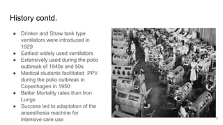
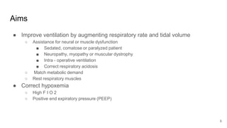
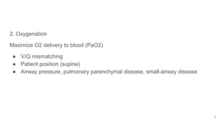
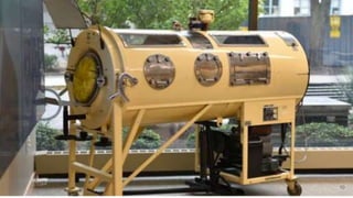
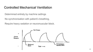
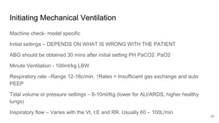
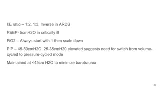
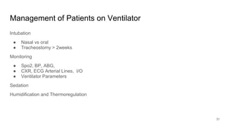
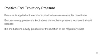
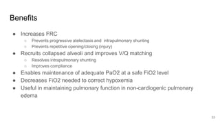
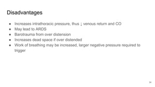
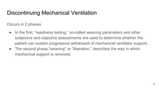
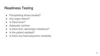
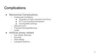
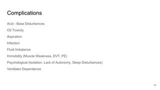
Artificial Ventilation is a method of assisting or replacing spontaneous breathing using a mechanical ventilator. It has a long history dating back to ancient times and was widely used during polio outbreaks. There are two main types - negative pressure ventilation which uses vacuum pressure and positive pressure ventilation which delivers gas via endotracheal tube. Modes include controlled mandatory ventilation where the machine does all breathing, assisted modes where the patient can trigger breaths, and pressure support where the patient determines breathing rate and depth. Key goals are to improve oxygenation and ventilation. Complications can include barotrauma, infections, and ventilator dependence. Proper use and monitoring can greatly impact patient outcomes.
3. Introduction
● Artificial or Mechanical ventilation is a method of artificially assisting or replacing
spontaneous breathing.
● A Mechanical ventilator is a machine that generates a controlled flow of gas into a
patient’s airways.
● Artificial Ventilation is a commonly used technique in the operating rooms and
intensive care unit (ICU) and knowledge and proper application can greatly impact
patient outcome
3
4. History
● Roman physician Galen first used mechanical breathing in the second century by
blowing air into the larynx of a dead animal using a reed.
● First officially documented by Andreas Wesele Vesalius in the 15th Century
“But that life may ... be restored to the animal,
an opening must be attempted in the trunk of the trachea,
in which a tube of reed or cane should be put;
you will then blow into this,
so that the lung may rise again and the animal take in air. ...
And as you do this, and take care that the lung is inflated in intervals,
the motion of the heart and arteries does not stop..." Vesalius 1543
● Author George Poe used a mechanical respirator to revive an asphyxiated dog.
4
5. History contd.
● Drinker and Shaw tank type
ventilators were introduced in
1929
● Earliest widely used ventilators
● Extensively used during the polio
outbreak of 1940s and 50s
● Medical students facilitated PPV
during the polio outbreak in
Copenhagen in 1950
● Better Mortality rates than Iron
Lungs
● Success led to adaptation of the
anaesthesia machine for
intensive care use 5
6. Aims
● Improve ventilation by augmenting respiratory rate and tidal volume
○ Assistance for neural or muscle dysfunction
■ Sedated, comatose or paralyzed patient
■ Neuropathy, myopathy or muscular dystrophy
■ Intra - operative ventilation
■ Correct respiratory acidosis
○ Match metabolic demand
○ Rest respiratory muscles
● Correct hypoxemia
○ High F I O 2
○ Positive end expiratory pressure (PEEP)
6
9. Negative Pressure Ventilation
A vacuum pump creates a negative pressure in the chamber, resulting in
chest expansion
This change reduces the intrapulmonary pressure and allows ambient air to
flow into the patient's lungs.
When the vacuum was terminated, the negative pressure applied to the chest
dropped to zero, and the elastic recoil of the chest and lungs permitted
passive exhalation.
Almost extinct
9
11. Types contd
● Disadvantages
○ Cumbersome
○ Significant patient discomfort
○ Pooling of blood in the lower limbs
○ Limited access
● Advantages
○ Does not require invasive methods
11
12. Positive Pressure Ventilation
● Modern day ventilator design
Airway pressure is applied at the patient's airway through an endotracheal or
tracheostomy tube.
The positive nature of the pressure causes the gas to flow into the lungs until the
ventilator breath is terminated.
As the airway pressure drops, elastic recoil of the chest accomplishes passive
exhalation by pushing the tidal volume out.
12
13. Non Invasive Positive Pressure Ventilation
Can be achieved using a facemask, nasal mask, helmet. It should be considered
first, if patient meets the criteria and has no contraindications
Indications
● Respiratory failure in the absence of haemodynamic instability, neurologic
compromise.
● Acute exacerbation of COPD.
● Acute cardiogenic pulmonary oedema.
● Post operative ventilatory failure.
13
14. Contraindications to NIPPV
● Cardiac or respiratory arrest
● Non respiratory organ failure
● GCS < 10
● Haemodynamic instability
● Facial surgeries/trauma/deformities
● Inability to clear secretions
● Un-cooperative patients
● Patients at risk of aspiration
14
15. Invasive Positive Pressure Ventilation
After instrumentation of the airway. Either via LMA, endotracheal tube or
tracheostomy tube
Indications
● Arterial oxygen tension <50 mm Hg on room air
● Arterial CO 2 tension >50 mm Hg in the absence of metabolic alkalosis
● Pa O 2 /F IO 2 ratio <300 mm Hg
● V D /V T >0.6
● Respiratory rate >35 cpm or < 5cpm
● Tidal volume <5 mL/kg
● Vital capacity <15 mL/kg
● Maximum inspiratory force > −25 cm H 2 O (eg, –15 cm H 2 O)
15
16. Indications for IPPV contd
● Drug overdose
● Following prolonged surgery
● Deteriorating LOC (GCS 8)
● Significant Chest Trauma
● Post cardiac arrest
● Use of NMB
● Thoracic surgery
● Inadequate ventilation via conventional methods
● Control/monitor arterial Paco2
16
17. Key Concepts
Control - How much to deliver
Trigger - When to Deliver
Cycling - How long to deliver for
17
18. Key Concepts contd
Control (How much to deliver)
1. Volume Controlled (volume limited, volume targeted) and Pressure Variable.
2. Pressure Controlled (pressure limited, pressure targeted) and Volume
Variable.
3. Dual Controlled (volume targeted (guaranteed) pressure limited) Newer
models.
18
19. Key Concepts contd
Cycling (How long to deliver)
1. Pressure-cycled ventilators cycle into the expiratory phase when airway
pressure reaches a predetermined level. V T and inspiratory time vary, being
related to airway resistance and pulmonary and circuit compliance.
2. Volume-cycled ventilators terminate inspiration when a preselected volume is
delivered. Many adult ventilators are volume cycled but also have secondary
limits on inspiratory pressure to guard against pulmonary barotrauma
3. Time-cycled ventilators cycle to the expiratory phase once a predetermined
interval elapses from the start of inspiration.
19
20. Key Concepts contd
Trigger (What triggers Inspiration)
1. Time: the ventilator cycles at a set frequency as determined by the controlled
rate.
2. Pressure: the ventilator senses the patient's inspiratory effort by way of a
decrease in the baseline pressure.
3. Flow: Ventilators deliver a constant flow around the circuit throughout the
respiratory cycle. A deflection in this flow, is monitored by the ventilator and it
delivers a breath. Requires less work than pressure triggering.
20
21. Modes of Mechanical Ventilation
Controlled Mandatory Ventilation
Assisted Control Mode
Intermittent Mandatory Ventilation
Synchronous Intermittent Mandatory Ventilation
Pressure Support Ventilation
Pressure Control Ventilation
Inverse I:E Ratio Ventilation
High Frequency Ventilation
21
23. ● Patient does not participate in ventilations
● Machine initiates inspiration, does work of breathing, controls tidal volume
and rate
● Useful in apneic or heavily sedated patients
● Useful when inspiratory effort contraindicated (flail chest)
● Patient must be incapable of initiating breaths
23
24. Assisted/Control
Can be used in spontaneously breathing patients
Delivers a set number of mandatory breaths
Patient triggers machine to deliver breaths but machine has preset backup rate
Patient initiates breath--machine delivers tidal volume
If patient does not breathe fast enough, machine takes over at preset rate
Each breath results in a pre-set flow rate and tidal volume
Tachypneic patients may hyperventilate dangerously
Can result in severe alkalosis and auto PEEP, So it is contraindicated on patients with potential for respiratory
alkalosis e.g patients with end-stage liver disease, hyperventilatory sepsis, and head trauma.
24
25. Intermittent Mandatory Ventilation
Patient breathes spontaneously, and the ventilator intermittently delivers positive
pressure breaths at a pre-set tidal volume and rate.
During spontaneous breaths, flow rate and tidal volume are patient determined
Less risk of barotrauma, not every breath is a positive pressure breath
Can result in stacking
25
26. Synchronous Intermittent Mandatory Ventilation
The ventilator delivers the set tidal volume, synchronizing the mandatory breaths
with the patients’ triggering efforts.
Spontaneous efforts between ventilator delivered breaths are unassisted.
26
27. Pressure Support Ventilation
Patient determines RR, inspiratory time – a purely spontaneous mode. Clinician
sets Pinsp and PEEP
● Triggered by patient’s breath
● Limited by pressure
● Affects inspiration only
Used in conjunction with other modes
Great for weaning
27
28. Other Ventilation Modes
Pressure control Ventilation
● Similar to PSV
● Does not guarantee Vt
Inverse I:E Ratio Ventilation
● Useful in patient with reduced FRC
● Requires Deep Sedation
High Frequency Ventilation
● HFPPV - 60-120cpm
● HFJV 120-600cpm
● HFO 180-3000cpm
28
29. Initiating Mechanical Ventilation
Machine check- model specific
Initial settings – DEPENDS ON WHAT IS WRONG WITH THE PATIENT
ABG should be obtained 30 mins after initial setting PH PaCO2, PaO2
Minute Ventilation - 100ml/kg LBW
Respiratory rate –Range 12-18c/min. ↑Rates = Insufficient gas exchange and auto
PEEP
Tidal volume or pressure settings – 6-10ml/Kg (lower for ALI/ARDS, higher healthy
lungs)
Inspiratory flow – Varies with the Vt, I:E and RR. Usually 60 – 100L/min
29
30. I:E ratio – 1:2, 1:3, Inverse in ARDS
PEEP- 5cmH2O in critically ill
FiO2 – Always start with 1 then scale down
PIP – 45-50cmH2O, 25-35cmH20 elevated suggests need for switch from volume-
cycled to pressure-cycled mode
Maintained at <45cm H2O to minimize barotrauma
30
31. Management of Patients on Ventilator
Intubation
● Nasal vs oral
● Tracheostomy > 2weeks
Monitoring
● Spo2, BP, ABG,
● CXR, ECG Arterial Lines, I/O
● Ventilator Parameters
Sedation
Humidification and Thermoregulation
31
32. Positive End Expiratory Pressure
Pressure is applied at the end of expiration to maintain alveolar recruitment
Ensures airway pressure is kept above atmospheric pressure to prevent alveoli
collapse
It is the baseline airway pressure for the duration of the respiratory cycle
32
33. Benefits
● Increases FRC
○ Prevents progressive atelectasis and intrapulmonary shunting
○ Prevents repetitive opening/closing (injury)
● Recruits collapsed alveoli and improves V/Q matching
○ Resolves intrapulmonary shunting
○ Improves compliance
● Enables maintenance of adequate PaO2 at a safe FiO2 level
● Decreases FiO2 needed to correct hypoxemia
● Useful in maintaining pulmonary function in non-cardiogenic pulmonary
edema
33
34. Disadvantages
● Increases intrathoracic pressure, thus ↓ venous return and CO
● May lead to ARDS
● Barotrauma from over distension
● Increases dead space if over distended
● Work of breathing may be increased, larger negative pressure required to
trigger
34
35. Discontinuing Mechanical Ventilation
Occurs in 2 phases
● In the first, “readiness testing,” so-called weaning parameters and other
subjective and objective assessments are used to determine whether the
patient can sustain progressive withdrawal of mechanical ventilator support.
● The second phase,“weaning” or “liberation,” describes the way in which
mechanical support is removed.
35
36. Readiness Testing
● Precipitating illness treated?
● Any organ failure?
● Is there fever?
● Adequate nutrition
● Is there fluid, electrolyte imbalance?
● Is the patient sedated?
● Is there any haemodynamic instability
36
37. Readiness Testing
Assess respiratory function
● PaO2 >60 on FiO2 < .5, PEEP <8, Ph >7.25
● RR < 35/min
● Tidal volume >5ml/kg
● Minute ventilation >6L and <10L
● Vital capacity >10-15ml/kg
● FRC >50% of predicted volume
● RSBI- Rapid Shallow Breathing Index (f/Vt) < 100
Intact Airway control
Cooperative patient
37
38. Weaning
SIMV
● In this weaning mode, the number of mandatory mechanical breaths is progressively
decreased
● Aim - IMV 2-4
PSV
● Accomplished by gradually decreasing the pressure support level by 2–3 cmH2O.
● Aim - pressure support level of 5–8 cm H2O
SBT/T Piece
● Τ -piece trials allow observation while the patient breathes spontaneously For patients on
prolonged ventilation, repeated trials may be required
● May Require CPAP
38
39. Complications of Mechanical Ventilation
● Barotrauma
○ Presence of extra alveolar air
○ This air may escape (usually due to alveolar or bleb rupture) into the:
■ pleura (pneumothorax)
■ mediastinum (pneumomediastinum)
■ pericardium (pneumopericardium)
■ under the skin (subcutaneous emphysema or crepitus)
○ May occur when the alveoli are over distended such as with positive pressure ventilation, high
tidal volumes or PEEP
○ Increased PIP, decreased breath sounds, tracheal shift, hypoxemia
○ Could worsen to tension pneumothorax
39
40. Complications
● Gastrointestinal
○ Stress ulcers
○ Hypomotility & paralytic ileus
○ Malnutrition: atrophy of respiratory muscles, protein,
albumin, immunity, surfactant production, impaired cellular oxygenation, and central
respiratory depression
● Cardiovascular
○ Decreased venous return and CO
40
41. Complications
● Mechanical Complications
○ Inadequate Ventilation
■ Intubation of right mainstem bronchus
■ ETT out of position/extubation
■ Incompatible settings
○ Operator error
○ Tracheal Damage/Necrosis
○ Leaks
● Artificial airway related
○ Sub Glottic Stenosis
○ Sinusitis
○ Otitis Media
○ Layngeal Oedema
41
43. Conclusion
Mechanical ventilation is becoming increasingly important in critical care and
extensive knowledge of this concept is important in Critical Care.
43