
Recommended
Recommended
More Related Content
What's hot
What's hot (20)
Similar to MASTOIDECTOMY PRESENTATION
Similar to MASTOIDECTOMY PRESENTATION (20)
Recently uploaded
Recently uploaded (20)
MASTOIDECTOMY PRESENTATION
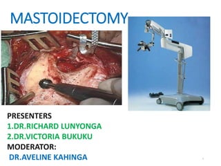
- 1. MASTOIDECTOMY PRESENTERS 1.DR.RICHARD LUNYONGA 2.DR.VICTORIA BUKUKU MODERATOR: DR.AVELINE KAHINGA 1
- 2. OUTLINE •Introduction •History •Surgical Anatomy •Types of Mastoidectomy • Indication of mastoidectomy •Surgical Techniques •Complications of Mastoidectomy •Controversies 2
- 3. 1.INTRODUCTION Mastoidectomy is a surgical procedure which opens up the mastoid cavity, cleans up the infected air cells and improves middle ear ventilation by widening the aditus. Prior to the advent of surgery and antibiotics, morbidity from acute mastoiditis was considerable higher. Mastoid surgery has evolved from simple trephination for acute infection, to the canalwall preserving mastoidectomy employed by most otologists today. 3
- 4. 2.HISTORY OF MASTOIDECTOMY. 1774 - John Luis Petit performed the first surgical trephination of the mastoid. Petit described exposing the mastoid cortex, performing a trephination, and then enlarging the surgically created fistula. 1873 - The first scholarly treatise on mastoid surgery for suppurative disease by Schwartze (cortical mastoidectomy). 1890 - Zaufal described removing the superior and posterior canal wall, tympanic membrane and lateral ossicular chain (radical mastoidectomy). 4
- 5. History of Mastoidectomy...... 1910 – Bondy recognized that disease limited to the pars flaccida could simply be exteriorized, leaving the uninvolved middle ear alone. His description of the “ ” or “Bondy procedure” represented one of the first reports addressing hearing function. 1938 - Lempert introduced the fenestration operation. 1950s - Zollner and Wullstein described tympanoplasty techniques. 1960s - Jansen, Sheehy, and others extended principles of restoring function and maintaining normal anatomy with the introduction of the intact canal wall mastoidectomy with facial recess approach. 5
- 6. FORMATION OF MASTOID BONE •The mastoid process is absent or rudimentary in the neonatal skull. •Mastoid is invisible and covered by a thin bony plate that extends to the squamous portion. • It forms during postnatal and starts to develop after 1- year-old as the sternocleidomastoid muscle develops and pulls on the bone. It usually finishes structural development by 2 years old. •Mastoid antrum becomes obvious at 5 years. •During puberty-mastoid thickness increases and become pneumatic and lined with mucosa. •20% of adults, their mastoid bone may not contain air cells. 6
- 7. 3.SURGICAL ANATOMY The temporal bone connects to the parietal, occipital, zygomatic, and sphenoid bones. It is a pyramidal bone with the apex pointing in the anteromedial direction. The temporal bone consists of four embryologically distinct components: Squamous part Tympanic part 7
- 10. Surgical Anatomy.... The mastoid part is a bulbous bony structure. It is shaped by the expansion of air-filled spaces within. The central air cell is called the antrum. Temporal line:estimates the location of the middle fossa floor 10
- 11. Surgical Anatomy.... Suprameatal spine of Henle Is a small bony protuberance found at the posterior superior lateral edge of the ear canal, which marks the level of the antrum of the mastoid Posterior to it is a group of small holes(Cribriform area).Lies within Macewen’s triangle. 11
- 12. Macewen’s triangle Is a surgical surface marking for mastoid antrum Borders • Superior: Temporal line. • Anterior: Postero-superior margin of bony EAC. • Posterior: . The mastoid antrum lie 12.5-15mm deep to the triangle. 12
- 13. Surgical Anatomy.... Anterior buttress is the point at which the posterior bony canal wall meets the tegmen. Posterior buttress marks the meeting of the posterior canal wall andthe floor of the EAC lateral to facial nerve. Removal of posterior buttress-floor of the EAC slops off gently into the mastoid tip. 13
- 14. Surgical Anatomy.... Facial bridge Is the portion of the posterosuperior bony meatal wall that bridges over the notch of Rivinus and overlies the ossicles. Facial ridge-part of the bony meatal wall that houses the posterior bend and vertical segment of the facial nerve. 14
- 15. Surgical Anatomy... • Citelli’s angle (Sinodural angle) is an angle between the sigmoid sinus and middle fossa dural plate. 15
- 16. Solid angle is an area where three bony semicircular canal meet. Subarcuate artery exits Trautmann’s triangle posterior SSC anteriorly, sigmoid sinus posteriorly, and superior petrosal sinus superiorly. Contain retrolabyrinthine tract that leads to the petrous apex, the endolymphatic sac, and the vestibular aqueduct Donaldson’s line is a line passing through the horizontal semicircular canal and bisects the posterior semicircular canal. This line is a landmark for the endolymphatic sac. 16
- 17. Surgical Anatomy... Facial recess The facial recess is the space bounded. •laterally by the chorda tympani nerve, •medially by the facial nerve, •superiorly by the fossa incudis. 17
- 18. VASCULAR SUPPLY OF TEMPORAL BONE External carotid artery: 1. Superior temporal artery 2. Stylomastoid artery The anterior inferior cerebellar artery gives rise to the internal auditory artery and subarcuate artery. The venous drainage is from inferior and superior petrosal veins into the jugular fossa of the skull base, and then into the internal jugular vein. 18
- 19. Traditionaly classified as 1. Simple (cortical, complete) mastoidectomy. 2. Radical mastoidectomy 3. Modified radical mastoidectomy 4. Tympanomastoidectomy ( ). 19
- 20. Classification of mastoidectomy… Broadly can be classified into two type; Open or Canal Wall Down Mastoidectomy Closed or Canal Wall Up Mastoidectomy 20
- 21. Classification of mastoidectomy… 21 Canal Wall Up Technique (CWU or ICW) Canal Wall Down Technique(CWD) Cortical mastoidectomy Radical mastoidectomy Tympanoplasty with intact CWM Modified radical (Bondy’s procedure) Canal wall reconstructive technique. Atticotomy Atticoantrostomy Mastoid obliteration
- 22. Types of mastoidectomy..... CORTICAL MASTOIDECTOMY Initial stage of any transmastoid surgery of the middle and inner ear and facial nerve. Involve removal of disease that is limited to the mastoid antrum and air cell system. Preserving the posterior bony EAC wall. The middle ear contents are not disturbed. Tympanostomy tube may be placed for improved ventilation. 22
- 24. Types of mastoidectomy..... Indications for cortical mastoidectomy. Coalescent Mastoiditis and Masked Mastoiditis. CSOM (tubo- tympanic) Active Refractory to antibiotics. Approach to: • Endolymphatic sac surgery. • Facial nerve decompression. • Vestibulo cochlear nerve section. • Translabyrinthine Approach for CP angle. • Cochlear implant surgery. • Combined Approach Tympanoplasty 24
- 25. Types of mastoidectomy..... TYMPANOPLASTY WITH ICW MASTOIDECTOMY An operation in which disease is removed from the mastoid and middle ear while preserving the posterior bony wall of the EAC. Often the mesotympanum is exposed by developing a posterior tympanotomy through the facial recess. primarily, but it is often staged in cholesteatoma cases. 25
- 26. Types of mastoidectomy..... The first operation is performed to remove all cholesteatoma and repair the tympanic membrane. 6 months later, the second operation is done to inspect the mastoid and middle ear for residual or recurrent cholesteatoma and to 26
- 27. Types of mastoidectomy..... Done to eradicate or exteriorize extensive middle ear disease by removing the posterior bony ear canal to open the middle ear, mastoid, and epitympanum into one common cavity. Remnants of the TM, malleus, and incus are removed leaving only the remaining portions of the stapes. The TM is not reconstructed, and the Eustachian tube may be left open or permanently obstructed with tissue grafts. 27
- 28. Canal wall down(CWD) mastoidectomy…. 28
- 29. Types of mastoidectomy..... Indications for radical mastoidectomy Unresectable cholesteatoma extending down the Eustachian tube or into the petrous apex. Promontory cochlear fistula caused by cholesteatoma Chronic perilabyrinthine osteitis or cholesteatoma that cannot be removed and must be cleaned or inspected periodically. Resection of temporal bone neoplasms with periodic monitoring. 29
- 30. Types of mastoidectomy.... An attempt is made to preserve or reconstruct the middle ear. Sometimes healthy TM and ossicular remnants are preserved. In the classic Bondy modified radical procedure, atticoantral cholesteatoma is exteriorized without disturbing the intact pars tensa of the TM or the intact ossicular chain. 30
- 31. MODIFIED RADICAL MASTOIDECTOMY Indications Absolute Indications Unresectable disease Unreconstructable Posterior canal wall. Failure of first stage CWU procedure because of poor ET function. Inadequate Patient Follow-up. Relative Indications Disease in only hearing ear or in a dead ear. Medical illness or severe otologic/CNS complications Neoplasms Poor E T function 31
- 32. MODIFIED RADICAL MASTOIDECTOMY Contraindications: Chronic otitis media without cholesteatoma Acute otitis media with coalescent mastoiditis, Persistent secretory otitis media, or Chronic allergic otitis media. Tuberculous otitis media. 32
- 33. Types of mastoidectomy.... ATTICOTOMY Removal of ear canal bone including the lateral wall (scutum) of the epitympanum to expose and exteriorize limited attic disease, usually lateral to healthy ossicles. 33
- 34. Types of mastoidectomy...... Done by entering the attic from the ear canal and then proceeding posteriorly, gradually removing posterior ear canal bone and exposing disease in the aditus and antrum until it is fully exteriorized. It is synonymous with modified radical mastoidectomy but is carried out from anterior to posterior ie. exposing the attic first and then proceeding posteriorly into the aditus and antrum. The surgeon’s intent is to exteriorize rather than resect the matrix of the cholesteatoma. 34
- 35. Types of mastoidectomy..... MASTOID OBLITERATION A procedure in which graft is used to obliterate a portion of the cavity following a canal wall down mastoidectomy. Mastoid reconstruction and obliteration procedures can be classified into two main categories: (a) Free grafts (b) local flaps. If successful, the size of the defect is minimized, which may avoid the need for long-term cavity care. 35
- 36. Types of mastoidectomy...... Intraoperative findings that may be indications for a CWD procedure include Labyrinthine fistula Unresectable disease on the facial nerve or stapes footplate A low-lying tegmen that limits access to the attic Unresectable sinus tympani disease. Unreconstructable posterior canal wall defect. 36
- 37. INTACT CANAL WALL UP MASTOIDECTOMY… Advantages •Physiological TM position. •No mastoid bowl. •Hearing aids easier to fit. Disadvantages •Technically difficult. •Residual disease harder to detect. •Second stage often required. •Periodic follow up is needed 37
- 38. CANAL WALL DOWN MASTOIDECTOMY…. Advantages •Residual cholesteatoma is visible on follow-up. •Recurrent cholesteatoma is rare. •Total exteriorization of facial recess. Disadvantages •Position of the pinna may be altered. •Mastoid bowl-life long problem •Hearing aids, difficult to fit. 38
- 39. Preoperative Assessment History Chronic otorrhoea Hearing loss Previous surgery Otoscopy TM perforation Retraction pockets Choleteatoma, polys 39
- 40. Preoperative Assessment Audiology PTA – Assess Hearing loss Tympanometry- assess the status of EAC and middle ear Speech Discrimination Test –Assess possibility of Middle ear reconstruction. CT scan – HRCT Diagnostic and surgical planning 40
- 41. HRCT of the temporal bones Normal Coalescent mastoiditis 41
- 42. Preoperative Assessment CT Scan – HRCT Can show temporal bone pneumatization, middle ear and mastoid air cells ventilation, EAC, sigmoid sinus, jugular bulb, tegmen tympany, facial nerve, extent of disease and status of ossicular chain MRI Non specific in COM Better for IC involvement 42
- 43. 5.SURGICAL TECHINIQUES Preparations: General anesthesia without paralytic agents and with continuous facial nerve monitoring. Patient is positioned in supine position with the head turned to the contralateral side to expose the diseased ear. “Pre-scrub" the ear and the entire side of the head, including hair, with betadine. 43
- 44. Surgical Technique.... Aseptic drapping of the surgical site. 44
- 45. Surgical Technique.... Tragus and postauricular skin are injected with 1% lidocaine with epinephrine (1: 100,000) to provide hemostasis and local anesthesia. 45
- 46. Surgical Technique.... Surgical approach to the ear in CWU a).Postaural/Retroauricul ar incision A C-shaped incision. Starts from the highest attachment of the pinna, follows the curve 0.8- 1cm behind the retro auricular groove, and ends at the mastoid tip. 46
- 48. Surgical Technique.... Slanting posteriorly in <2years children due to underdeveloped mastoid with the superficial facial nerve. 48
- 49. Surgical Technique.... b). Endaural incision approach Lempert I incision. The semicircular incision from 12 o’clock to 6 o’clock position in the posterior meatal wall at the bony- cartilaginous junction. Lempert II Starts from the first incision at 12 o clock and then passes upward curvilinear between tragus and crus of helix through incisura terminalis. 49
- 51. Surgical Technique.... Elevate the skin flap •Towards the ex-ternal ear canal. •Cut through the post- auricular muscle to reach the correct plane just superficial to temporalis fascia. •large rake can be used to retract the pinna forward. 51
- 52. Surgical Technique.... An anteriorly based musculoperiosteal flap is developed, about 1.5cm in length A T-shaped incision is made in the mastoid periosteum to expose the mastoid cortex • The 1st incision-Along the linea temporalis to the level of the underlying bone. • The 2nd incision-Perpendicular to the linear temporalis down to the mastoid tip. 52
- 53. Surgical technique..... Periosteal incisions are made, the periosteum elevated a lampert elevator and retracted forward with the auricle. 53
- 54. Surgical Technique.... 54 •Elevation of the flap from the bone is done until the spine of Henlé and the entrance to bony canal come into view.
- 55. Surgical Technique.... •In an adult two self- retaining retractors are placed between the skin edges and soft tissue for exposure. •One self-retaining retractor is usually sufficient in a child. 55
- 57. Surgical Techniques...... Initial Drilling: When the mastoid cortex becomes fully exposed The first bur cut is made along the temporal line, which approximates the level of the middle cranial fossa dural plate The second bur cut is made perpendicular to this and tangential to the bony EAC It should be carried inferiorly to the mastoid tip. 57
- 60. Surgical Technique.... Appropriate irrigation is necessary To clear bone dust from the field of dissection, To prevent excessive heat transfer to underlying structures (especially the facial nerve), and To maintain a clean cutting surface on the bur. 60
- 61. Surgical Techniques....... •As the dissection is carried medially and the antrum is approached, a bony septum (Körner’s septum) may be encountered. •This plate is a remnant of the petrous- squamous septum. 61
- 62. Surgical Techniques....... A key landmark in performing mastoid surgery is the antrum with the dome of the horizontal semicircular canal (HSCC) along its floor. 62
- 64. Surgical Technique.... 64 Key principles that assist in locating the antrum include saucerization, identification of the tegmen plate, and thinning of the posterior canal wall. Consideration should also be made during posterior dissection where the sigmoid sinus is located.
- 67. Surgical Techniques.... Drilling tips: Avoid keyhole surgery; work through a wide space. The tip of the drill should always be visible Never drill behind edges of bone. Drilling should always be parallel to any structure you are trying to preserve e.g. facial nerve, sigmoid sinus. 67
- 68. Surgical Techniques.... Drilling tips: When drilling deeper in the mastoid cavity the burr needs to be lengthened. However, one cannot lengthen a cutting burr as this will cause the drill to jump with the risk of injuring structures. Therefore if it is necessary to lengthen the burr, then change to a rough diamond or smooth diamond burr. 68
- 69. Surgical Techniques..... Facial Nerve Identification: Identifying the facial nerve is fundamental to performing good mastoid surgery. The most important landmarks for the facial nerve are the HSCC, the short process of the incus, and the posterior bony EAC. 69
- 70. Surgical Techniques.... The genu and proximal portion of the mastoid segment of the facial nerve lie anterior and just medial to the dome of the HSCC The mastoid segment facial nerve also lies medial to the plane of the short process of the incus at the base of the posterior canal wall. 70
- 71. Surgical Techniques.... Opening the Facial Recess: provides access to the middle ear from the mastoid. 71
- 72. Surgical Techniques.... The promontory, round window niche, stapes, long process of the incus, cochleariform process, medial side of the tympanic membrane and malleus handle, and eustachian tube all are well seen. 72
- 73. Surgical Techniques.... The facial recess can be extended superiorly and inferiorly, providing a large “posterior tympanotomy.” Sacrificing the chorda tympani nerve permits additional dissection inferiorly with good exposure of the hypotympanum. 73
- 74. Surgical Techniques........ Opening the Epitympanum Smaller, diamond burs are required in the epitympanic dissection Involves thinning dura bone and the superior canal wall. The Cog: landmark from tegmen towards malleus head and separates the epitympanum into ant. And post. portion 74
- 75. Surgical Techniques.... Facial nerve takes a slight medial course in the epitympanum as it is traced anteriorly from the mastoid genu to the geniculate ganglion. It passes superior to the oval window and cochleariform process. 75
- 76. Surgical Techniques...... Canal Wall Down Mastoidectomy Removing the posterior bony canal wall to the level of the facial nerve: only a thin shell of bone remains over the nerve, creating a smooth, gently curving transition from the anterior epitympanum to the anterior canal wall. 76
- 77. Surgical Techniques.... This dissection is continued toward the stylomastoid foramen until there is no bony spur (inferior or posterior buttress) between the floor of the external bony canal and the mastoid cavity. In a similar manner, the anterior extent of the superior canal wall (anterior buttress) is completely removed. 77
- 78. Surgical Techniques.... Canal wall down mastoidectomy… 78
- 79. Canal wall down mastoidectomy… 79
- 81. 81
- 82. Postoperative Care Mastoid pressure dressing is preferred Mastoid dressings are typically removed 48 - 72 hours after surgery. Patients are typically instructed to keep the operative ear dry. 82
- 83. Postoperative Care.. Monitor of vital signs (BP, PR, T, RR) Appropriate analgesia within the first 5-7 days. Patients can start topical antibiotic drops the following day after surgery for several days before the initial postoperative visit. Remove packing in the ear canal 1-2 weeks after surgery. Depends merely on the type of ear surgery done. 83
- 84. Postoperative Care The topical antibiotic drops serve a dual purpose of decreasing the risk of a post-surgical infection and keeping the packing moist to ease removal at their initial postoperative visit Long term monitoring Patients with cholesteatoma need to be followed long term. Access for Recurrence. 84
- 85. 6.COMPLICATIONS OF MASTOIDECTOMY Facial Nerve Injury Facial nerve paralysis is the most dreaded complication of mastoidectomy. The risk of iatrogenic facial nerve injury increase in: Revision surgery Extensive disease Facial nerve dehiscence Poor operator experience or misadventure with the drill 85
- 86. Complications of mastoidectomy… Facial nerve injury…. Minimal injury intra-op •-Decompress the fallopian canal proximal and distal to the site. Partial transection. •Anastomose the separated fassicles. •Decompress the fallopian canal proximal and distal to the site. Complete transection-intra-op •Attempt primary anastomosis without tension, cable graft if necessary. Immediate post OP early facial weakness -Reassess after 4hours, allow the anesthesia to wear off. Mild paresis-observe, give steroid. Severe paresis-Return to the operating room for exploration and repair. More than 8HRS post.OP Mild paresis-Observe, give steroid. Severe paresis-Observe, give steroid. 86
- 87. Complications of Mastoidectomy.... Dural injury A dural tear or significant abrasion with herniation of arachnoid tissue with or without a cerebrospinal fluid leak requires repair. Dural defects are best repaired with a layered closure using soft tissue such as fascia or perichondrium combined with a more rigid support material such as bone or cartilage. 87
- 88. Complications of Mastoidectomy.... Firmly packing the mastoid (or epitympanum) with absorbable gelatin sponge (Gelfoam) (with or without fibrin glue) can be used to support the repair as needed Instituting a broadspectrum antibiotic with cerebrospinal fluid penetration should also be considered. 88
- 89. Complications of Mastoidectomy...... Vascular injury (sigmoid sinus/ jugular bulb) The sigmoid sinus and jugular bulb (variable anatomy). These low-pressure, but high-volume, venous structures is initially treated with digital pressure. For small tears, bone wax may suffice For larger rents, cellulose-type surgical packing is required A significant injury to the sigmoid sinus can result in thrombosis of that vessel. 89
- 90. Complications of Mastoidectomy...... Hearing loss A temporary CHL is very common as blood, serous fluid and packing fill the middle ear space. A significant SNHL is rarely encountered in patients undergoing surgical intervention for COM. SNHL may arise from the high-speed drill contacting an intact ossicular chain, labyrinthine fistula or noise exposure from the drill. 90
- 91. Complications of Mastoidectomy.... Horizontal Semicircular Fistula Iatrogenic injury to the HSCC predisposes the patient to bacterial labyrinthitis with resulting vertigo and severe sensorineural hearing loss. Immediate closure, usually with bone wax is required. A short course of a broad-spectrum antibiotic and steroids can be considered. 91
- 92. Complications of Mastoidectomy... Change in taste Chorda tympani nerve may need to be sacrificed if it is encased in cholesteatoma or inflammatory tissue especially in patients undergoing revision surgery or a canal wall down procedure. Patients typically notice an altered sensation of taste, typically described as a metallic or sour taste on the affected side. This sensation may be persistent but often resolves over a period of months. 92
- 93. 7.CONTROVERSIES. The choice for preserving or removing the posterior wall of the EAC, ie, ICW versus CWD mastoidectomy, has been extensively debated. Preservation of the canal wall is preferred vs Canal wall down leads to a ‘safe’ and technically less demanding. Judgment depends on the patient's reliability, and the surgeon's experience, and often decision is made during surgery. Mastoid obliteration-Cartilage and hydroxyapatite vs bone dust Schapola et al, India,2014
- 94. References Paul W. Flint et al, Cummings Otolaryngology-Head & Neck Surgery, Fifth Edition John Jacob Ballenger, ‘Ballenger’s Otorhinolaryngology-Head and Neck Surgery’ Sixteenth Edition Bailey, Byron J.; Johnson, Jonas T.; Newlands, Shawn D. Head & Neck Surgery - Otolaryngology, 4th Edition • Professor Tuncay Ulug, MD Istanbul University Atlas of temporal bone surgery,2010 • Glasscock-Shambaugh, Surgery of the Ear, 5th Edition Leliever, W C (1983), Temporal Bone Surgical Dissection Manual. Archives of Otolaryngology-Head and Neck Surgery Eugene N. Myers, MD, FACS, FRCS Edin (Hon), Head and neck surgery,volume 1, 2014. 94
- 95. 95
Editor's Notes
- Cymba concha is the soft tissue anatomical landmark for the mastoid antrum. Dissecting at this margins of triangle is safer because of vital neurovascular structure are absent Cribriform-passage of vessels
- NOTE: other important landmarks and structures will be highlighted with the surgical steps.
- stylomastoid artery give rises to posterior tympani arter
- free graft can be biological (bone chips) or non-biological(hydroxyopatite) local flap-pedicle flap or musculoperiostal flap-palva
- An otologist’s road map though has poor specificity with mass lesions e.g cholesteatoma
- This plate is a remnant of the petrous squamous septum, and simply separates more superficial air cells from deeper ones
- Tympanoplasty need to pack the eac 7-10 days