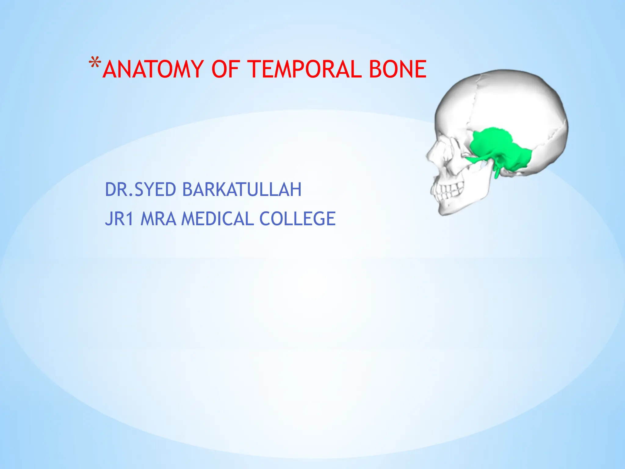
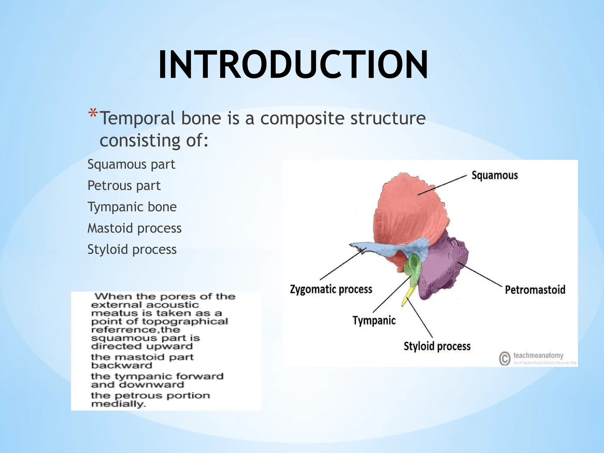
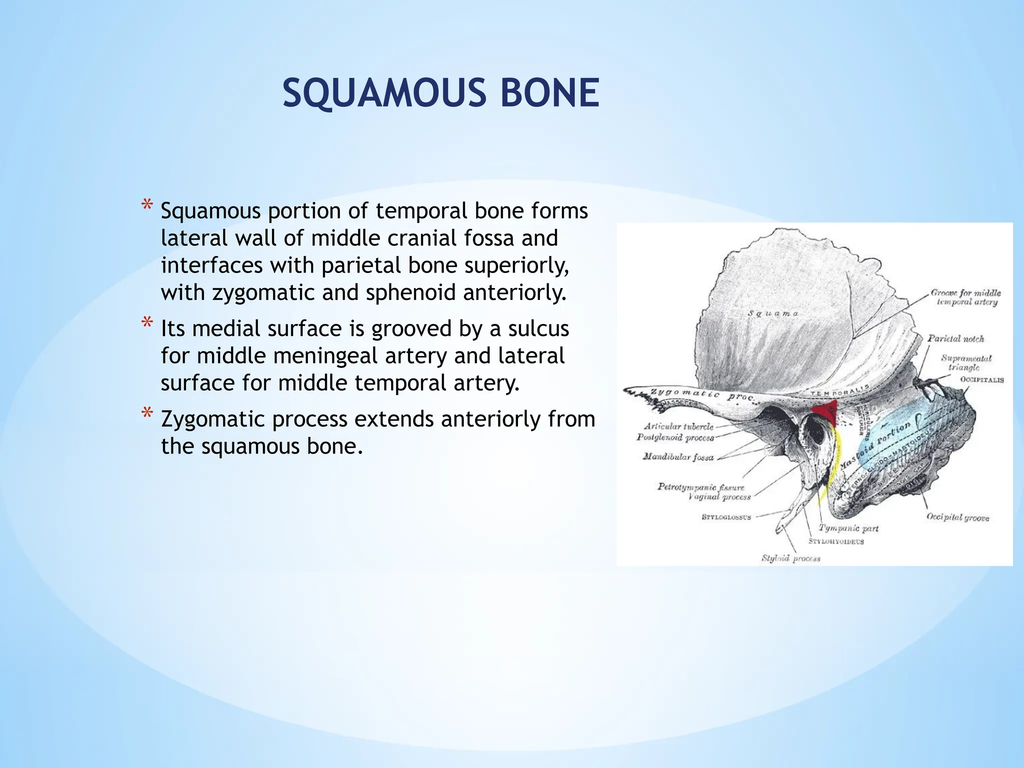
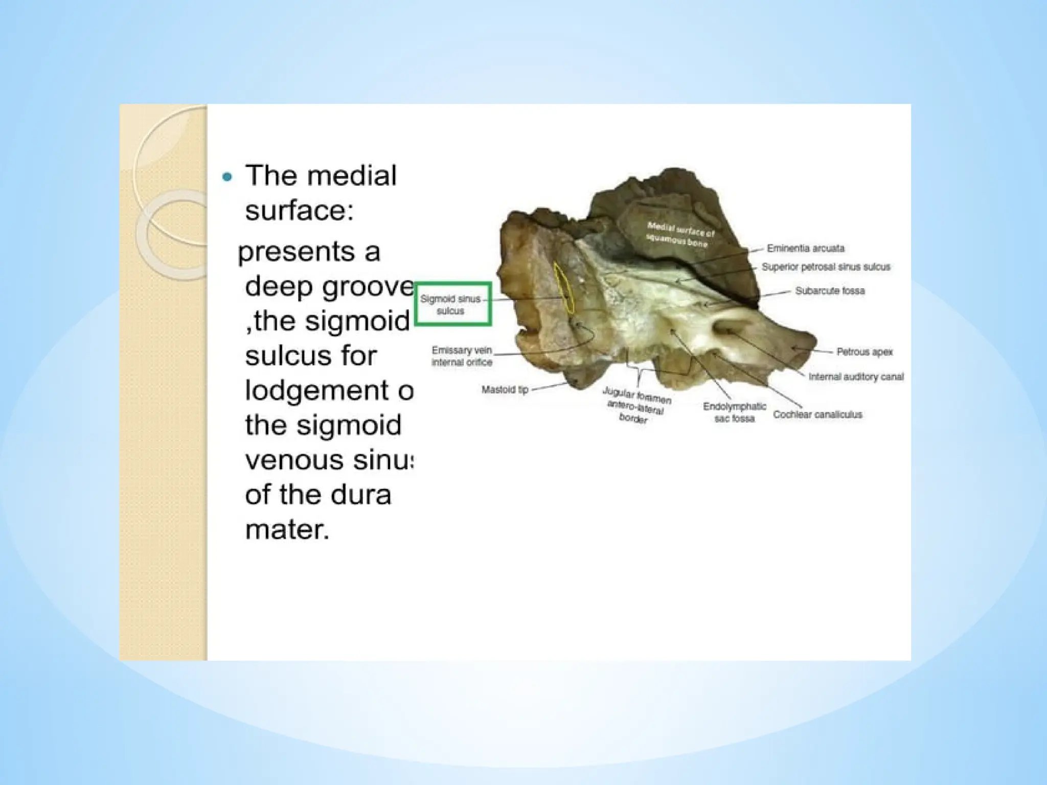
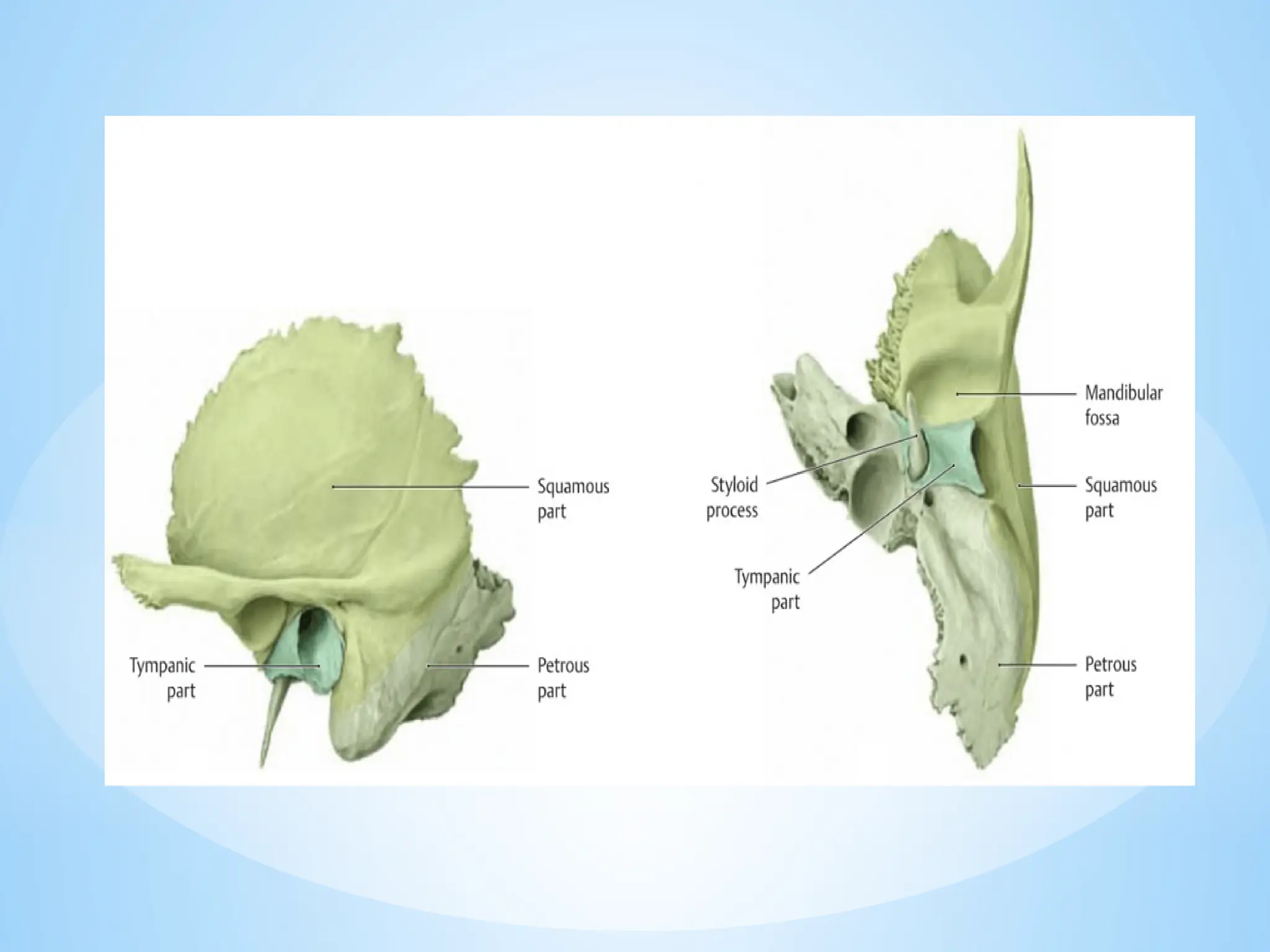
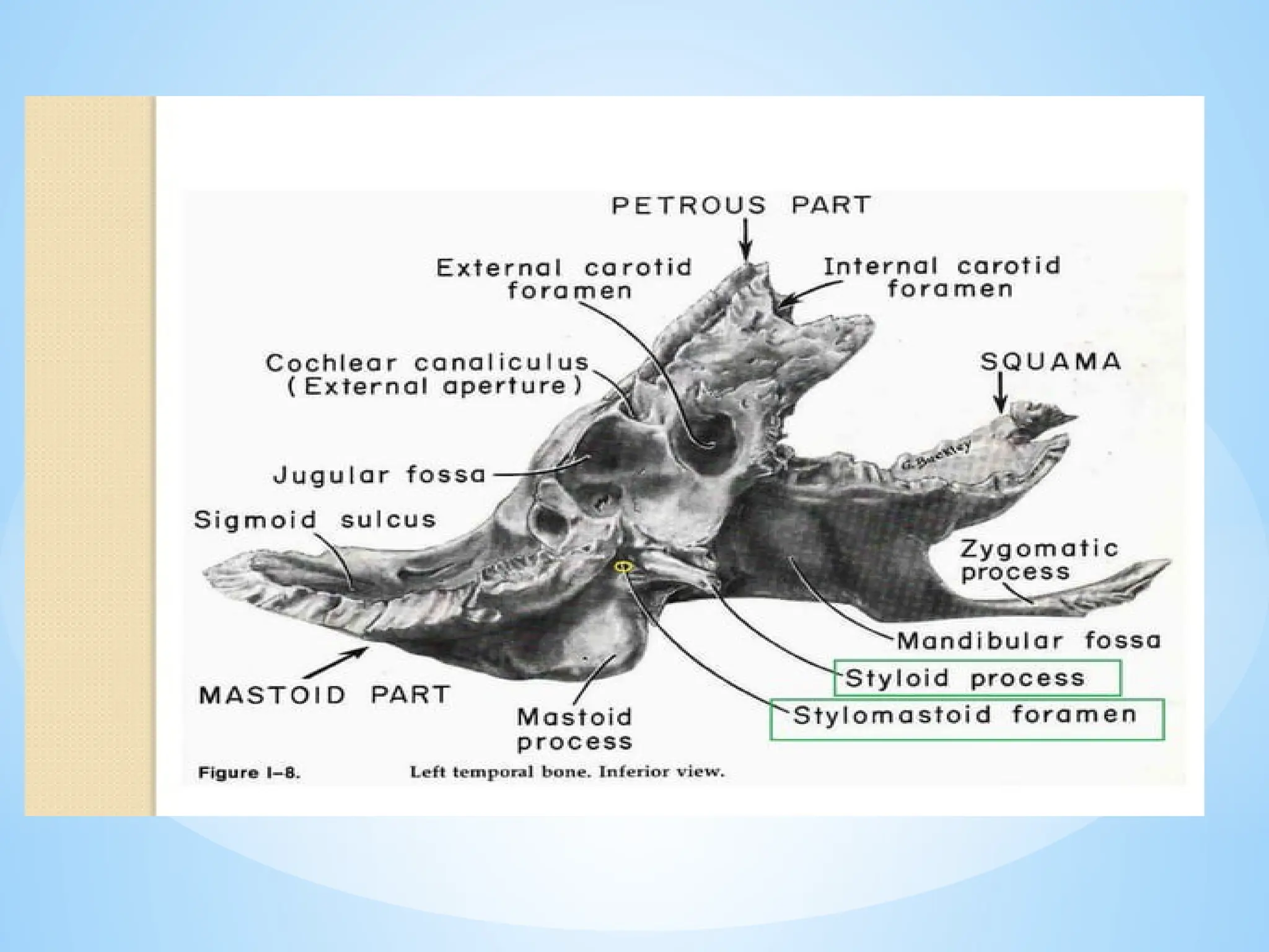
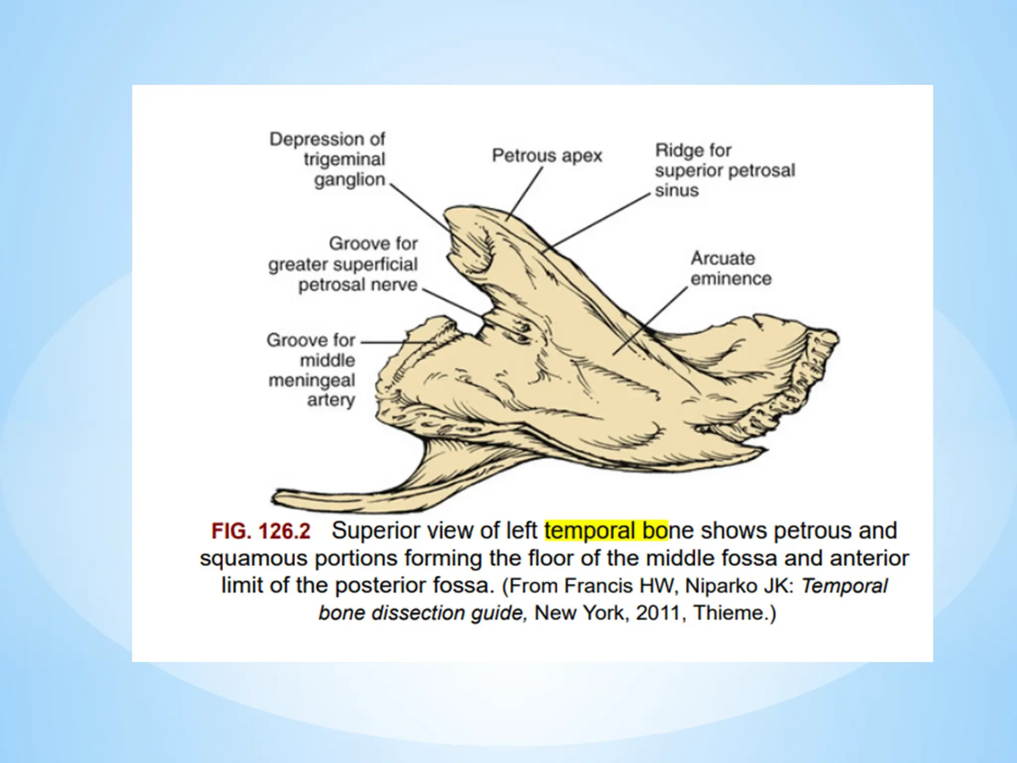
*ANATOMY OF TEMPORAL BONE
DR.SYED BARKATULLAH
JR1 MRA MEDICAL COLLEGE
INTRODUCTION
*Temporal bone is a composite structure
consisting of:
Squamous part
Petrous part
Tympanic bone
Mastoid process
Styloid process
EMBRYOLOGY
* Temporal bone is formed by the fusion and growth of five bones, however, of these, the mastoid
and styloid processes do not fully develop until after birth.
* The squamous part of temporal bone develops from intramembranous ossification that appears
during 8th
week of gestation.
* The development of upper and lower halves of the squamous bone primordium differs:
The upper part is flat and thin and will become the vertical portion.
The lower part, due to the presence of the tympanic bone by the 16th
week, bulges and grows
rapidly in three directions: anterior, middle and posterior.
Tympanic bone is a C shaped bone formed by the intramembranous
ossification by the 8th
week.
A full development of tympanic ring is recognised by 11th
week of
gestation. However, it never closes superiorly, giving rise to a notch
called as Rivinus Notch.
The styloid process is derived from Reichert’s cartilage.
It develops from two parts:
1. Proximal part named as, tymanohyale situated close to tympanic bone.
Its ossification center appears before birth and continues to grow until
the age of 4 years.
2. Distal part, stylohyale starts its ossification only after birth. It fuses
with the proximal part only after birth.
* The petrous bone is represented by the
cartilaginous otic capsule.
Between 8th
-9th
week the otic capsule budge in
the base of cartilagenous cranium.
Later the otic capsule becomes surrounded by
membranous layers which later becomes
extracapsular part of petrous bone.
Shortly after this, the lateral and superior
boundaries of otic capsule begin to form
mastoid process and tegmen tympani.
A cartilaginous flange grows from lateral and
superior part of otic capsule – forms tegmen
tympani and lateral wall of ET tube.
Whereas, a cartilaginous flange grows from
lateral and inferior part of otic capsule– forms
jugular plate and the floor of tympanic cavity.
Antermedially another periosteal layer grows to
form petrous apex.
Ossification of otic capsule is completed shortly
after birth.
Anatomy
TEMPORAL BONE ( LATERAL SURFACE)
* The four parts visible are:
1. Squamous bone
2. Tympanic bone
3. Styloid bone
4. Mastoid bone
* Squamous portion of temporal bone forms
lateral wall of middle cranial fossa and
interfaces with parietal bone superiorly,
with zygomatic and sphenoid anteriorly.
* Its medial surface is grooved by a sulcus
for middle meningeal artery and lateral
surface for middle temporal artery.
* Zygomatic process extends anteriorly from
the squamous bone.
SQUAMOUS BONE
MASTOID PORTION OF TEMPORAL BONE
* It is the poster-inferiorly extending projection seen
on the lateral surface of the temporal bone.
* It is composed of a squamous portion (laterally) and
a petrous portion (medially) seperated by Korner’s
(petro-squamous) septum.
* The fossa mastoidea ( MacEwen’s triangle) is defined
by the temporal line superiorly , the postero-
superior margin of the EAC and a tangent to the
posterior margin of the EAC posteriorly.
* It is a cribrose (cribriform) area, having numerous,
perforating small blood vessels.
* It is an important surgical landmark as it laterally
overlies the mastoid antrum. Therefore the site
where mastoid drilling ordinarily commences.
* The mastoid foramen located posteriorly on the
mastoid process, is traversed by the mastoid
emissary vein and one or two mastoid arteries.
* Mastoid process serves laterally for the attachment of SCM
muscle and medially to the posterior belly of digastric.
* Mastoid tip is easily palpable and is landmark for positioning of
post-auricular incisions.
* Mastoid process in children is not fully developed, thus cannot be
palpated easily.
* Hence, post-auricular incision in children should be given more
horizontally to prevent injury to the facial nerve.
TYMPANIC BONE
* It forms the anterior, inferior and the posterior wall of the external
auditory canal.
* It interfaces with 1)the squama at the tympanosqamous suture.
2) the mastoid at the tympanomastoid suture.
3) the petrosa at the petrotympanic fissure.
* The chorda tympani nerve , the anterior process of malleus, anterior
tympanic artery traverse the petro-tympanic fissure.
* Laterally, the tympanic bone borders the cartilaginous EAC, whereas
medially it bears a circular groove, the annular sulcus.
* The annular sulcus houses the annulus of tympanic membrane except
superiorly where it is deficient, known as the notch of Rivinus.
* Anteriorly , the tympanic ring seperates the EAC from the glenoid
fossa, which lies beneath the root of zygoma.
Tympano-mastoid suture is traversed by Arnold’s nerve.
Tympano-squamous and tympano-mastoid sutures are the landmarks for
the vascular ring incisions used in taympano-mastoid surgery.
STYLOID PROCESS
* Normal length – 2.0-2.5cm
* It’s a slender pointed piece of bone just below the ear.
* Projects down and forwards and medially from the
inferior surface of the temporal bone.
* Its proximal part(TYMPANOHYAL) is ensheathed by the
vaginal process of the tympanic bone.
* Its distal part(STYLOHYAL) gives attachment to the
following:
Ligaments: stylohyoid and stylomandibular.
Muscles: styloglossus, Stylohyoid and Stylopharyngeus.
*Stylomandibular ligament is a specialised band of cervical
fascia which extends from anterior surface of apex of
styloid process to the posterior surface of the angle of
mandible.
*This ligament separates parotid gland from
submandibular gland and medial pterygoid muscle.
*Eagle syndrome is characterised by recurrent pain in the
oropharynx and face due to elongated styloid process or
calcified stylohyoid ligament which interferes with the
adjacent anatomical structures giving rise to pain.
PETROUS BONE
* Petrosa can be seen in superior, medial and posterior views.
* It houses the inner ear, internal carotid artery, fallopian canal and
the major part of middle ear.
* It is shaped like a pyramid that projects anteromedially forming an
angle of 45 degree with the transverse axis.
* Its has a postero lateral base ( the mastoid) and the anteromedial
summit (the petrous apex).
* Its superior surface participates in the formation of middle cranial
fossa floor and posterosuperior surface forms the anterolateral wall
of posterior cranial fossa.
MEDIAL SURFACE OF
TEMOPRAL BONE
* It features the Porus of IAC
* ICA exits at petrous apex through internal
carotid foramen
* Sigmoid portion of lateral venous sinus runs
in the deep sulcus seen posteriorly.
* Whereas, superior petrosal sinus runs in the
sulcus located at the junction of posterior
and middle fossa faces of the temporal bone.
* The petrous portion of the temporal bone
houses part of the middle ear (ossicles) and
inner ear (cochlear and vestibular end
organs).
* The following landmarks are visible on the medial
surface:
* Cochlear aqueduct: a fine canal that connects the scala
tympani in the basal turn to the CSF space around the
brain.
* Vestibular aqueduct: a bony passage that runs from the
vestibule to the subarachnoid space. It transmits the
endolymphatic duct.
* Internal auditory meatus: a bony opeing through which
the 7th
and 8th
cranial nerves and internal auditory
artery enters the petrous part of temporal bone.
* Mastoid process
* Sigmoid sinus sulcus: s- shaped depression, located
posterior to the operculum and vestibular aqueduct
* Styloid process
* Jugular fossa marks the location of jugular bulb.
POSTERIOR SURFACE
* It is formed exclusively by the petrous part.
* It represents the anterolateral wall of the posterior cranial fossa.
* Lies between superior and inferior petrosal sinuses.
* Laterally it presents the sigmoid sinus sulcus and the internal
orifice of the emissary vein canal.
 The internal auditory meatus, which lies in the center of this
surface midway between the apex and the anterior border of
the sigmoid sinus sulcus.
* The endolymphatic sac situated at the lateral part of this surface,
sac lies medial to the level of the posterior semicircular canal.
* The Internal Auditory Canal penetrates the posterior surface of
the petrous ridge, runs antero-medially to postero-latrally and
contains the cochlear, vestibular and facial nerves along with
their blood supply.
* At the fundus, the canal is divided into an upper and a lower
portion by the transverse crest -CRISTA FALCIFORMIS
* The inferior compartment contains the cochlear nerve anteriorly
and inferior vestibular nerve posteriorly.
* A vertical crest of bone BILL’S BAR separates superior portion of
canal into an anterior compartment, occupied by the facial
nerve and a posterior compartment containing the superior
vestibular nerve.
INFERIOR SURFACE
* Separates the upper neck from skull base, pyramidal in shape
and lies horizontally.
* Many vital neurovascular structures traverse this surface:
* Anteriorly and medially: carotid foramen present from which
the ICA enters the temporal bone.
* Posteriorly, a ridge of bone, the jugulo carotid crest,
separates the carotid canal from the jugular foramen.
* Jugular foramen (formed with occipital bone) has been
divided into a posterolateral pars venosa, occupied by
jugular vien and an anteromedial pars nervosa traversed by
IX, X and XI nerves.
* Lateral to the jugular foramen is the styloid process.
* Immediately posteromedial to the styloid process is the stylo-
mastoid foramen, by which the facial nerve exits the
temporal bone.
* A triangular opening of the cochlear aqueduct is located
medial to the jugular foramen.
ANTERIOR SURFACE
* The petrous apex is the wedge of
bone that seperates greater wing of
sphenoid from occipital bone.
* The most prominent feature of this
surface is the internal carotid
foramen through which carotid artery
exits temporal bone.
* The impression for the trigeminal
ganglion is located on lateral surface
of petrous apex.
* The semi canal for tensor tympani is
lateral to carotid canal. Bony portion
of eustachian tube runs inferior and
parallel to tensor tympani muscle.
*VASCULAR ANATOMY
Several large dural venous sinuses are intimately
associated with temporal bone and comprise the
principal venous drainage of brain and cranial
vault.
1. The superior sagittal sinus and straight sinus
merge at the internal occipital protuberance.
2. Right transverse sinus is primarily continuation
of superior sagittal sinus and left transverse
sinus is continuation of straight sinus.
Transverse sinuses lie just inferior to tentorium
and parallel its course.
3. Anteriorly the superior petrosal sinus joins
transverse sinus and this junction marks
beginning of sigmoid sinus.
4. Sigmoid sinus is the posterior boundary
of the mastoid cavity. Middle fossa dura
approximates the superior portion of
sigmoid sinus at sinodural angle of Citelli.
At its inferior extent the sigmoid sinus rises
to the jugular bulb.
5. Inferior petrosal sinus arises from medial
aspect of jugular bulb and run
anteromedial tp cavernous sinus.
6. The jugular vein exits skull through
jugular foramen, accompanied by vagus,
glossopharyngeal and spinal accessory
nerves.
* the internal carotid artery also travels through
temporal bone. Its entrance, the carotid foramen is
medial to the styloid process and anterior to the
jugular foramen. ICA travels superiorly until it
encounters dense bone of cochlea where it makes
90degree bend to run anteriorly and medially.
* Emissary veins are drainage of dural venous sinuses
through skull that communicates with superficial veins
of scalp. A fairly constant emissary vein the mastoid
emissary vein can be found at the junction of
temporal and occipital bones and usually
communicates with occipital vein.
Cranial nerves in relation to
temporal bone
TRIGEMINAL NERVE-
SIXTH NERVE-
EIGHT NERVE-
NINTH NERVE-
TENTH NERVE-
ELEVENTH NERVE-
SEVENTH NERVE-
Innervates structure derived from Reichert’s cartilage.
• Facial nerve enters temporal bone through internal auditory
canal which it exits at meatal foramen to travel anteriorly to
geniculate ganglion.
COURSE OF FACIAL NERVE IS DIVIDED INTO 5
SEGMENTS:
• Intracranial segment- 24mm from pons to
IAC.
• Intracanalicular segment- traverses IAC,
occupies anterosuperior quadrant at
fundus where it is joined by nervous
intermedius.
• Intralabyrinthine segment-
shortest,4mm,from beginning of fallopian
canal to the geniculate ganglion.
• Tympanic segment- 13mm long, courses
in the medial wall of the tympanic
cavity, superior to the cochleariform
process and oval window.
• Mastoid segment- 20mm, from 2nd
genu
to stylomastoid foramen.
THANK YOU

Surgical anat Temporal bone in detail...

  • 1.
    *ANATOMY OF TEMPORALBONE DR.SYED BARKATULLAH JR1 MRA MEDICAL COLLEGE
  • 4.
    INTRODUCTION *Temporal bone isa composite structure consisting of: Squamous part Petrous part Tympanic bone Mastoid process Styloid process
  • 6.
    EMBRYOLOGY * Temporal boneis formed by the fusion and growth of five bones, however, of these, the mastoid and styloid processes do not fully develop until after birth. * The squamous part of temporal bone develops from intramembranous ossification that appears during 8th week of gestation. * The development of upper and lower halves of the squamous bone primordium differs: The upper part is flat and thin and will become the vertical portion. The lower part, due to the presence of the tympanic bone by the 16th week, bulges and grows rapidly in three directions: anterior, middle and posterior.
  • 7.
    Tympanic bone isa C shaped bone formed by the intramembranous ossification by the 8th week. A full development of tympanic ring is recognised by 11th week of gestation. However, it never closes superiorly, giving rise to a notch called as Rivinus Notch. The styloid process is derived from Reichert’s cartilage. It develops from two parts: 1. Proximal part named as, tymanohyale situated close to tympanic bone. Its ossification center appears before birth and continues to grow until the age of 4 years. 2. Distal part, stylohyale starts its ossification only after birth. It fuses with the proximal part only after birth.
  • 8.
    * The petrousbone is represented by the cartilaginous otic capsule. Between 8th -9th week the otic capsule budge in the base of cartilagenous cranium. Later the otic capsule becomes surrounded by membranous layers which later becomes extracapsular part of petrous bone. Shortly after this, the lateral and superior boundaries of otic capsule begin to form mastoid process and tegmen tympani. A cartilaginous flange grows from lateral and superior part of otic capsule – forms tegmen tympani and lateral wall of ET tube. Whereas, a cartilaginous flange grows from lateral and inferior part of otic capsule– forms jugular plate and the floor of tympanic cavity. Antermedially another periosteal layer grows to form petrous apex. Ossification of otic capsule is completed shortly after birth.
  • 9.
  • 10.
    TEMPORAL BONE (LATERAL SURFACE) * The four parts visible are: 1. Squamous bone 2. Tympanic bone 3. Styloid bone 4. Mastoid bone
  • 11.
    * Squamous portionof temporal bone forms lateral wall of middle cranial fossa and interfaces with parietal bone superiorly, with zygomatic and sphenoid anteriorly. * Its medial surface is grooved by a sulcus for middle meningeal artery and lateral surface for middle temporal artery. * Zygomatic process extends anteriorly from the squamous bone. SQUAMOUS BONE
  • 14.
    MASTOID PORTION OFTEMPORAL BONE * It is the poster-inferiorly extending projection seen on the lateral surface of the temporal bone. * It is composed of a squamous portion (laterally) and a petrous portion (medially) seperated by Korner’s (petro-squamous) septum. * The fossa mastoidea ( MacEwen’s triangle) is defined by the temporal line superiorly , the postero- superior margin of the EAC and a tangent to the posterior margin of the EAC posteriorly. * It is a cribrose (cribriform) area, having numerous, perforating small blood vessels. * It is an important surgical landmark as it laterally overlies the mastoid antrum. Therefore the site where mastoid drilling ordinarily commences. * The mastoid foramen located posteriorly on the mastoid process, is traversed by the mastoid emissary vein and one or two mastoid arteries.
  • 15.
    * Mastoid processserves laterally for the attachment of SCM muscle and medially to the posterior belly of digastric. * Mastoid tip is easily palpable and is landmark for positioning of post-auricular incisions. * Mastoid process in children is not fully developed, thus cannot be palpated easily. * Hence, post-auricular incision in children should be given more horizontally to prevent injury to the facial nerve.
  • 19.
    TYMPANIC BONE * Itforms the anterior, inferior and the posterior wall of the external auditory canal. * It interfaces with 1)the squama at the tympanosqamous suture. 2) the mastoid at the tympanomastoid suture. 3) the petrosa at the petrotympanic fissure. * The chorda tympani nerve , the anterior process of malleus, anterior tympanic artery traverse the petro-tympanic fissure. * Laterally, the tympanic bone borders the cartilaginous EAC, whereas medially it bears a circular groove, the annular sulcus. * The annular sulcus houses the annulus of tympanic membrane except superiorly where it is deficient, known as the notch of Rivinus. * Anteriorly , the tympanic ring seperates the EAC from the glenoid fossa, which lies beneath the root of zygoma. Tympano-mastoid suture is traversed by Arnold’s nerve. Tympano-squamous and tympano-mastoid sutures are the landmarks for the vascular ring incisions used in taympano-mastoid surgery.
  • 21.
    STYLOID PROCESS * Normallength – 2.0-2.5cm * It’s a slender pointed piece of bone just below the ear. * Projects down and forwards and medially from the inferior surface of the temporal bone. * Its proximal part(TYMPANOHYAL) is ensheathed by the vaginal process of the tympanic bone. * Its distal part(STYLOHYAL) gives attachment to the following: Ligaments: stylohyoid and stylomandibular. Muscles: styloglossus, Stylohyoid and Stylopharyngeus.
  • 22.
    *Stylomandibular ligament isa specialised band of cervical fascia which extends from anterior surface of apex of styloid process to the posterior surface of the angle of mandible. *This ligament separates parotid gland from submandibular gland and medial pterygoid muscle. *Eagle syndrome is characterised by recurrent pain in the oropharynx and face due to elongated styloid process or calcified stylohyoid ligament which interferes with the adjacent anatomical structures giving rise to pain.
  • 23.
    PETROUS BONE * Petrosacan be seen in superior, medial and posterior views. * It houses the inner ear, internal carotid artery, fallopian canal and the major part of middle ear. * It is shaped like a pyramid that projects anteromedially forming an angle of 45 degree with the transverse axis. * Its has a postero lateral base ( the mastoid) and the anteromedial summit (the petrous apex). * Its superior surface participates in the formation of middle cranial fossa floor and posterosuperior surface forms the anterolateral wall of posterior cranial fossa.
  • 24.
    MEDIAL SURFACE OF TEMOPRALBONE * It features the Porus of IAC * ICA exits at petrous apex through internal carotid foramen * Sigmoid portion of lateral venous sinus runs in the deep sulcus seen posteriorly. * Whereas, superior petrosal sinus runs in the sulcus located at the junction of posterior and middle fossa faces of the temporal bone. * The petrous portion of the temporal bone houses part of the middle ear (ossicles) and inner ear (cochlear and vestibular end organs).
  • 25.
    * The followinglandmarks are visible on the medial surface: * Cochlear aqueduct: a fine canal that connects the scala tympani in the basal turn to the CSF space around the brain. * Vestibular aqueduct: a bony passage that runs from the vestibule to the subarachnoid space. It transmits the endolymphatic duct. * Internal auditory meatus: a bony opeing through which the 7th and 8th cranial nerves and internal auditory artery enters the petrous part of temporal bone. * Mastoid process * Sigmoid sinus sulcus: s- shaped depression, located posterior to the operculum and vestibular aqueduct * Styloid process * Jugular fossa marks the location of jugular bulb.
  • 26.
    POSTERIOR SURFACE * Itis formed exclusively by the petrous part. * It represents the anterolateral wall of the posterior cranial fossa. * Lies between superior and inferior petrosal sinuses. * Laterally it presents the sigmoid sinus sulcus and the internal orifice of the emissary vein canal.  The internal auditory meatus, which lies in the center of this surface midway between the apex and the anterior border of the sigmoid sinus sulcus. * The endolymphatic sac situated at the lateral part of this surface, sac lies medial to the level of the posterior semicircular canal.
  • 28.
    * The InternalAuditory Canal penetrates the posterior surface of the petrous ridge, runs antero-medially to postero-latrally and contains the cochlear, vestibular and facial nerves along with their blood supply. * At the fundus, the canal is divided into an upper and a lower portion by the transverse crest -CRISTA FALCIFORMIS * The inferior compartment contains the cochlear nerve anteriorly and inferior vestibular nerve posteriorly. * A vertical crest of bone BILL’S BAR separates superior portion of canal into an anterior compartment, occupied by the facial nerve and a posterior compartment containing the superior vestibular nerve.
  • 32.
    INFERIOR SURFACE * Separatesthe upper neck from skull base, pyramidal in shape and lies horizontally. * Many vital neurovascular structures traverse this surface: * Anteriorly and medially: carotid foramen present from which the ICA enters the temporal bone. * Posteriorly, a ridge of bone, the jugulo carotid crest, separates the carotid canal from the jugular foramen. * Jugular foramen (formed with occipital bone) has been divided into a posterolateral pars venosa, occupied by jugular vien and an anteromedial pars nervosa traversed by IX, X and XI nerves. * Lateral to the jugular foramen is the styloid process. * Immediately posteromedial to the styloid process is the stylo- mastoid foramen, by which the facial nerve exits the temporal bone. * A triangular opening of the cochlear aqueduct is located medial to the jugular foramen.
  • 42.
    ANTERIOR SURFACE * Thepetrous apex is the wedge of bone that seperates greater wing of sphenoid from occipital bone. * The most prominent feature of this surface is the internal carotid foramen through which carotid artery exits temporal bone. * The impression for the trigeminal ganglion is located on lateral surface of petrous apex. * The semi canal for tensor tympani is lateral to carotid canal. Bony portion of eustachian tube runs inferior and parallel to tensor tympani muscle.
  • 47.
    *VASCULAR ANATOMY Several largedural venous sinuses are intimately associated with temporal bone and comprise the principal venous drainage of brain and cranial vault. 1. The superior sagittal sinus and straight sinus merge at the internal occipital protuberance. 2. Right transverse sinus is primarily continuation of superior sagittal sinus and left transverse sinus is continuation of straight sinus. Transverse sinuses lie just inferior to tentorium and parallel its course. 3. Anteriorly the superior petrosal sinus joins transverse sinus and this junction marks beginning of sigmoid sinus.
  • 48.
    4. Sigmoid sinusis the posterior boundary of the mastoid cavity. Middle fossa dura approximates the superior portion of sigmoid sinus at sinodural angle of Citelli. At its inferior extent the sigmoid sinus rises to the jugular bulb. 5. Inferior petrosal sinus arises from medial aspect of jugular bulb and run anteromedial tp cavernous sinus. 6. The jugular vein exits skull through jugular foramen, accompanied by vagus, glossopharyngeal and spinal accessory nerves.
  • 49.
    * the internalcarotid artery also travels through temporal bone. Its entrance, the carotid foramen is medial to the styloid process and anterior to the jugular foramen. ICA travels superiorly until it encounters dense bone of cochlea where it makes 90degree bend to run anteriorly and medially. * Emissary veins are drainage of dural venous sinuses through skull that communicates with superficial veins of scalp. A fairly constant emissary vein the mastoid emissary vein can be found at the junction of temporal and occipital bones and usually communicates with occipital vein.
  • 50.
    Cranial nerves inrelation to temporal bone TRIGEMINAL NERVE- SIXTH NERVE- EIGHT NERVE- NINTH NERVE- TENTH NERVE- ELEVENTH NERVE-
  • 51.
    SEVENTH NERVE- Innervates structurederived from Reichert’s cartilage. • Facial nerve enters temporal bone through internal auditory canal which it exits at meatal foramen to travel anteriorly to geniculate ganglion.
  • 52.
    COURSE OF FACIALNERVE IS DIVIDED INTO 5 SEGMENTS: • Intracranial segment- 24mm from pons to IAC. • Intracanalicular segment- traverses IAC, occupies anterosuperior quadrant at fundus where it is joined by nervous intermedius. • Intralabyrinthine segment- shortest,4mm,from beginning of fallopian canal to the geniculate ganglion. • Tympanic segment- 13mm long, courses in the medial wall of the tympanic cavity, superior to the cochleariform process and oval window. • Mastoid segment- 20mm, from 2nd genu to stylomastoid foramen.
  • 53.